





Bile Duct Cancer (Cholangiocarcinoma) (2026): Symptoms, Types, Diagnosis & Treatment
Filters & Insights
Written By :
Medically Reviewed By :

Shifam Health Medical Team
Shifam Health - Medical Reviews Department
10+ Years Of Experience
Comprehensive guide to bile duct cancer (cholangiocarcinoma) types, symptoms, diagnosis, staging, surgery, chemotherapy, immunotherapy, targeted therapy, and treatment in India.
📅
Last updated: August 25, 2026

Written by: Shifam Health Editorial Team — Oncology Content Division Medically reviewed by: Shifam Health Clinical Advisory Panel Published: January 2026 | Last updated: January 2026 Sources: National Cancer Institute, NCCN Guidelines, ESMO Clinical Practice Guidelines, ASCO, peer-reviewed hepatobiliary oncology literature
Medical disclaimer: This article is intended for educational purposes only. It does not constitute medical advice, diagnosis, or a treatment recommendation for any individual. Always consult a qualified oncologist or hepatobiliary specialist for assessment and guidance specific to your situation.
Bile duct cancer (cholangiocarcinoma) is a malignant tumor that develops in the bile ducts — the tube-like channels that transport bile from the liver and gallbladder to the small intestine. The term “cholangiocarcinoma” and “bile duct cancer” refer to the same disease, though cholangiocarcinoma also encompasses tumors arising within the liver itself (intrahepatic type).
Bile is produced by the liver and plays an essential role in digesting dietary fats. It travels through a branching network of ducts — first through small intrahepatic ducts within the liver, then through progressively larger ducts, eventually reaching the common bile duct, which empties into the duodenum (the first part of the small intestine). Tumors can develop at any point along this system.
Cholangiocarcinoma is relatively uncommon compared with cancers such as colorectal or lung cancer, though its incidence has been rising in several regions over recent decades. It tends to be diagnosed at an advanced stage because early symptoms are often absent or non-specific, and because the bile duct system is not easily visualised without dedicated imaging.
Understanding where in the bile duct system a tumor has developed is not a minor anatomical detail, it determines which symptoms a patient develops, which imaging and staging system applies, which type of surgery (if any) might be appropriate, and ultimately how the disease is managed.
Table of Contents
ToggleTypes of Cholangiocarcinoma
Cholangiocarcinoma is classified into three anatomical subtypes — intrahepatic, perihilar, and distal each of which has distinct clinical behavior, staging systems, surgical approaches, and treatment considerations. Perihilar cholangiocarcinoma is the most common type overall.
Comparison Table: The Three Types
| Feature | Intrahepatic | Perihilar (Klatskin Tumor) | Distal |
|---|---|---|---|
| Location | Bile ducts within the liver substance | At the junction of the right and left hepatic ducts (hilum of the liver) | In the common bile duct below the cystic duct, toward the small intestine |
| Proportion of Cases | ~15–20% of cholangiocarcinomas | ~50–60% | ~20–25% |
| Common Early Symptoms | May remain silent longer; abdominal discomfort and weight loss may occur. | Jaundice is often an earlier feature. | Jaundice, typically progressive. |
| Staging System | Distinct staging applies, similar to intrahepatic mass lesions. | Specific perihilar staging; Bismuth-Corlette classification is commonly used. | Distal bile duct staging. |
| Surgical Approach | Liver resection, which may be major. | Complex hepatobiliary resection, often with vascular reconstruction. | Pancreaticoduodenectomy (Whipple procedure). |
| Liver Transplant Considered | Rarely. | Selected cases under specific protocols. | Not typically. |
Why Location Matters
The anatomical type isn’t merely a classification exercise. A perihilar tumor sits at the confluence of major bile ducts and blood vessels at the liver’s hilum — a surgically complex region where achieving clear margins requires highly specialized hepatobiliary expertise. An intrahepatic tumor may behave more similarly to a primary liver mass and is staged differently. A distal tumor, situated near the pancreatic head, is typically addressed surgically through a Whipple procedure.
When you encounter the term Klatskin tumor, it refers specifically to perihilar cholangiocarcinoma — named after the physician who described the entity. The term is still widely used in clinical and pathology settings.
Symptoms of Bile Duct Cancer
The most common and recognizable symptom of bile duct cancer is jaundice — yellowing of the skin and whites of the eyes caused by bile duct obstruction. However, symptoms vary significantly by tumor location; intrahepatic tumors may cause no jaundice at all until late in their course, while perihilar and distal tumors often obstruct bile flow relatively early.
Common Symptoms
- Jaundice (yellowing of the skin and sclera — whites of the eyes)
- Dark urine — caused by bilirubin being diverted into the bloodstream and excreted by the kidneys
- Pale or clay-coloured stools — caused by bile not reaching the intestine
- Itching (pruritus) — often related to bile salt deposition in the skin; can be severe and distressing
- Abdominal pain — often in the right upper quadrant; may be dull and persistent
- Unexplained weight loss
- Loss of appetite
- Fatigue
- Fever — can occur, particularly if bile duct obstruction leads to cholangitis (infection of the bile ducts)
- Nausea
Why Symptoms Differ by Location
Intrahepatic tumors develop within the liver substance, away from the main bile ducts, and may grow to a significant size before causing symptoms that bring them to medical attention. Weight loss, vague abdominal discomfort, and fatigue may precede any jaundice.
Perihilar tumors sit at the junction of the major hepatic ducts and tend to obstruct bile flow relatively early — making jaundice a more common presenting symptom at an earlier stage. Distal tumors similarly obstruct the common bile duct, causing progressive jaundice.
When Symptoms Require Urgent Assessment
Important: The following symptoms in combination — particularly in someone with existing jaundice or liver disease — warrant prompt medical evaluation rather than a “wait and see” approach:
- Worsening or rapidly deepening jaundice
- Fever with jaundice (which may indicate acute cholangitis — a serious infection)
- Severe or worsening abdominal pain
- Confusion or altered mental state (which may indicate liver-related encephalopathy)
- Vomiting that prevents adequate hydration
Jaundice, fever, and right upper abdominal pain occurring together (Charcot’s triad) is a recognised presentation of acute cholangitis and should be assessed urgently, as it can progress to septic shock.
These symptoms do not automatically indicate cancer many conditions can cause jaundice or abdominal pain. However, new or worsening jaundice always warrants medical investigation.
Causes and Risk Factors
Cholangiocarcinoma does not have a single identifiable cause in most patients. Established risk factors include chronic inflammatory or obstructive conditions of the bile ducts, certain parasitic infections in endemic regions, and some liver diseases but the majority of patients diagnosed in Western countries have none of the known major risk factors.
Established and Recognised Risk Factors
| Risk Factor | Notes |
|---|---|
| Primary Sclerosing Cholangitis (PSC) | A chronic inflammatory disease of the bile ducts and one of the strongest known risk factors. People with PSC have a significantly elevated lifetime risk compared with the general population. |
| Choledochal Cysts | Congenital cystic malformations of the bile ducts associated with increased risk, particularly when not surgically corrected. |
| Liver Fluke Infection | Parasitic infections such as Opisthorchis viverrini and Clonorchis sinensis are endemic in parts of Southeast and East Asia and are strongly associated with cholangiocarcinoma in these regions. |
| Hepatitis B and C Infection | Associated with increased risk, likely through chronic liver inflammation. |
| Cirrhosis | Cirrhosis, regardless of its underlying cause, is associated with an elevated risk. |
| Chronic Biliary Inflammation | Conditions causing persistent inflammation of the bile ducts may increase the risk of cholangiocarcinoma. |
| Biliary Stone Disease | Intrahepatic stones (hepatolithiasis) are associated with intrahepatic cholangiocarcinoma, particularly in Asian populations. |
| Thorotrast Exposure | Historical exposure to this contrast agent, which is no longer used, is a recognised but now rare risk factor. |
Important distinction: Having a risk factor means elevated statistical risk compared with the general population — it does not mean cancer will develop. The majority of people with primary sclerosing cholangitis, for example, do not develop cholangiocarcinoma. These risk factors help clinicians decide on surveillance strategies; they should not be a source of certainty or panic for patients who have them.
In a meaningful proportion of cholangiocarcinoma cases, no predisposing condition is identified.
How Bile Duct Cancer Is Diagnosed
Diagnosis of cholangiocarcinoma typically requires a combination of blood tests, cross-sectional imaging, specialised bile duct imaging, and — where feasible — tissue confirmation. No single test is sufficient, and the diagnostic approach is shaped by tumor location and whether surgical resection might be considered.
Initial Evaluation
Medical history and physical examination: Clinicians will assess the pattern and duration of symptoms, any risk factors for bile duct disease, and signs including jaundice, liver enlargement, or a palpable abdominal mass.
Liver function tests: Elevated bilirubin (which causes jaundice), raised alkaline phosphatase, and other liver enzyme abnormalities are common findings and prompt further investigation.
Tumor markers: CA 19-9 is the most commonly used tumor marker in bile duct cancer, though it is neither sensitive nor specific enough to diagnose or rule out cholangiocarcinoma on its own. It may be elevated in biliary obstruction from other causes and can be normal in some patients with confirmed cholangiocarcinoma. CEA may also be measured. These markers have more value in monitoring treatment response than in initial diagnosis.
Imaging, Biopsy, and the Diagnostic Pathway
Cross-sectional imaging (CT and MRI/MRCP) is central to diagnosis and staging. Tissue confirmation, while important, can be technically challenging in bile duct cancers, and the approach to obtaining tissue depends on tumor location, respectability assessment, and whether surgery is being planned.
Imaging Studies
| Investigation | Role |
|---|---|
| Abdominal Ultrasound | Often the first imaging study; may detect bile duct dilation, a liver mass, or bile duct abnormality, but is limited for detailed assessment. |
| Contrast-Enhanced CT Scan | Provides detailed assessment of tumor extent, vascular involvement, lymph nodes, and distant disease; essential for staging. |
| MRI with MRCP | Provides excellent detail of bile duct anatomy and tumor involvement along the duct; particularly important for perihilar tumors. |
| PET/CT | May be used in selected patients to assess for distant metastatic disease that is not visible on CT; it is not universally required. |
| ERCP | Can directly visualise the bile duct, obtain brushings for cytology, and place a biliary stent to relieve obstruction. It is primarily diagnostic and therapeutic. |
| Endoscopic Ultrasound (EUS) | Can assess the bile duct wall, surrounding structures, and lymph nodes, and may guide biopsy in selected cases. |
Tissue Diagnosis: Why It Can Be Challenging
Obtaining a tissue biopsy from a bile duct tumor is technically more challenging than biopsying a solid liver or lung mass. The bile duct wall is thin, and tumors may be primarily along the duct rather than forming a bulky, easily accessible mass.
Methods for obtaining tissue include:
- Brush cytology obtained during ERCP — adequate in some patients, but sensitivity is limited
- Intraductal biopsy via ERCP or cholangioscopy (direct visual inspection of the bile duct)
- Image-guided percutaneous biopsy (CT or ultrasound-guided) — often appropriate for intrahepatic tumors
- Endoscopic ultrasound-guided biopsy — particularly useful for perihilar and distal lesions
An important clinical note: For patients being evaluated for potentially curative surgery, some specialist hepatobiliary centres may proceed to resection without a pre-operative tissue biopsy if imaging is highly characteristic and the patient is a good surgical candidate. This is because, in certain situations, the route of biopsy may affect subsequent surgery (e.g., risk of tract seeding in patients being evaluated for liver transplantation protocols). This is a nuanced decision made by a multidisciplinary team — not a reason for patients to avoid seeking tissue diagnosis in general.
Molecular and Pathological Assessment
When tissue is obtained, pathological analysis includes:
- Histopathology — confirming the cancer type (adenocarcinoma is most common in cholangiocarcinoma)
- Immunohistochemistry — may help distinguish cholangiocarcinoma from other tumor types
- Molecular profiling — increasingly important for treatment planning in advanced disease (see Section 13)
Staging and Resectability
Cholangiocarcinoma staging differs by anatomical subtype — there are separate staging systems for intrahepatic, perihilar, and distal bile duct cancers. However, a concept that is equally and in many ways more important than formal stage number is respectability: whether the tumor can potentially be surgically removed.
Staging Principles
All three types are staged using systems that consider:
- T stage: Size, extent of local involvement, and for perihilar tumors, the level of bile duct involvement (Bismuth-Corlette classification is widely used alongside formal TNM staging)
- N stage: Whether regional lymph nodes are involved
- M stage: Whether distant metastatic disease is present
The Critical Concept: Resectability
Stage numbers provide a broad framework, but the most clinically decisive question for a newly diagnosed patient is not “what stage am I?” — it is “is this tumor potentially removable by surgery?”
| Term | What It Means |
|---|---|
| Resectable | Imaging and surgical assessment suggest the tumor can be completely removed (R0 resection) while preserving adequate liver function. Surgery offers the main potential for long-term disease control. |
| Borderline Resectable / Locally Advanced | The tumor involves critical structures, such as major blood vessels or extensive bile duct involvement, making complete removal technically complex or uncertain. Treatment response may be assessed, and specialist surgical opinion is often important. |
| Unresectable | The tumor cannot be completely removed because of extensive local disease, such as bilateral hepatic duct involvement with vascular invasion, inadequate liver reserve, or the patient’s overall fitness for major surgery. |
| Metastatic | Distant spread, such as to the peritoneum, lungs, or distant lymph nodes, is present. This generally places the disease outside the range of curative surgical intent. |
Respectability is not the same thing as stage. A Stage III perihilar tumor at one center may be deemed unrespectable, while at a highly specialized hepatobiliary center with greater technical expertise, the same tumor might be approached surgically. This is one of the key reasons why specialist hepatobiliary surgical assessment matters in cholangiocarcinoma.
Treatment Overview
Treatment for cholangiocarcinoma depends primarily on whether the disease is resectable, locally advanced, or metastatic — and on tumor biology, particularly molecular characteristics in advanced disease. Surgery remains the only approach with curative potential for localized disease; systemic treatment (chemotherapy, immunotherapy, targeted therapy) is the mainstay for unrespectable and metastatic disease.
The management of cholangiocarcinoma has advanced considerably in recent years, particularly in the systemic treatment of advanced disease, with immunotherapy combinations and molecularly targeted therapies now available for selected patients. Treatment planning is best conducted by a multidisciplinary team including hepatobiliary surgery, medical oncology, radiation oncology, gastroenterology, interventional radiology, and pathology/molecular diagnostics.
Surgery
Surgical resection offers the main realistic chance of long-term disease control in cholangiocarcinoma — but it is only an option for a minority of patients whose tumor is resectable, whose liver reserve is adequate, and whose overall health permits major surgery. The type of surgery differs substantially depending on tumor location.
Surgery by Tumor Type
Intrahepatic cholangiocarcinoma: Liver resection (hepatectomy) — removing the portion of the liver containing the tumor, with clear margins. The extent of resection (e.g., left or right hepatectomy) depends on tumor size and position. Adequate future liver remnant is essential; if the anticipated remaining liver is insufficient, portal vein embolisation may be performed beforehand to stimulate growth of the remaining liver.
Perihilar cholangiocarcinoma: This is one of the most technically demanding operations in hepatobiliary surgery. It typically involves resection of the bile duct confluence, often with resection of part of the liver (usually the caudate lobe and one hepatic lobe), bile duct reconstruction, and sometimes vascular reconstruction. The complexity means it should be performed at centres with substantial experience in major hepatobiliary surgery.
Distal cholangiocarcinoma: Surgical removal typically requires a pancreaticoduodenectomy (Whipple procedure) — an operation that removes the head of the pancreas, the duodenum, part of the bile duct, and sometimes part of the stomach, followed by reconstruction. It is a major operation but is performed at high-volume pancreatic surgery centres with well-defined outcomes.
Lymph Node Assessment
Regional lymph node sampling (lymphadenectomy) is performed as part of curative-intent surgery. Lymph node involvement affects staging and prognosis, and some guidelines specify a minimum number of lymph nodes to be assessed for adequate staging.
What Patients Should Know Before Surgery
- Not every patient is a surgical candidate, regardless of whether they want surgery
- A second surgical opinion at a specialist hepatobiliary centre is appropriate and does not represent a failure of the original assessment
- Preoperative nutritional optimisation and biliary drainage (if jaundiced) are often part of preparation
- Achieving clear margins (no cancer cells at the cut edge of the specimen — R0 resection) is the primary surgical goal and significantly influences outcome
Biliary Drainage and Stenting
Biliary drainage typically through stent placement during ERCP or percutaneously under imaging guidance — is used to relieve bile duct obstruction caused by cholangiocarcinoma. It is not a cancer treatment; it is a critically important intervention to manage the consequences of obstruction, improve liver function, and in some patients, prepare for other treatments.
Why Biliary Drainage Matters
Bile duct obstruction causes bilirubin to back up into the bloodstream, producing jaundice and its accompanying symptoms (itching, dark urine, pale stools). If prolonged or severe, obstruction can impair liver function to a degree that prevents the patient from tolerating chemotherapy or surgery. Relieving the obstruction restores bile flow and can markedly improve symptoms.
Methods
| Method | Approach | Typical Use |
|---|---|---|
| ERCP with Stent Placement | An endoscope is passed through the mouth to access the bile duct, where a plastic or metal stent is placed to keep the duct open. Brush cytology can also be performed. | Distal and selected perihilar obstructions; also useful for tissue sampling. |
| Percutaneous Transhepatic Cholangiography (PTC) with Drainage | A needle is passed through the skin into the bile duct under imaging guidance, followed by placement of a drain or stent. | Used when ERCP is not technically feasible or has failed. |
| Surgical Biliary Bypass | A surgical bypass is created to provide an alternative route for bile drainage. | Less common; may be considered in selected unresectable cases when endoscopic or percutaneous drainage cannot provide adequate long-term relief. |
Stent Types
Plastic stents are often used initially; self-expanding metal stents (SEMS) have longer patency and are preferred when the patient is not a surgical candidate and prolonged drainage is anticipated.
Chemotherapy
Chemotherapy is the foundational systemic treatment for cholangiocarcinoma in patients who are not surgical candidates or have recurred after surgery. The combination of gemcitabine and cisplatin has been a standard first-line regimen for advanced biliary tract cancer, and this backbone has been enhanced in recent years with the addition of immunotherapy.
First-Line Systemic Treatment
The ABC-02 trial established gemcitabine plus cisplatin as a standard of care for advanced biliary tract cancers (which includes cholangiocarcinoma and gallbladder cancer). This regimen improved overall survival compared with gemcitabine alone and became the global reference standard.
More recently, the TOPAZ-1 trial demonstrated that adding durvalumab (an anti-PD-L1 immunotherapy) to gemcitabine/cisplatin improved outcomes in patients with previously untreated advanced biliary tract cancer. This combination has gained regulatory approval in multiple regions and is now incorporated into leading guidelines as a first-line option. (See Section 12 for immunotherapy detail.)
Adjuvant Chemotherapy (After Surgery)
For patients who have undergone surgery, the BILCAP trial established capecitabine as an option for adjuvant (post-operative) chemotherapy. This is now a consideration for patients who have had resection, to reduce recurrence risk.
Second-Line Chemotherapy
Options after first-line failure are more limited and remain an area of active research. FOLFOX (oxaliplatin, leucovorin, and 5-fluorouracil) has demonstrated modest benefit in the second-line setting and is an option for eligible patients.
Immunotherapy
Immunotherapy — specifically immune checkpoint inhibitors — now has an established role in the first-line treatment of advanced biliary tract cancers when combined with chemotherapy. Not all cholangiocarcinoma patients respond equally, and eligibility and benefit depend on disease setting, prior treatment, and increasingly on biomarker characteristics.
Durvalumab Plus Gemcitabine/Cisplatin
The TOPAZ-1 phase III trial showed statistically significant improvement in overall survival with the addition of durvalumab to standard gemcitabine/cisplatin chemotherapy in previously untreated advanced biliary tract cancer. This combination is approved in multiple jurisdictions and is now a recommended first-line option per major guidelines.
MSI-H and dMMR Tumors
Tumors with high microsatellite instability (MSI-H) or deficient mismatch repair (dMMR) — regardless of cancer type — may be eligible for treatment with pembrolizumab (anti-PD-1). This is a biomarker-driven indication, and MSI-H/dMMR is uncommon in cholangiocarcinoma (more common in intrahepatic than other subtypes, but still a minority). Molecular testing identifies patients who may benefit.
What Patients Should Know
Immunotherapy does not work for every patient, and responses when they occur vary in duration. Side effects differ from chemotherapy: immune-related adverse events (e.g., inflammation of the lungs, liver, colon, or endocrine glands) are the key safety consideration and require prompt recognition and management.
Targeted Therapy and Molecular Testing
Molecular testing of tumor tissue is now standard practice for advanced cholangiocarcinoma because a meaningful subset of patients carry targetable genetic alterations most notably FGFR2 fusions and IDH1 mutations for which approved targeted therapies exist. Molecular profiling should be performed early in the management of advanced disease to inform treatment sequencing.
Why Molecular Testing Matters
Unlike many cancer types where systemic treatment follows a standard algorithm, advanced cholangiocarcinoma is now understood to be a molecularly heterogeneous disease. Approximately 10–15% of cholangiocarcinomas harbour FGFR2 fusions or rearrangements (more common in intrahepatic subtype), and approximately 10–20% carry IDH1 mutations (also more common in intrahepatic). Patients with these alterations have approved targeted therapy options available.
Key Targetable Alterations
| Alteration | Frequency | Approved Therapeutic Option | Predominant Subtype |
|---|---|---|---|
| FGFR2 Fusion / Rearrangement | ~10–15% | Pemigatinib (FDA approved), infigratinib, and futibatinib — FGFR inhibitors. | Intrahepatic |
| IDH1 Mutation | ~10–20% | Ivosidenib — an FDA-approved IDH1 inhibitor. | Intrahepatic |
| BRAF V600E Mutation | ~5% | BRAF/MEK inhibitor combinations, as used in other BRAF V600E-positive tumors. | All Subtypes |
| HER2 Amplification / Overexpression | ~5% | HER2-directed therapies are under investigation and available in some approved combinations. | All Subtypes |
| NTRK Fusion | Rare | Larotrectinib or entrectinib, which have tumor-agnostic approvals. | All Subtypes |
| MSI-H / dMMR | Rare (<5%) | Pembrolizumab — a tumor-agnostic immunotherapy option. | All Subtypes |
Verification note: Regulatory approvals for targeted therapies evolve. The approvals listed reflect established approvals available as of the knowledge cutoff period; patients should confirm current approved indications with their oncologist and the hospital’s pharmacy/formulary.
When to Test and What Testing Involves
Molecular profiling typically through next-generation sequencing (NGS) of a tumor sample should be initiated early in the management of advanced disease, as results may take 2–4 weeks and should be available when second-line treatment decisions are being made (or first-line, if targeted therapy is being considered from the outset in selected cases).
Testing requires adequate tumor tissue either from the original biopsy or surgical specimen, or from a fresh biopsy if no adequate preserved sample is available. Liquid biopsy (analysis of circulating tumor DNA in blood) is an emerging alternative when tissue is insufficient.
Radiation Therapy
Radiation therapy does not have a standard role in the routine management of resectable cholangiocarcinoma, but it is considered in selected situations — particularly for locally advanced unresectable disease, as part of stereotactic body radiotherapy protocols, and sometimes for symptom management.
Where Radiation Is Used
- Locally advanced, unresectable cholangiocarcinoma: External beam radiotherapy — including stereotactic body radiotherapy (SBRT) — may be used to achieve local disease control in patients who cannot have surgery. This is typically combined with systemic treatment.
- Postoperative (adjuvant) radiation: Its benefit is less clearly established than adjuvant chemotherapy, and it is not universally recommended; practice varies by centre.
- Palliative intent: Radiation can be used to manage local symptoms (e.g., pain from hepatic capsule stretch, compression symptoms) in patients with advanced disease.
- Brachytherapy: Intraductal radiation delivery (via ERCP or percutaneous approach) is used at some centres for local control of biliary obstruction.
Liver Transplantation
Liver transplantation for cholangiocarcinoma is an option for a highly selected subset of patients — specifically, those with unresectable perihilar cholangiocarcinoma meeting strict criteria, under specialised neoadjuvant protocols. It is not an option for most patients with cholangiocarcinoma, and it requires evaluation at centres with dedicated transplant and hepatobiliary oncology programs.
The Mayo Clinic protocol combining neoadjuvant chemoradiation with transplantation for patients with unresectable perihilar cholangiocarcinoma meeting specific staging criteria — has produced outcomes that established transplantation as a legitimate option in this narrow setting. Most transplant centres with experience in this approach have strict selection criteria, including absence of extrahepatic disease, tumor size parameters, and patient fitness for the multi-step process.
Patients interested in transplantation evaluation should be referred to a centre with specific experience in this protocol, as not all transplant centres offer this approach for cholangiocarcinoma.
Treatment by Disease Setting
Treatment strategy in cholangiocarcinoma is determined primarily by whether the disease is resectable, locally advanced, or metastatic and increasingly by molecular tumor characteristics. The table below provides an educational overview; individual treatment plans require specialist multidisciplinary assessment.
| Clinical Setting | Common Treatment Strategy |
|---|---|
| Potentially Resectable Disease | Surgery with curative intent, followed by adjuvant chemotherapy such as capecitabine in appropriate candidates. |
| Resectable with Adverse Pathological Features | Adjuvant chemotherapy; adjuvant radiotherapy may be considered in selected settings. |
| Borderline Resectable / Locally Advanced | Systemic treatment first, such as chemotherapy ± immunotherapy, followed by reassessment for resectability. SBRT may be considered in selected cases. |
| Unresectable, Locally Advanced | Systemic chemotherapy ± immunotherapy, SBRT, and biliary drainage when obstruction is present. Liver transplantation may be considered in highly selected perihilar cases. |
| Metastatic Disease | First-line treatment commonly includes gemcitabine/cisplatin + durvalumab or gemcitabine/cisplatin alone. Second-line options may include FOLFOX or molecularly matched therapy when an actionable alteration is identified. |
| Biomarker-Positive Disease | Targeted or immunotherapy matched to biomarkers such as FGFR2, IDH1, BRAF, NTRK, or MSI-H. Treatment timing depends on the clinical setting and available evidence. |
| Recurrent Disease After Surgery | Systemic treatment similar to that used for advanced or metastatic disease. Re-resection may be considered in rare, carefully selected cases. |
This table is educational. Individual treatment planning requires multidisciplinary specialist review.
Prognosis — What Affects Outcomes
Prognosis in cholangiocarcinoma is highly variable and depends on tumor location, stage, whether surgical resection is achievable, lymph node involvement, metastatic spread, molecular characteristics, and individual patient factors. Providing a single survival figure for “cholangiocarcinoma” is not medically meaningful because outcomes differ substantially across these variables.
Factors That Influence Prognosis
Favourable factors include:
- Surgically resectable disease with clear (R0) margins
- Absence of lymph node involvement
- Absence of vascular invasion
- Smaller tumor size
- Earlier stage at diagnosis
- Presence of a targetable molecular alteration (FGFR2, IDH1, or others) in the era of available targeted therapy
Unfavourable factors include:
- Unresectable or metastatic disease at presentation
- Positive surgical margins (R1 resection) after surgery
- Lymph node involvement
- Perihilar location (historically the most challenging location surgically)
- Poor performance status
How to Interpret Survival Statistics
Published survival statistics for cholangiocarcinoma exist in the medical literature, but they come with important limitations:
- They reflect populations from specific time periods and settings, not individual patients
- They may not account for newer treatments (immunotherapy combinations, targeted therapies) that have improved outcomes for selected patients
- They represent averages across heterogeneous groups — some patients do substantially better, some worse
It is not appropriate to present a specific percentage and say “this is your prognosis.” An oncologist who knows the individual patient’s tumor characteristics, treatment response, molecular profile, and overall health can give more meaningful — though still uncertain — guidance.
The introduction of molecularly targeted therapies and immunotherapy combinations has improved outcomes for some patients with advanced disease, and this is an area where clinical trial participation, where available, may be worth discussing with your oncology team.
Bile Duct Cancer Treatment Cost in India
Cholangiocarcinoma treatment cost in India varies substantially based on disease stage, the procedures required, hospital tier, and treatment duration. No single figure applies to all patients — a patient requiring complex hepatobiliary surgery with subsequent chemotherapy will face very different costs from one managed with systemic treatment alone.
Cost Components
| Treatment Component | What Drives the Cost |
|---|---|
| Diagnostic Imaging | CT, MRI/MRCP, PET/CT — multiple scans may be needed during staging and follow-up. |
| Biopsy and Pathology | Includes molecular/NGS testing, which adds cost but may help identify targeted therapy options. |
| Surgery | Major hepatobiliary procedures such as liver resection or Whipple surgery are among the more costly components. ICU care, anaesthesia, and hospital stay also contribute. |
| Biliary Stenting | Includes ERCP and stent placement. Metal stents generally cost more than plastic stents. |
| Chemotherapy | Costs depend on treatment duration. Gemcitabine/cisplatin is generally less expensive than targeted therapies or immunotherapy combinations. |
| Immunotherapy | Durvalumab can add significant cost per cycle, with cumulative costs increasing over several months of treatment. |
| Targeted Therapy | FGFR inhibitors such as pemigatinib and IDH1 inhibitors such as ivosidenib are specialist oncology medicines and are typically more expensive than standard chemotherapy. |
| Radiation Therapy | SBRT is generally more expensive than conventional radiotherapy, with costs varying according to the number of treatment fractions. |
| Hospitalisation and ICU | Particularly relevant for major surgery and complex post-operative care. |
Approximate Ranges (International Patient Guidance)
The following ranges are approximate guidance for international patients and should be verified directly with the hospital at the time of treatment planning. Indian domestic pricing is significantly lower than international patient pricing.
- Diagnostic workup (imaging + pathology + molecular testing): Approximately USD 2,000–6,000 depending on tests required
- Major hepatobiliary surgery (liver resection): Approximately USD 8,000–18,000+ (hospital, surgical, anaesthesia, ICU combined — varies by complexity)
- Whipple procedure: Approximately USD 10,000–20,000+ (given the complexity and typically longer hospital stay)
- Chemotherapy (per cycle — gemcitabine/cisplatin): Approximately USD 300–600 per cycle; multiple cycles required
- Immunotherapy addition: Adds several hundred to over a thousand USD per infusion
- Targeted therapy (monthly oral): Varies significantly by drug; confirm with hospital oncology pharmacy
Important note: These ranges are for general orientation only. Individual hospital estimates based on your specific reports are the only reliable source of cost information. Shifam Health can facilitate a formal cost estimate from partner hospitals.
Choosing a Hospital for Cholangiocarcinoma
Cholangiocarcinoma management particularly surgical cases requires a multidisciplinary team at a center with specific hepatobiliary surgical expertise, oncology, interventional gastroenterology, molecular diagnostics, and ICU support. For patients considering treatment in India, comparing hospitals on the basis of relevant capabilities is more meaningful than general hospital rankings.
What a Suitable Cholangiocarcinoma Centre Should Have
| Capability | Why It Matters |
|---|---|
| Dedicated Hepatobiliary Surgical Team | Complex liver, bile duct, and pancreatic surgery requires specialist experience; case volume matters. |
| Medical Oncology | Chemotherapy, immunotherapy, and targeted therapy prescription and management. |
| Interventional Gastroenterology | ERCP for stenting, tissue sampling, and biliary drainage. |
| Interventional Radiology | Percutaneous drainage, image-guided biopsy, and arterial procedures. |
| Molecular Diagnostics / NGS | In-house or reliable external molecular profiling is critical for advanced disease management. |
| Radiation Oncology | SBRT capability for selected locally advanced cases. |
| Multidisciplinary Tumor Board | Regular case discussion involving all relevant specialties. |
| ICU and Complex Care | Essential for major post-operative management and complex cases. |
| International Patient Department | Facilitates medical record review, cost estimates, visa letters, and patient coordination. |
For international patients, practical questions worth asking any hospital include: How many hepatobiliary resections for cholangiocarcinoma does this team perform per year? Is molecular testing performed in-house or sent externally? How is the case reviewed before treatment begins?
International Patient Journey
For international patients considering cholangiocarcinoma treatment in India, the typical coordination pathway involves:
- Share medical records — imaging (CD/digital files), pathology reports, biopsy results, blood tests — with the hospital’s international patient department
- Specialist review — hepatobiliary surgeon, medical oncologist, and relevant specialists review records and provide a treatment recommendation
- Second opinion if needed — patients with complex cases may request opinions from more than one centre before deciding
- Treatment recommendation and cost estimate — hospital provides a written estimate covering planned investigations, procedures, and hospitalisation
- Medical visa application — hospital invitation letter is issued; patient applies for Indian Medical Visa with visa assistance from Shifam Health (see our Medical Visa guide)
- Travel planning — flights, accommodation near the hospital, airport coordination
- Hospital admission — formal diagnostic workup, multidisciplinary review in India, treatment initiation
- Treatment — surgery, systemic treatment, biliary drainage, or combination depending on disease
- Post-treatment monitoring — response assessment imaging, clinic reviews
- Return-home planning — treatment summary, follow-up plan, communication with home oncologist
- Telemedicine follow-up — ongoing communication between Indian treating team and home physicians
Shifam Health coordinates steps 1–6 and can facilitate ongoing communication. Clinical decisions throughout remain with the treating medical team.
[Speak With Our International Patient Team →]
Connect With Shifam Health for Cholangiocarcinoma Coordination
If you or a family member has been diagnosed with bile duct cancer and you are exploring treatment options in India, the first step is sharing your medical records for specialist review.
Shifam Health coordinates:
- Medical record and imaging review with hepatobiliary and oncology specialists
- Treatment recommendation and cost estimate facilitation
- Medical visa documentation assistance (see our complete visa guide)
- Travel, accommodation, and arrival coordination
- Post-treatment follow-up communication between Indian and home-country physicians
[Share Your Medical Reports for a Free Specialist Review →]
Frequently Asked Questions
What are the first symptoms of bile duct cancer?
Symptoms depend on tumor location. Jaundice (yellowing of the skin and eyes), dark urine, pale stools, and itching are common when obstruction of bile flow occurs — more typical of perihilar and distal types at earlier stages. Intrahepatic tumors may first cause abdominal discomfort, weight loss, or fatigue before jaundice develops.
How many types of cholangiocarcinoma are there?
Three: intrahepatic (within the liver), perihilar (at the confluence of the major bile ducts — also called Klatskin tumor), and distal (in the lower bile duct near the pancreas/small intestine). Each has different clinical characteristics, surgical approaches, and staging.
What is a Klatskin tumor?
Klatskin tumor is another name for perihilar cholangiocarcinoma — a tumor at the junction of the right and left hepatic ducts. It is the most common type of bile duct cancer overall.
What is the Whipple procedure?
The Whipple procedure (pancreaticoduodenectomy) is a major surgical operation that removes the head of the pancreas, the duodenum, part of the bile duct, and sometimes part of the stomach — used for distal cholangiocarcinoma. It is followed by reconstruction of the digestive tract.
What chemotherapy is used for bile duct cancer?
Gemcitabine combined with cisplatin has been the standard first-line regimen for advanced biliary tract cancers, often now combined with durvalumab (immunotherapy) based on TOPAZ-1 trial results. Capecitabine is used adjuvantly after surgery in appropriate patients. FOLFOX is an option in the second-line setting.
What is FGFR2 in cholangiocarcinoma?
FGFR2 (Fibroblast Growth Factor Receptor 2) fusions or rearrangements are targetable genetic alterations found in approximately 10–15% of cholangiocarcinomas, predominantly the intrahepatic subtype. Patients with FGFR2 fusions may be eligible for FGFR inhibitor therapy (e.g., pemigatinib).
People Ask Further
Is immunotherapy used for cholangiocarcinoma?
Yes. Durvalumab plus gemcitabine/cisplatin is now a first-line option for advanced biliary tract cancers based on the TOPAZ-1 trial. Pembrolizumab may be used in the small subset with MSI-H/dMMR tumors. Immunotherapy does not benefit all patients equally, and eligibility depends on disease setting, biomarker status, and current treatment guidelines.
Is liver transplant an option for bile duct cancer?
Liver transplantation may be considered for a small, carefully selected group of patients with unresectable perihilar cholangiocarcinoma who meet strict criteria (no extrahepatic disease, specific tumor size parameters, response to neoadjuvant treatment), under specialised protocols at experienced transplant centres. It is not appropriate for most patients with cholangiocarcinoma.
Can cholangiocarcinoma spread to the liver?
Intrahepatic cholangiocarcinoma originates within the liver. Perihilar and distal cholangiocarcinomas can extend into or metastasise to the liver. Additionally, other cancers can metastasise to the liver — which is why accurate pathological diagnosis of any liver tumor is important.
What does Stage 4 cholangiocarcinoma mean?
Stage 4 generally indicates distant spread — metastases to organs beyond the liver and regional lymph nodes (e.g., peritoneum, lungs). Staging criteria differ between intrahepatic, perihilar, and distal subtypes. Stage 4 is generally not surgically resectable with curative intent; treatment focuses on systemic therapy and symptom management.
Related Reads:



Popular Posts From Last Week
- October 23, 2026
- shifamhealth
A fluttering chest, a sudden racing heartbeat or a skipped beat can be frightening, and it is hard…
- October 19, 2026
- shifamhealth
Coronary artery disease is one of the most common and serious heart conditions, yet it can develop quietly…
- October 17, 2026
- shifamhealth
If your child has been told they have pulmonary stenosis, you may be anxious about the word “stenosis”…
- October 22, 2026
- shifamhealth
A remote medical second opinion from India allows patients living in the USA, UK and other countries to…
- October 21, 2026
- shifamhealth
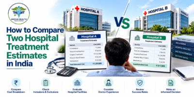
When you receive treatment estimates from two hospitals in India, do not compare only the final amounts. To…
- October 22, 2026
- shifamhealth
An itemized hospital quote in India should clearly explain the expected cost of treatment by separating major charges,…


