



Discitis: Causes, Symptoms, Diagnosis and Treatment (2026 Guide)
Filters & Insights
Discover the causes, symptoms, diagnosis, MRI findings, treatment options, antibiotics, surgery, and recovery for spinal disc infection.

Medically reviewed content · Published 2026 · Last reviewed September 2026
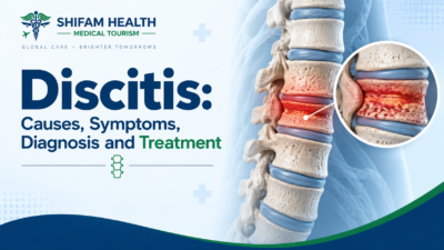
Discitis is inflammation or infection involving an intervertebral disc — the cushion-like structure between spinal vertebrae. In adults, discitis very frequently occurs together with infection of the adjacent vertebral bone (vertebral osteomyelitis), and the combined condition is often called spondylodiscitis, since the disc and adjacent bone share blood supply and pathology in this setting and are, in practice, usually part of the same disease process rather than separate problems.
This is not ordinary mechanical back pain, and it’s not the same thing as degenerative disc disease or a herniated disc, even though all three can cause significant back pain and can look confusingly similar on early imaging. Discitis is genuinely uncommon — it affects roughly 1 in 200,000 to 250,000 people — but it matters disproportionately to its rarity because delayed diagnosis can allow infection to progress to spinal instability, abscess formation, or neurological injury, some of which may not be fully reversible if treatment is delayed.
The reassuring side of this: when identified and treated promptly, outcomes are generally good, and many patients recover with antibiotic treatment alone, without needing surgery.
This guide explains how discitis differs from common mechanical back conditions, who’s at higher risk, how the diagnosis is actually established (it’s not simply “an abnormal MRI”), current antibiotic and surgical treatment approaches, and what evaluation and treatment in India can look like for international patients. This is general medical education, not a diagnosis, and does not replace urgent medical evaluation for suspected spinal infection.
Table of Contents
ToggleDiscitis, Spondylodiscitis, and Vertebral Osteomyelitis: One Spectrum, Not Three Diseases
These three terms describe overlapping, often simultaneous parts of the same underlying process rather than three genuinely separate conditions:
- Discitis refers to inflammation/infection centered on the intervertebral disc.
- Vertebral osteomyelitis refers to infection involving the vertebral bone itself.
- Spondylodiscitis is the term often used when both the disc and adjacent vertebral endplates/bodies are involved together — which, in adults, is the far more common pattern, since infection reaching the disc from the bloodstream typically seeds the richly vascularized region near the vertebral endplate first and spreads from there.
Different clinicians, radiologists, and research studies use these terms somewhat interchangeably, and you may see all three applied to what is, clinically, the same patient’s condition — this isn’t inconsistency so much as a reflection of genuinely overlapping pathology. Pediatric discitis can behave somewhat differently from the adult pattern, partly due to differences in disc blood supply at younger ages, and is discussed separately below.
Infectious vs. Noninfectious Discitis
The overwhelming clinical concern with a discitis diagnosis is infection, but it’s worth knowing that a small number of noninfectious inflammatory or degenerative conditions can produce imaging findings that superficially resemble infection — this is discussed further in the section on Modic changes below. When clinical features suggest infection is a real possibility, it needs to be actively investigated and excluded, not assumed away based on imaging alone — this asymmetry (treating suspected infection seriously until ruled out, rather than the reverse) is appropriate given how much delayed diagnosis can cost a patient.
Causes and Types of Infectious Discitis
Pyogenic (bacterial) discitis is the most common infectious type in most populations. Staphylococcus aureus is the single most frequently identified organism (accounting for roughly a third to two-thirds of cases in published series), though gram-negative organisms, streptococcal species, and others can also be responsible depending on the clinical setting — no single organism should be assumed universal, which is exactly why identifying the actual causative organism matters so much for treatment, discussed below.
Tuberculous spinal infection (spinal TB, historically called Pott disease) is a genuinely important and distinct category, particularly relevant for Shifam’s international patient base given how prevalent TB infection remains in India and several other countries patients travel from. Spinal TB tends to present more gradually than acute bacterial infection — often with weeks to months of symptoms (back pain, low-grade evening fever, weight loss, loss of appetite, night pain) rather than the more acute, dramatic presentation typical of pyogenic infection — and it requires a genuinely different, longer treatment course, covered in its own section below.
Fungal discitis is uncommon and generally occurs in specific higher-risk populations — significant immunosuppression, certain chronic diseases, intravenous drug use, or extensive prior healthcare exposure — and requires organism identification and antifungal treatment guided by infectious disease expertise. Brucellar infection and other less common organisms occur in specific epidemiological contexts and are considered when relevant exposure history is present, rather than screened for universally.
How does infection reach the spine?
Most adult vertebral osteomyelitis/discitis arises from hematogenous spread — bacteria traveling through the bloodstream from another site of infection or from transient bacteremia (bloodstream infection) and seeding the richly vascularized region near the vertebral endplate. Other routes include direct inoculation during a spinal procedure or surgery, and, less commonly, direct extension from an adjacent infected structure. In some patients, despite thorough investigation, the original source of the bacteremia is never definitively identified.
Risk factors
Diabetes, older age, immunosuppression (from disease or medication), cancer, chronic kidney disease, liver disease, intravenous drug use, recent bacteremia or another recent infection, recent spinal surgery or spinal injections/procedures, indwelling vascular catheters, malnutrition, and TB exposure or risk are all recognized risk factors. A person without any of these risk factors can still develop discitis — these describe increased likelihood, not a requirement for the diagnosis to be considered.
Children vs. Adults
Pediatric discitis differs from the adult pattern in several ways: the disc in young children retains some direct blood supply that regresses with age, which may partly explain why the disease pattern and typical age of presentation differ; children more often present with vaguer, less specific symptoms than adults — refusal to walk or bear weight, irritability, back stiffness, or a limp, sometimes without dramatic pain complaints a younger child can clearly articulate. This can lead to pediatric discitis being mistaken for a hip problem, abdominal pain, general back strain, or simply behavioral change, particularly in very young children who can’t describe their symptoms clearly.
Pediatric discitis should not be assumed to be automatically mild just because children overall tend to have a somewhat different and sometimes more indolent course than adults — persistent refusal to walk or bear weight in a child, especially with fever or elevated inflammatory markers, warrants prompt pediatric evaluation.
Symptoms
Symptoms can be variable and, in some patients, genuinely subtle — this is part of why diagnosis is sometimes delayed. Common features include persistent back or neck pain (often worse with movement), localized spinal tenderness, stiffness, reduced mobility, and muscle spasm. Systemic symptoms — fever, chills, malaise, fatigue, and, in more prolonged or TB-related cases, weight loss — can occur but are genuinely inconsistent. Fever may be absent, particularly in some adult patients, and its absence should not be used to reassure yourself or dismiss the possibility of infection if other features are concerning.
Neurological symptoms
Numbness, tingling, weakness, gait disturbance, balance problems, radiating (radicular) pain, or bowel or bladder dysfunction can indicate that the infection has caused an epidural abscess, direct spinal cord or nerve-root compression, spinal instability, or another serious complication requiring urgent assessment — these are not simply “worse back pain” and should be treated with the seriousness described in the next section.
Emergency Red Flags: When Is Discitis an Emergency?
Suspected spinal infection combined with any neurological deficit is an urgent medical situation, not something to monitor at home or address at a routine appointment. Seek immediate evaluation for: new or progressive limb weakness, difficulty walking, new loss of coordination, new bowel or bladder dysfunction, numbness in the saddle area, severe or rapidly worsening spinal pain, fever with severe back pain, chills or other signs of systemic illness (possible sepsis), altered mental status in someone who appears significantly unwell, known recent bacteremia with new severe spinal pain, recent spinal surgery or procedure with worsening pain or new fever, or significant immunosuppression combined with concerning spinal symptoms.
Do not wait for a routine specialist appointment if these features are present — neurological deterioration from an untreated spinal infection, particularly from an epidural abscess, can progress over hours to days and isn’t always reversible once it occurs.
Why Discitis Gets Misdiagnosed
Discitis symptoms genuinely overlap with far more common, benign conditions — mechanical back strain, degenerative disc disease, a herniated disc, sciatica, spinal stenosis, osteoporosis-related pain, and, less commonly, spinal tumors or inflammatory back disease can all cause similar pain patterns, and discitis may present without the dramatic fever or systemic illness that might otherwise prompt suspicion of infection early on.
| Feature | Discitis/Spondylodiscitis | Degenerative Disc Disease | Herniated Disc |
|---|---|---|---|
| Underlying Process | Infection/inflammation | Age-related structural change | Disc material displaced beyond its normal boundary |
| Fever | May occur, but often absent | Not expected | Not expected |
| Inflammatory Blood Markers (ESR/CRP) | Often elevated, though variable | Usually normal or not infection-pattern | Usually normal |
| MRI Appearance | Characteristic disc/endplate/marrow changes, often with enhancement | Dehydration, height loss, bulging without infection markers | Focal disc material displacement, may compress a nerve root |
| Typical Onset | Days to weeks (bacterial) or weeks to months (TB) | Gradual, chronic | Often sudden, sometimes after a specific movement or strain |
| Primary Treatment | Antimicrobial therapy | Activity, physiotherapy, symptom management | Often conservative; surgery for persistent nerve compression |
| Role of Surgery | Selected complicated cases (abscess, instability, neurological compromise, treatment failure) | Selected structural/neurological cases | Selected cases with persistent nerve compression |
The genuinely important takeaway isn’t that any single feature definitively separates these conditions — it’s that when clinical suspicion for infection exists (based on risk factors, symptom pattern, or inconclusive response to typical mechanical-pain treatment), it should be actively investigated with the specific tests described below, rather than assumed to be ordinary degenerative or mechanical back pain by default.
How Discitis Is Diagnosed
Diagnosis combines history (pain duration and pattern, fever, recent infections or procedures, recent surgery, known bacteremia, immunosuppression, TB exposure or risk, cancer history, weight loss, and any neurological symptoms) with physical examination (spinal tenderness, range of motion, and a full neurological exam — strength, sensation, reflexes, gait).
Blood tests and blood cultures
Inflammatory markers — ESR (erythrocyte sedimentation rate) and CRP (C-reactive protein) — are often elevated in discitis and are useful for supporting the diagnosis, establishing a baseline, and later tracking response to treatment. A normal ESR or CRP does not completely rule out discitis — these markers are supportive evidence, not a definitive test in either direction. A complete blood count may show other abnormalities.
Blood cultures matter enormously because identifying the actual causative organism directly determines antibiotic selection — treating “blindly” with broad-spectrum antibiotics, without ever knowing the specific organism, is a genuinely worse starting position than having a confirmed organism to target treatment against. Blood cultures should generally be obtained before starting antimicrobial therapy when it’s clinically safe to do so, since antibiotics can reduce the chance of a positive culture. This must not be read as “always delay treatment to get cultures first” — in a patient who is unstable, septic, or has significant neurological compromise, starting empiric antibiotics promptly takes priority over waiting for culture results, and the treating team will weigh this balance for each specific patient.
MRI: the central imaging test
MRI is the most important imaging test for suspected discitis. It can show disc-space abnormalities, vertebral endplate changes, bone marrow edema in the adjacent vertebrae, abnormal enhancement (when contrast is used), paraspinal soft-tissue involvement, and — critically — whether an epidural abscess or significant neural compression is present. MRI with contrast is often used when infection is genuinely suspected, though this isn’t an absolute universal requirement for every single patient in every clinical circumstance — the specific protocol depends on clinical judgment.
X-rays and CT
Plain X-rays are frequently normal in early discitis — significant bony changes typically take time to develop and appear on X-ray, so an early normal X-ray does not rule out the diagnosis. CT is useful for detailed assessment of bony destruction, structural planning, and guiding image-guided biopsy, but it is not interchangeable with MRI for evaluating the disc, soft tissue, and neural structures — the two provide different, complementary information.
Is a biopsy needed?
When blood cultures have already identified a likely causative organism and the clinical picture is consistent, biopsy may not be necessary. When blood cultures are negative or the picture remains unclear, CT-guided biopsy of the disc or adjacent bone can provide tissue for culture, histopathology, and, where relevant, molecular testing. Biopsy yield is genuinely imperfect — a meaningful proportion of biopsies don’t identify an organism even when infection is truly present, particularly if antibiotics were started before the biopsy was performed (which can reduce culture yield). A negative biopsy does not completely exclude infection — it’s one piece of evidence considered alongside the full clinical and imaging picture, not an automatic “ruled out” result. Testing (aerobic and anaerobic bacterial cultures, mycobacterial testing for TB, fungal cultures, and molecular diagnostics) is selected based on clinical context, epidemiology, immune status, and exposure history — not every test is run on every patient by default.
Modic changes: a genuine source of imaging confusion
Modic type 1 changes — a specific pattern of vertebral bone marrow signal change seen on MRI, associated in some cases with degenerative disc disease — can genuinely resemble the marrow edema pattern seen in early infection. Distinguishing the two relies on the full clinical picture (fever, inflammatory markers, symptom pattern and duration, distribution and evolution of the findings) and specialist radiological interpretation, not the MRI appearance in isolation. This is a real, documented diagnostic challenge, not a rare edge case — it’s one of the more important reasons discitis can be misdiagnosed as, or confused with, degenerative disease on imaging alone.
Treatment Overview
Treatment follows a general framework: confirm or strongly suspect infection, identify the causative organism where possible, determine the extent of disease (disc alone, adjacent bone, abscess, neural involvement), assess neurological status and spinal stability, provide targeted antimicrobial treatment, monitor the response, and proceed to surgery if specific indications are present. The treatment is not simply “antibiotics” as a single undifferentiated step — it’s this whole coordinated process, generally involving both a spine specialist and an infectious disease specialist together.
Antibiotic treatment
For bacterial discitis, antibiotics are the central treatment. Initial (“empiric”) therapy — started before the organism is confirmed, in patients who need treatment promptly — is later adjusted to targeted therapy once culture results identify the specific organism and its antibiotic susceptibility, which generally produces better-directed, often narrower treatment than continuing broad empiric coverage indefinitely. Duration is genuinely variable, not a fixed universal number — current evidence, including a landmark randomized trial, found 6 weeks of treatment was not inferior to 12 weeks for many patients with pyogenic vertebral osteomyelitis, and current practice generally uses a minimum of roughly 6 to 8 weeks of antimicrobial therapy, adjusted based on the organism, whether an abscess or significant bone involvement is present, neurological status, immune status, and how well the patient is responding — this decision is made by the treating team, generally with infectious disease input, not fixed in advance by a generic rule.
IV vs. oral antibiotics: Treatment often begins with intravenous (IV) antibiotics, particularly in the initial period, though current evidence has increasingly supported transitioning to appropriate oral antibiotics earlier than was traditionally practiced, when the specific antibiotic has good oral bioavailability, the organism’s susceptibility supports it, and the patient is clinically stable — a large trial specifically found no difference in outcomes for patients discharged on oral antibiotics compared to continued IV therapy in appropriate circumstances. Oral antibiotics are not inherently inferior when these conditions are met, but not every patient or every organism is a safe candidate for early oral transition — this is an individualized decision.
Tuberculous discitis: a genuinely separate treatment course
Spinal TB requires multidrug anti-tuberculosis therapy, following standard TB treatment protocols, generally continued for a considerably longer duration than typical bacterial discitis treatment — commonly cited durations run 6 to 9 months or longer depending on response and current guideline recommendations, genuinely different from the roughly 6-to-8-week course typical for pyogenic infection. Microbiological or pathological confirmation is pursued where feasible, drug resistance considerations are increasingly important given rising drug-resistant TB globally, and surgery may be needed for neurological compromise, significant deformity (historically associated with Pott disease specifically), or instability. TB-related spinal infection should never be treated using an ordinary bacterial discitis antibiotic regimen — the drugs, duration, and monitoring are genuinely different.
Fungal discitis
Fungal infection is uncommon and should not be assumed as a routine possibility for most patients — it’s specifically considered in higher-risk populations described above, requires organism identification, and is treated with antifungal therapy under infectious disease guidance, with surgery considered when needed for source control or structural reasons.
Supportive care: pain management, bracing, and rehabilitation
Appropriate pain medication supports mobilization and comfort but does not treat the underlying infection — this bears stating plainly, since pain improvement alone shouldn’t be mistaken for the infection resolving. Bracing may be used for pain control, temporary structural support, or postoperative support in selected patients, but a brace does not treat the infection either. Prolonged immobilization or extended bed rest is not the treatment and is generally avoided beyond what’s clinically necessary for pain or stability — gradual, guided mobilization and physiotherapy support functional recovery once the infection is being appropriately treated. Spinal manipulation is not an appropriate treatment for suspected or confirmed spinal infection and should be avoided in this setting.
When Is Surgery Needed?
Many appropriately treated infectious cases are managed successfully without surgery — this is genuinely the majority outcome for early, uncomplicated cases identified and treated promptly. Surgery may be needed for: a neurological deficit correlating with imaging findings, spinal cord compression, an epidural abscess with neurological compromise, significant spinal instability, progressive deformity, severe structural bone destruction, infection that persists or worsens despite appropriate antimicrobial therapy, situations needing surgical source control (draining an abscess that antibiotics alone won’t adequately address), or, in selected cases, diagnostic uncertainty where tissue sampling requires a surgical rather than percutaneous approach. Not every abscess and not every case of discitis automatically requires surgery — either or progressive neurological deficit specifically is the indication most consistently associated with urgent surgical need, per current guidelines.
What surgery involves
Depending on the specific clinical problem, surgery may include drainage or debridement of infected/abscessed tissue, decompression of compressed neural structures, and stabilization — sometimes with instrumented fixation (screws, rods) when instability or significant bone destruction is present. A common, outdated misconception is that spinal implants can never be used in the presence of active infection — current evidence and practice have moved past this blanket prohibition: instrumentation and stabilization can be appropriate even in an infected field when combined with adequate debridement and appropriate antimicrobial therapy, and this hasn’t been shown to impede successful treatment in current evidence. The specific procedure chosen depends on the location and extent of infection, neurological status, degree of instability and bone destruction, the causative organism, prior surgery, and the patient’s overall health.
Spinal epidural abscess: a specific high-priority concern
An epidural abscess — a collection of infected material in the space around the spinal cord or nerve roots — can occur alongside discitis/spondylodiscitis and represents one of the more urgent scenarios in this entire topic. Neurological deterioration from a growing or compressing epidural abscess can occur rapidly and, if allowed to progress, may not be fully reversible — this is precisely why the emergency red flags described earlier deserve to be taken seriously rather than treated as routine worsening back pain, and why urgent MRI and, where indicated, urgent surgical evaluation are pursued promptly when this is suspected.
Complications
Potential complications include epidural abscess, neurological deficits (from mild to severe), spinal cord or nerve-root compression, sepsis, vertebral bone destruction, spinal instability, progressive kyphotic deformity (particularly associated with untreated spinal TB), chronic pain, persistent or recurrent infection, and, in more severe or delayed cases, lasting functional impairment. This list shouldn’t be read as the expected outcome — early diagnosis and appropriate treatment substantially improve the chance of avoiding these more severe complications, which is the central, actionable message of this entire guide.
Monitoring Treatment Response and Follow-Up Imaging
Response to treatment is assessed through a combination of clinical improvement (pain, function, resolution of fever and systemic symptoms), neurological status, trends in CRP/ESR over time, and, where clinically relevant, follow-up imaging — not any single one of these in isolation. An important, commonly misunderstood point: MRI abnormalities frequently persist for a considerable time even after successful treatment — bone and disc changes don’t normalize on imaging as quickly as a patient clinically improves, and a still-abnormal-looking follow-up MRI in a patient who is otherwise improving clinically should not automatically be interpreted as treatment failure. Routine repeat MRI is not necessarily required for every patient who is clinically improving as expected — it becomes more relevant specifically when there’s a poor clinical response, worsening symptoms, new neurological findings, or concern for a new abscess or other complication, rather than as an automatic fixed-interval check for everyone.
Recovery and Prognosis
Recovery depends on the specific organism, extent of infection at diagnosis, presence and severity of any neurological involvement, degree of bone destruction, whether surgery was needed, overall health and immune status, and significantly — how promptly treatment began relative to symptom onset.
This guide won’t promise a fixed recovery timeline, since these factors genuinely vary the course from patient to patient; recovery can reasonably span weeks for straightforward, promptly-treated cases to many months, particularly for TB-related infection or cases involving significant structural damage requiring surgical reconstruction and staged rehabilitation.
Many patients recover well with appropriate, prompt treatment. Delayed diagnosis is associated with worse outcomes, and neurological deficits, if severe or present for a prolonged period before treatment begins, may not fully recover even with appropriate subsequent care — this is precisely why early recognition matters as much as it does throughout this guide, and it’s an honest limitation rather than a reason to withhold hope: many patients, including some with neurological symptoms at diagnosis, do recover substantially with timely, appropriate treatment.
Prevention
Realistic, evidence-supported measures include prompt treatment of bloodstream infections generally (reducing the chance of hematogenous spread to the spine), appropriate infection-control and sterile technique during spinal procedures and surgery, careful management of diabetes and other risk factors that increase infection susceptibility, appropriate postoperative monitoring after spinal surgery, and TB prevention and control measures in relevant settings and populations. Discitis cannot always be prevented — in a meaningful proportion of cases, no clear preventable cause or missed opportunity is identified, even in retrospect.
Discitis Treatment Cost in India
Reliable, independently verifiable cost figures specific to discitis treatment in India are genuinely difficult to establish, and this is worth stating plainly rather than manufacturing a range that would look more authoritative than the underlying data supports. Unlike a defined single procedure (a hip replacement or a specific spinal fusion), discitis treatment cost depends on an unusually wide range of variables that genuinely can’t be estimated in advance: the causative organism and whether it’s identified quickly or requires extended workup, whether treatment stays purely medical (antibiotics) or requires surgery, the duration of IV therapy and associated hospitalization, whether ICU-level care is needed, and — for TB-related disease specifically — a treatment course lasting many months rather than weeks.
What can be said responsibly about the components involved:
- Initial consultation, blood tests, and blood cultures
- MRI (and CT where used for planning or biopsy guidance)
- Image-guided biopsy and microbiology/pathology processing, where performed
- Infectious disease specialist consultation
- Hospitalization, including IV antibiotic administration (potentially requiring a PICC line or similar vascular access for extended IV therapy)
- Surgery, where clinically indicated (drainage, decompression, and/or stabilization)
- ICU care, if required
- Extended oral antibiotic or, for TB, extended multidrug therapy over months
- Rehabilitation and follow-up consultations and imaging
Given how much these components vary by the specific clinical course, a written, itemized estimate from the treating hospital, based on your actual diagnosis, suspected or confirmed organism, and treatment plan, is essential — far more so than for most other topics in this content series, since a general cost range here would be more likely to mislead than inform.
Discitis Treatment in India for International Patients
Appropriate discitis care requires genuine multidisciplinary coordination: a spine specialist (orthopedic spine surgeon or neurosurgeon), an infectious disease specialist, microbiology laboratory capability, radiology (MRI, CT, and image-guided biopsy), and, where surgery is needed, appropriate surgical and ICU capability. Not every hospital offers this full combination — verifying that a specific center has genuine infectious disease specialist involvement, not just spine surgery capability alone, matters particularly for this diagnosis, since antibiotic selection and duration decisions benefit substantially from dedicated infectious disease expertise working alongside the surgical team.
The international patient journey
- Collect and share previous medical records, imaging (MRI/CT/X-ray), and any available blood test or culture results
- Specialist (spine and, where appropriate, infectious disease) reviews the case remotely
- Clarify suspected organism or cause, and determine whether additional testing or biopsy is needed
- Assess neurological status and spinal stability from available information
- Develop a treatment plan and, where possible, a cost estimate, understanding these will likely need refinement after in-person evaluation
- Arrange medical visa documentation, if travel is appropriate for the clinical situation
- Travel to India for in-person evaluation and any needed additional investigations
- Begin or confirm antimicrobial treatment; proceed to surgery if clinically indicated
- Complete rehabilitation and receive medical documentation
- Coordinate ongoing follow-up, particularly important given how long TB or complicated bacterial treatment courses can run, with your home-country physician
A remote record review cannot replace an urgent in-person assessment when neurological compromise, signs of sepsis, or suspected spinal instability are present — for these situations, appropriate emergency evaluation wherever the patient currently is should always come before travel planning.
Questions to Ask Your Doctor
- Is this definitely discitis or spondylodiscitis, or could it be a mimic like degenerative disease?
- Is the suspected cause bacterial, tuberculous, fungal, or another cause?
- Is there vertebral osteomyelitis, and is there an epidural or paraspinal abscess?
- Is there spinal instability, and are my nerves or spinal cord affected?
- Have blood cultures been obtained, and is an image-guided biopsy needed?
- What organism is suspected or confirmed, and what antibiotic is being recommended and why?
- Will I need IV antibiotics, and for how long before transitioning to oral treatment, if appropriate?
- How will treatment response be monitored, and do I need repeat MRI?
- Under what circumstances would surgery become necessary in my case?
- What signs should prompt me to seek urgent reassessment during treatment?
- How will follow-up continue once I return home?
How Shifam Health Helps International Patients
Shifam Health is a medical tourism facilitator/coordinator, not a hospital, infectious disease physician, spine surgeon, or treating medical team — we don’t diagnose discitis, identify the causative organism, prescribe antibiotics, or perform surgery. What we do is help international patients navigate the practical side of accessing discitis evaluation and treatment in India: coordinating the sharing of medical records and imaging with appropriate spine and infectious disease specialists, helping identify hospitals with genuine multidisciplinary capability for this specific diagnosis, assisting with medical visa documentation, arranging airport pickup and accommodation, providing interpreter support where needed, and staying in touch for follow-up communication once you return home. Clinical decisions — diagnosis, organism identification, antibiotic selection, and any decision about surgery — remain entirely with your treating medical team.
If you or someone you’re caring for has a suspected or confirmed discitis diagnosis and you’re exploring evaluation or treatment options in India, share your available reports and imaging with our team on WhatsApp or through a quick inquiry form — there’s no obligation, and we typically respond within 24 hours. For any suspected acute infection with neurological symptoms, please seek emergency evaluation wherever you currently are before reaching out to us.
Frequently Asked Questions
What is discitis?
Discitis is inflammation or infection of an intervertebral disc, often occurring with infection of the adjacent vertebral bone, known as spondylodiscitis.
Is discitis an infection?
Usually, yes. Most clinically significant cases are bacterial, although tuberculosis and fungal infections can also cause discitis.
Is discitis serious?
Yes. Untreated infection can cause abscesses, spinal instability, neurological injury, or sepsis, although outcomes are often good with timely treatment.
Can discitis occur without fever?
Yes. Fever may be absent, particularly in adults, so its absence does not exclude infection.
What are the symptoms?
Persistent back or neck pain is common. Fever, weakness, numbness, or other neurological symptoms may occur.
What is spondylodiscitis?
Spondylodiscitis refers to infection involving both the intervertebral disc and adjacent vertebral bone.
Can tuberculosis cause discitis?
Yes. Spinal tuberculosis, or Pott disease, can cause gradual vertebral and disc infection and requires prolonged treatment.
Who is at risk?
Risk factors include diabetes, older age, immunosuppression, cancer, intravenous drug use, recent bloodstream infection, spinal procedures, and TB exposure.
How is discitis diagnosed?
Evaluation may include examination, blood tests, blood cultures, MRI, and sometimes image-guided biopsy.
Is a biopsy always necessary?
No. If blood cultures identify the causative organism and findings are consistent, biopsy may not be needed. It is considered when cultures are negative or diagnosis remains uncertain.
How long are antibiotics needed?
Bacterial discitis commonly requires around 6–8 weeks of treatment, although duration varies. Tuberculosis usually requires treatment for several months.
Can discitis cause paralysis?
Severe or delayed infection causing spinal cord or nerve compression can result in paralysis, making neurological symptoms urgent.
Conclusion
Discitis is not ordinary back pain, and it shouldn’t be approached or treated as such — but this doesn’t mean every case is a catastrophe either. Infectious discitis requires proper multidisciplinary evaluation combining spine and infectious disease expertise; early diagnosis meaningfully improves outcomes; treatment depends heavily on the specific cause and severity, with many patients recovering with antibiotics alone and others requiring biopsy, drainage, or spinal stabilization; and any neurological symptom deserves urgent, not routine, assessment.
If you have a suspected or confirmed discitis diagnosis, connecting with a genuine spine-and-infectious-disease team — not simply a general back-pain clinic — is the most important step, in India or anywhere else.
This article is for general medical education and does not replace urgent medical evaluation or individualized advice from a qualified spine surgeon and infectious disease specialist. It is not a diagnosis, treatment recommendation, or guarantee of any outcome. For suspected spinal infection with any neurological symptoms, fever, or signs of serious illness, seek prompt or emergency medical evaluation.



Popular Posts From Last Week
- September 10, 2026
- shifamhealth
Medically reviewed content · Published 2026 · Last reviewed August 2026 Blount’s disease, also called tibia vara, is…
- September 9, 2026
- shifamhealth
A cervical herniated disc occurs when the soft inner material of a disc in the neck pushes through…
- September 10, 2026
- shifamhealth
Chronic back pain is one of the most common reasons people seek medical care, and also one of…
- September 11, 2026
- shifamhealth
Myelopathy means dysfunction of the spinal cord itself — not a single disease, but a syndrome that can…
- September 9, 2026
- shifamhealth
Medically reviewed content · Published 2026 · Last reviewed September 2026 Disc degeneration seen on an MRI does…
- September 12, 2026
- shifamhealth
Medically reviewed content · Published 2026 · Last reviewed September 2026 Discitis is inflammation or infection involving an…
- September 12, 2026
- shifamhealth
A femoral hernia occurs when tissue often part of the bowel or fatty tissue — pushes through the…
- September 11, 2026
- shifamhealth
Kümmell’s disease refers to a delayed collapse of a vertebra — a nonunion or failure to heal properly…
- September 7, 2026
- shifamhealth
Ductal carcinoma in situ (DCIS) is a non-invasive breast condition in which abnormal cells are confined to the…
- September 8, 2026
- shifamhealth
Written by: Shifam Health Editorial Team — Neuro-Oncology Content Division Medically reviewed by: Shifam Health Clinical Advisory Panel…
- September 8, 2026
- shifamhealth
Medically reviewed content · Published 2026 · Last reviewed August 2026 Olfactory neuroblastoma also called esthesioneuroblastoma (ENB) —…
- September 7, 2026
- shifamhealth
Medically reviewed content · Published 2026 · Last reviewed August 2026 Oligodendroglioma is a diffuse glioma of the…