



Baastrup’s Disease (Kissing Spine) (2026): Symptoms, Causes, Diagnosis & Treatment
Filters & Insights
Learn about Baastrup’s Disease (Kissing Spine), including symptoms, causes, diagnosis, MRI findings, treatment options, and when surgery may be needed.

If a lumbar MRI report mentions “kissing spinous processes” or “Baastrup’s disease,” it’s worth pausing before assuming this explains your back pain or worse, before agreeing to a procedure based on that phrase alone. This is a real, describable finding with a real name, but it’s also one of the more commonly over-interpreted findings in spine imaging. This guide explains what’s actually happening anatomically, when it genuinely causes symptoms, and — just as importantly — when it’s an incidental finding that has little to do with why your back hurts.
What Is Baastrup’s Disease?
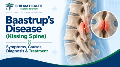
Baastrup’s disease, commonly called kissing spine syndrome, refers to abnormally close approximation or direct contact between adjacent spinous processes — the bony projections you can feel running down the middle of your back — most often in the lower lumbar spine, particularly between the L4 and L5 vertebrae. It develops from degenerative changes that narrow the space between these bones, sometimes causing irritation, inflammation, and a fluid-filled sac (bursa) to form between them.
First described by Danish radiologist Christian Baastrup in 1933, it can cause localized midline back pain that characteristically worsens with backward bending (extension) and eases with forward bending (flexion). Critically, this exact imaging appearance can also be found in people with no back pain at all — which is why a diagnosis requires matching the imaging to a specific clinical pattern, not simply noting the finding on a report.
Baastrup’s Disease ≠ Every Case of “Touching” Spinous Processes
This distinction matters more than almost anything else in this article. Spinous process contact visible on imaging is not automatically “Baastrup’s disease” in any clinically meaningful sense, and it is absolutely not automatically the explanation for a patient’s back pain. Studies of the general population have found spinous process contact or close approximation in a meaningful percentage of people who have no relevant back pain whatsoever — reported prevalence figures range from roughly 8% in some MRI-based population studies up to considerably higher rates in specific groups (one study of heavy-vehicle drivers found it in 13% of the tested group). The condition is more frequently identified in people over 70, with no clear predilection by sex.
Baastrup’s disease as a genuine clinical diagnosis specifically means: close spinous-process approximation plus a characteristic clinical pain pattern plus, ideally, supporting evidence (imaging changes consistent with active inflammation, and/or symptom relief with a diagnostic injection) that this specific finding — not something else — is actually generating the patient’s pain. Baastrup’s disease is also not synonymous with all chronic low-back pain — it’s one specific, uncommon-relative-to-its-imaging-frequency cause among many.
The Anatomy: What’s Actually Happening
To understand this condition, it helps to picture the relevant structures simply. Each vertebra has a spinous process — the bony bump you can feel along your spine — projecting backward from the vertebral arch. Between adjacent spinous processes sit the interspinous ligaments, and running along the tips of the spinous processes is the supraspinous ligament. Further forward, each vertebral level also has paired facet joints (which guide and limit spinal movement) and an intervertebral disc (the cushion between vertebral bodies), with the spinal canal and its neural contents running through the center of the whole structure.
With age-related spinal degeneration, several changes can narrow the interspinous space: loss of disc height brings adjacent vertebrae closer together vertically, facet joint degeneration can alter spinal segment mechanics, and increased lumbar lordosis (inward curve of the lower back) — though evidence on this specific link is genuinely mixed, with at least one MRI-based study finding no significant correlation, possibly because supine MRI shows less lordosis than an upright, weight-bearing posture would. No single mechanism explains every patient — this is typically the end result of several degenerative processes converging at one spinal level, most commonly L4-L5.
What Actually Happens Pathologically
The proposed sequence, based on current imaging and pathology literature, generally runs: adjacent spinous processes become unusually close → repetitive mechanical contact and stress occurs between them → the interspinous soft tissues become irritated → degenerative and sometimes inflammatory changes develop → reactive bone changes (sclerosis, flattening, enlargement) occur at the contact points → in some, though not all, patients, this becomes a source of pain.
On imaging, this can show as: narrowing or loss of the normal interspinous space, direct bone-on-bone contact, sclerosis (bone density increase at contact points), flattening and enlargement of the articulating spinous-process surfaces, cystic or degenerative changes, and — on MRI specifically — edema-like signal changes (fluid/inflammation signal, often seen with STIR sequences) indicating more active, inflammatory disease. A fluid-filled adventitious bursa (essentially a new, abnormal cushioning sac formed in response to repetitive friction) frequently develops at the contact point, sometimes referred to as interspinous bursitis. In rare cases, this bursa can extend into the epidural space, occasionally producing a cyst that contributes to spinal stenosis — a genuine but uncommon complication.
The essential caveat, worth repeating: these imaging abnormalities can be found in people without symptoms attributable to this specific finding. An abnormal-looking spinous process contact point on your MRI does not, by itself, prove it’s the source of your pain.
Causes and Risk Factors
| Factor | How It May Contribute | Strength of Evidence |
|---|---|---|
| Age-Related Degeneration | Disc height loss, facet changes narrow the interspinous space over time | Well-established; strongly age-associated (more common over 70) |
| Lumbar Lordosis/Spinal Alignment | Proposed to increase mechanical loading on the interspinous region | Mixed — at least one MRI study found no significant correlation |
| Degenerative Disc Disease | Reduced disc height brings adjacent vertebrae closer together | Commonly cited mechanism, biologically plausible |
| Facet Joint Degeneration | Coexisting degeneration can alter segmental spine mechanics | Frequently coexists; causal role less clearly isolated |
| Repetitive Mechanical Loading | Occupational/activity-related loading proposed as a contributor | Supported by occupational studies (e.g., professional drivers) showing elevated prevalence |
| Obesity/Body Habitus | Association reported in some studies | Limited, not established as a direct cause in every patient |
| Coexisting Spinal Disease | Spondylosis, spondylolisthesis, scoliosis can coexist and complicate the picture | Well-documented co-occurrence, complicating diagnosis |
Symptoms
The characteristic symptom pattern, when Baastrup’s disease is genuinely the pain generator, is fairly specific: localized, midline low-back pain that worsens with lumbar extension (bending backward, prolonged standing, walking downhill) and eases with flexion (bending forward, sitting). Pain is often reproducible with direct finger pressure over the affected spinous processes — a genuinely useful bedside clue. Some patients report associated stiffness.
Symptom presentation does vary between patients, and not every case follows this textbook pattern precisely — which is exactly why clinical correlation matters more than pattern-matching a single symptom description found online.
What Baastrup’s Disease Usually Does NOT Explain
This is worth stating directly and clearly: isolated Baastrup’s disease generally does not explain significant radicular (nerve-root) pain radiating down the leg in a dermatomal pattern, numbness following a specific nerve distribution, progressive muscle weakness, major neurological deficits, bowel or bladder dysfunction, or classic neurogenic claudication (leg pain/weakness brought on by walking and relieved by sitting). This does not mean Baastrup’s disease can never coexist with nerve compression or other spinal disease — it genuinely can, particularly when an associated bursa or cyst extends into the spinal canal — but when these more serious symptoms are present, clinicians need to actively investigate other or additional spinal and neurological causes rather than attributing everything to spinous-process contact.
Red Flags Requiring Urgent Evaluation
The following symptoms warrant prompt medical evaluation for a possible alternative or additional diagnosis, rather than routine follow-up:
- New bowel or bladder dysfunction
- Saddle anesthesia (numbness in the groin/inner thigh area)
- Progressive neurological weakness
- Severe or rapidly worsening pain
- Fever accompanying severe back pain
- Unexplained, significant weight loss
- A history of cancer with new, concerning back pain
- Major trauma preceding the pain
- Immunosuppression with concerning new symptoms
These signs point toward possible infection, fracture, malignancy, or significant neurological compromise — situations requiring urgent assessment for conditions other than, or in addition to, Baastrup’s disease.
Baastrup’s Disease vs. Other Causes of Back Pain
| Condition | Typical Pain Pattern | Key Clue | Neurological Symptoms |
|---|---|---|---|
| Baastrup’s Disease | Midline, worse with extension, eased with flexion | Tenderness directly over spinous processes | Typically absent unless a cyst/canal extension is present |
| Lumbar Facet Joint Pain | Often paramedian (off-center), worse with extension | Tenderness more lateral to midline | Typically absent |
| Discogenic Pain | Often worse with flexion/sitting | Pain with forward bending, prolonged sitting | Typically absent unless disc herniation compresses a nerve |
| Lumbar Spinal Stenosis | Leg pain/weakness with walking | Relief with sitting/forward bending (neurogenic claudication) | Often present |
| Lumbar Radiculopathy | Pain radiating down the leg in a nerve distribution | Dermatomal pattern, positive nerve-tension signs | Present |
| Sacroiliac Joint Pain | Lower, often one-sided, near the SI joint | Positive SI provocation tests | Typically absent |
| Vertebral Compression Fracture | Often sudden onset, worse with movement | History of trauma, osteoporosis, or minimal trauma in older patients | Depends on severity |
Symptoms alone often cannot reliably distinguish these conditions from each other — several frequently coexist in the same patient, particularly in older adults with widespread degenerative spinal changes, which is exactly why careful clinical correlation and, sometimes, diagnostic injection are used rather than relying on pattern-matching alone.
How Baastrup’s Disease Is Diagnosed
Diagnosis rests on a combination of medical history, physical examination, the specific symptom pattern described above, imaging, assessment for competing or coexisting diagnoses, and sometimes a diagnostic injection. There is no single imaging finding that should automatically be treated as proof that Baastrup’s disease is the sole or primary cause of a patient’s back pain — this bears repeating because it’s the crux of avoiding overdiagnosis.
Physical Examination
A clinician will typically assess for tenderness localized directly over the spinous processes (rather than more laterally, which would suggest facet involvement), pain reproduced or worsened with lumbar extension, range of motion, and — importantly — a full neurological examination (strength, sensation, reflexes, gait) to identify or rule out signs suggesting nerve-root irritation or another neurological process. Hip and sacroiliac joint examination may also be performed, since these are common mimics.
X-ray
Plain radiographs can show the characteristic “kissing” approximation of spinous processes, associated sclerosis, and overall spinal alignment, and can screen for other bony abnormalities like spondylolisthesis. X-rays are not definitive for diagnosing symptomatic Baastrup’s disease — they show the anatomical finding but can’t confirm it’s an active source of pain.
MRI
MRI provides the most detailed evaluation, assessing the interspinous soft tissues, bone marrow signal changes (edema-like signal on STIR/T2 sequences suggesting more active, inflammatory disease rather than old, quiescent degenerative change), interspinous bursitis where present, and — critically — the disc, facet joints, spinal canal, and nerve roots, which helps identify or exclude competing causes of pain at the same visit. It is particularly useful when the clinical picture is unclear or when multiple potential pain generators need to be evaluated together. An abnormal MRI finding does not necessarily mean that finding is the actual pain generator — this is the single most important interpretive principle in this entire diagnostic process.
CT
CT offers detailed bony anatomy and can be useful for surgical planning in selected cases, but is generally less useful than MRI for evaluating the soft tissues and neural structures that often matter most for distinguishing competing diagnoses.
Diagnostic Injections
Image-guided (fluoroscopic or ultrasound-guided) injection of local anesthetic, sometimes combined with corticosteroid, into the interspinous region can serve both a diagnostic and, potentially, short-term therapeutic purpose. If a patient’s pain is substantially and reproducibly relieved by an injection targeted precisely at the suspected interspinous level, this supports (though doesn’t absolutely prove) that this specific site is genuinely the pain source. Published data from one series of 55 patients receiving interspinous corticosteroid/anesthetic injections reported meaningful pain reduction and improved mobility, with roughly 22% requiring a repeat injection within 7-10 days. A positive response to injection should be interpreted alongside the full clinical picture, not treated as automatic proof of a permanent-cure pathway — temporary relief from a diagnostic block confirms a pain generator location; it doesn’t guarantee that treating that location will produce lasting relief.
Imaging Finding ≠ Diagnosis ≠ Pain Generator
This is worth its own section because it’s the single most protective concept in this entire article. A patient can simultaneously have spinous-process contact, general degenerative disc changes, some facet arthritis, and mild spinal stenosis — all visible on the same MRI, all common with age, and none of them individually proven to be causing that specific patient’s pain just because they appear on the report. Clinicians need to determine, through the combination of history, exam, and sometimes diagnostic injection, which finding — if any — is actually clinically relevant to the symptoms being experienced.
Finding something on imaging is not the same as diagnosing the cause of pain, and diagnosing a finding is not the same as identifying the true pain generator. If a treatment recommendation — especially an invasive one — is based solely on an MRI report showing spinous process contact, without this deeper clinical correlation, that’s worth questioning directly with your treating physician or seeking a second opinion.
Treatment: An Honest, Ranked Overview
Treatment options here genuinely differ enormously in evidence quality, invasiveness, and appropriate use — they should not be presented as equally reasonable choices to pick between.
First-Line: Conservative Management
Most patients are appropriately managed conservatively first, and this is where the evidence is most consistent, if still not extensive for this specific condition.
Activity modification — reducing or modifying activities that reliably provoke symptoms (particularly sustained extension postures) while maintaining overall movement and activity, rather than complete rest.
Physical therapy — including core and lumbar stabilization exercise, hip and trunk conditioning, flexibility work, and movement/postural retraining, individualized to the patient. It’s worth being honest here: published literature on physiotherapy specifically for Baastrup’s disease is mixed — some case reports and small studies show good outcomes, while broader reviews note physiotherapy is “not much effective” per some published assessments, with satisfactory outcomes reported in only some studies. Physical therapy will not “cure” every case, but it remains a reasonable, low-risk first approach, and case reports do describe patients achieving significant improvement with physiotherapy alone over several months.
Medication — NSAIDs or acetaminophen/paracetamol are commonly used for symptom control; this article does not prescribe specific medications or doses, and any medication choice should account for your individual health history and other medications under your clinician’s guidance. No medication is established as disease-modifying for Baastrup’s disease specifically — pain medications control symptoms; they don’t reverse the underlying anatomical change.
Injections
Interspinous corticosteroid and local-anesthetic injections are commonly used both diagnostically and for potential short-term therapeutic relief, as described above. Evidence supports meaningful short-term pain reduction in a proportion of patients, though a notable minority need repeat injection relatively soon afterward, and durability of benefit beyond the short term is less well established in the literature. Repeated injections without reassessment of the underlying diagnosis are generally not appropriate — if injections aren’t providing meaningful, durable relief, that’s a signal to reconsider whether the diagnosis or treatment target is correct, not simply to repeat the same injection.
Radiofrequency Ablation — Evidence Is Limited for This Specific Indication
Radiofrequency ablation is well-established for facet-joint-mediated pain, but evidence specifically supporting its use for Baastrup’s disease is limited. It should not be assumed that evidence from facet-joint radiofrequency procedures automatically transfers to this different anatomical target. If this procedure is proposed for you, it’s reasonable to ask directly what evidence specifically supports it for interspinous/Baastrup’s disease, rather than facet pain generally.
Regenerative Medicine (PRP, Stem-Cell Injections) — An Honest Assessment
Platelet-rich plasma (PRP), stem-cell therapy, and other biologic injections are sometimes marketed for spinal degenerative conditions including Baastrup’s disease. Being direct about this: there is not strong, specific evidence supporting these treatments for Baastrup’s disease. Available studies in this specific area are generally small, treatments are not standardized between providers, and these approaches are not established or regulatory-approved specifically for this indication in most jurisdictions. This doesn’t mean regenerative medicine has no role in any spinal condition — but for Baastrup’s disease specifically, it should be understood as experimental and insufficiently supported by current evidence, not as an advanced, proven alternative to established care. Be appropriately skeptical of any clinic marketing these treatments for this specific condition without disclosing the limited evidence base.
Surgery — Reserved for Highly Selected Patients
Surgery is generally considered only after appropriate conservative treatment has failed, when symptoms are persistent and clearly focal, and — critically — when there’s genuine clinical evidence (not just an imaging finding) that the interspinous region is actually responsible for the pain, with competing pain generators reasonably excluded or addressed.
Procedures used include excision of the interspinous bursa, partial or complete resection of the affected spinous process(es), and, in some published case series, minimally invasive or endoscopic approaches (one 2021 study described an endoscopic “interspinous plasty” technique). In cases with coexisting sagittal spinal imbalance, more extensive procedures like spinal fusion may occasionally be considered as part of broader spinal realignment surgery — a different and more significant undertaking than isolated Baastrup’s-disease-directed surgery.
On the evidence: surgical interventions for Baastrup’s disease specifically have limited published evidence of effectiveness compared with well-studied spine procedures — small case series rather than large randomized trials. Surgery carries genuine risks including recurrence or persistent pain, and removing a spinous process does compromise the integrity of supporting ligament and muscle attachments at that level, which is a real trade-off to understand before proceeding.
Surgery Should Not Be the Automatic Answer
To state this as plainly as possible: a patient with chronic back pain, an MRI showing “kissing” spinous processes, and mild degenerative changes does not automatically need surgery. This combination is common in the general population and frequently doesn’t require any procedure at all, let alone an operation. Treatment decisions should never be based solely on an imaging report without proper clinical correlation — confirming the symptomatic level, excluding competing diagnoses, and generally exhausting appropriate conservative care first.
Recovery and Prognosis
Realistically: outcomes vary considerably between patients. Some improve substantially with conservative treatment, sometimes over a period of months; others have more persistent symptoms. Outcomes depend heavily on whether Baastrup’s disease is genuinely the dominant pain generator (versus one of several coexisting findings), the presence of other degenerative spinal disease, age, overall fitness, occupational demands, and individual healing response. There is no universal recovery timeline — claims like “patients recover completely in X weeks” don’t hold up against the genuine variability in this condition, and should be treated skeptically wherever you encounter them.
Can Baastrup’s Disease Be Prevented?
Maintaining regular physical activity, healthy body weight, core and trunk conditioning, and reasonable lifting mechanics are generally sensible for spinal health broadly, and may plausibly reduce some contributing mechanical stress. However, there is no guaranteed way to prevent age-related spinal degeneration or Baastrup’s disease specifically — this is fundamentally a degenerative process linked strongly to aging, and lifestyle measures should be understood as generally supportive of spine health rather than a specific, proven prevention strategy for this particular finding.
When Should You See a Spine Specialist?
Specialist assessment is reasonable for persistent focal midline back pain, symptoms that meaningfully interfere with daily activities, failure of an adequate trial of conservative care, any concerning neurological symptoms (see red flags above), an unclear diagnosis despite initial evaluation, significant imaging abnormalities requiring interpretation, recurrent symptoms despite prior treatment, or if injections or surgery are genuinely being considered.
Questions to Ask Your Doctor
- Is Baastrup’s disease definitely causing my symptoms, or is this an incidental imaging finding?
- Could another condition — facet, disc, or nerve-related — be responsible instead or as well?
- Does my MRI show active, inflammatory changes, or an old, likely-incidental finding?
- Would physical therapy be an appropriate first step for me?
- What specific movements or activities should I modify in the meantime?
- Would a diagnostic injection help confirm the actual pain source before committing to further treatment?
- What are the realistic risks of that injection?
- How long should conservative treatment reasonably be tried before reconsidering the plan?
- Is surgery genuinely necessary, or being proposed based mainly on the imaging report?
- What specific evidence supports the exact procedure being proposed for my case?
- What happens if this treatment doesn’t work?
- What symptoms should prompt me to seek urgent reassessment?
Baastrup’s Disease Treatment in India
International patients researching evaluation in India will find relevant specialists across orthopedic spine surgery, neurosurgery, spine-focused rehabilitation medicine, musculoskeletal radiology, pain medicine, and physiotherapy at major hospitals. A reasonable evaluation pathway generally moves from spine specialist consultation and imaging review, through confirmation of whether Baastrup’s disease is genuinely clinically relevant to your symptoms (as opposed to an incidental finding), assessment of alternative or coexisting diagnoses, and — only where appropriate — progression through conservative care, diagnostic/therapeutic injection, and, rarely, surgery.
Cost — What We Can and Cannot Tell You
Costs vary significantly by hospital, city, specialist, the specific procedures and investigations needed, and individual patient complexity, so a fixed price cannot be responsibly quoted without clinical evaluation. We’re not going to invent a number here, and any source offering you a single confident “Baastrup’s disease treatment cost” without having reviewed your case should be treated with real skepticism — this is exactly the kind of condition where cost depends entirely on whether you end up needing a consultation and physiotherapy, or imaging plus an injection, or the far less common surgical pathway.
What we can outline is the general components involved, each priced individually rather than as one package: initial spine specialist consultation, X-ray and/or MRI imaging, physiotherapy sessions (typically a course rather than a single visit), image-guided diagnostic/therapeutic injection if pursued, and, only in the uncommon surgical scenario, hospital charges, anesthesia, surgical fees, and post-operative rehabilitation. Not every patient requires every item on this list — many patients are managed entirely through the first two or three components. Get an itemized estimate specific to your recommended treatment plan after clinical evaluation, not before.
International Patient Journey
- Share existing medical records, including any prior imaging reports
- Have MRI/X-ray images (not just reports) reviewed by a specialist, where possible
- Obtain a specialist opinion on whether Baastrup’s disease is clinically relevant to your specific symptoms
- Assessment of alternative or coexisting diagnoses
- Development of a treatment plan, generally conservative-first
- An estimated cost specific to that plan, once clinically appropriate
- Medical travel documentation coordination, if treatment abroad is genuinely pursued
- Travel and accommodation coordination
- Consultation and treatment, only where clinically appropriate — not simply because travel has already been arranged
- Rehabilitation and follow-up
- Documentation for continued care with your home physician
It’s worth being direct here: travelling internationally is not necessary for a condition that can often be evaluated and managed conservatively through local care. If your situation does warrant more specialized evaluation, we can help coordinate that — but we won’t suggest international travel is required when it may not be.
How Shifam Health Can Help
Shifam Health is a medical tourism facilitator — not a hospital, doctor, or clinic — and we don’t diagnose or treat Baastrup’s disease ourselves. What we can do is help you share your existing medical records and imaging with appropriate spine specialists, coordinate appointments, help build a transparent, itemized cost estimate once a treatment plan is actually established, assist with medical visa documentation where relevant, and coordinate travel, accommodation, and follow-up communication.
We won’t tell you India offers a “guaranteed cure” or that treatment will “always be affordable” — what we can offer is honest coordination and connection to specialists who can give your specific case the clinical correlation it actually needs, rather than a treatment plan built from an MRI report alone.
Reach out on WhatsApp or submit a quick inquiry — our team responds within 24 hours, with no obligation to proceed.
Frequently Asked Questions
What is Baastrup’s disease?
Baastrup’s disease is a condition where adjacent spinal spinous processes come unusually close or touch, sometimes causing localized back pain.
Why is it called kissing spine syndrome?
The term describes adjacent spinous processes appearing to “kiss” or touch on imaging.
What causes Baastrup’s disease?
Age-related spinal degeneration, disc-height loss, facet changes, spinal alignment, and repetitive mechanical stress may contribute.
What are the symptoms?
Typical symptoms include midline lower-back pain and tenderness, often worse with backward bending and relieved by forward bending.
Can Baastrup’s disease cause sciatica?
Usually not. Radiating leg pain or sciatica may indicate another or additional spinal condition.
Is Baastrup’s disease visible on X-ray?
Yes. X-rays can show close approximation of the spinous processes and related bony changes, but imaging alone cannot confirm that they cause pain.
Is MRI better than X-ray?
MRI provides greater detail of soft tissues and inflammation and can identify other possible causes of back pain.
Can kissing spine be an incidental finding?
Yes. Spinous-process contact can occur in people without related symptoms, so clinical findings must match imaging.
Can it improve without surgery?
Yes. Many patients improve with activity modification, physical therapy, and other conservative treatments.
Are PRP or stem-cell treatments proven?
No. Evidence specifically supporting PRP or stem-cell injections for Baastrup’s disease remains limited.
Is surgery necessary?
Rarely. Surgery is generally considered only for carefully selected patients with persistent, clearly localized pain despite appropriate non-surgical treatment.
Can Baastrup’s disease recur?
Yes. Symptoms may persist or return as underlying spinal degeneration progresses.


Popular Posts From Last Week
- September 10, 2026
- shifamhealth
Medically reviewed content · Published 2026 · Last reviewed August 2026 Blount’s disease, also called tibia vara, is…
- September 9, 2026
- shifamhealth
A cervical herniated disc occurs when the soft inner material of a disc in the neck pushes through…
- September 13, 2026
- shifamhealth
If a lumbar MRI report mentions “kissing spinous processes” or “Baastrup’s disease,” it’s worth pausing before assuming this…
- September 10, 2026
- shifamhealth
Chronic back pain is one of the most common reasons people seek medical care, and also one of…
- September 11, 2026
- shifamhealth
Myelopathy means dysfunction of the spinal cord itself — not a single disease, but a syndrome that can…
- September 9, 2026
- shifamhealth
Medically reviewed content · Published 2026 · Last reviewed September 2026 Disc degeneration seen on an MRI does…
- September 12, 2026
- shifamhealth
Medically reviewed content · Published 2026 · Last reviewed September 2026 Discitis is inflammation or infection involving an…
- September 12, 2026
- shifamhealth
A femoral hernia occurs when tissue often part of the bowel or fatty tissue — pushes through the…
- September 11, 2026
- shifamhealth
Kümmell’s disease refers to a delayed collapse of a vertebra — a nonunion or failure to heal properly…
- September 7, 2026
- shifamhealth
Ductal carcinoma in situ (DCIS) is a non-invasive breast condition in which abnormal cells are confined to the…
- September 8, 2026
- shifamhealth
Written by: Shifam Health Editorial Team — Neuro-Oncology Content Division Medically reviewed by: Shifam Health Clinical Advisory Panel…
- September 8, 2026
- shifamhealth
Medically reviewed content · Published 2026 · Last reviewed August 2026 Olfactory neuroblastoma also called esthesioneuroblastoma (ENB) —…