




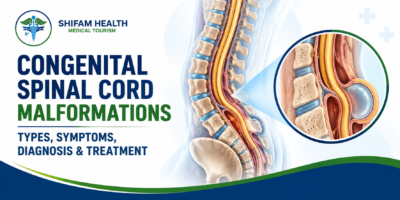
Congenital Spinal Cord Malformations (2026): Types, Symptoms, Diagnosis & Treatment
Filters & Insights
Written By :
Medically Reviewed By :

Shifam Health Medical Team
Shifam Health - Medical Reviews Department
10+ Years Of Experience
Learn about congenital spinal cord malformations, including their types, symptoms, diagnosis, and treatment options for children and adults.
📅
Last updated: October 13, 2026

What are congenital spinal cord malformations? Congenital spinal cord malformations are developmental conditions, present from birth, that affect the spinal cord, the bones of the spine, the coverings around the cord, or nearby tissues. They include spina bifida, tethered cord, split cord malformation and other forms of spinal dysraphism. Symptoms and severity range widely, from none to major neurological impairment.
Do congenital spinal cord malformations always require surgery? No. Some, such as an open myelomeningocele, are usually repaired surgically, but others, including some closed malformations found without symptoms, are monitored. Surgery is considered when there is an open defect, progressive neurological change or a symptomatic malformation, and the decision depends on the specific diagnosis and a specialist’s assessment.
- “Congenital spinal cord malformation” is an umbrella term for several different conditions. Getting the exact diagnosis matters because treatment differs a lot.
- Open defects such as myelomeningocele are visible at birth. Closed (skin-covered) ones, such as lipomas, tethered cord or split cord malformation, may be found later, sometimes because of a skin marker or new symptoms.
- Warning signs include leg weakness, changes in walking, new bladder or bowel problems, foot or spine deformity and new pain.
- MRI is the main test. Not every malformation needs surgery, and surgery cannot reverse damage that has already happened.
- Rapidly worsening weakness, sudden loss of function or signs of serious infection need urgent care.
Finding out that your baby or child has a problem with the spine or spinal cord is frightening, and the terminology is confusing: spina bifida, dysraphism, tethered cord, split cord, lipoma, dermal sinus. This guide explains what each of these is, how doctors find them, what causes them, and how treatment decisions are made, in plain language. It is general education and cannot diagnose you or your child. Because several of these conditions overlap and share names, an accurate specialist diagnosis is the essential first step.
Table of Contents
ToggleWhat Are Congenital Spinal Cord Malformations?
“Congenital” means present at birth, formed during development in the womb. The spine is made of bones (vertebrae) that form a canal protecting the spinal cord, a bundle of nerves that carries messages between the brain and the body. The cord is surrounded by protective coverings (the meninges) and bathed in cerebrospinal fluid (CSF).
Early in pregnancy a flat strip of tissue folds into a tube (the neural tube), which becomes the brain and spinal cord, and the surrounding bone and skin close over it. If any step goes differently, the result can be a malformation. The neural tube normally closes in about the third to fourth week after conception, which is often before a pregnancy is recognised.
It helps to separate two ideas:
- A defect in the spinal column (bones that do not fully form or fuse) does not always involve the cord. Some, such as spina bifida occulta, are very common and harmless.
- An abnormality of the spinal cord itself (for example, a cord that is split, tethered or covered with fatty tissue) can affect nerve function.
These conditions can affect movement, sensation, bladder and bowel control and, indirectly, growth of the spine and limbs. Some are found before birth, some in infancy or childhood, and some only in adolescence or adulthood.
What Causes Congenital Spinal Cord Malformations?
Most arise from differences in how the neural tube and surrounding structures form in early pregnancy. In many cases no single cause is identified, and the causes differ between conditions.
- Neural tube development: failure of the neural tube to close fully leads to open defects such as myelomeningocele.
- Formation or separation of spinal structures: differences in how the cord and spine separate or form lead to conditions such as split cord malformation, dermal sinus or lipomas.
- Genetic and environmental influences in some conditions, and multifactorial causes in many, meaning several small factors acting together.
- Associated abnormalities in other organs or the brain in some children.
Folic acid and prevention: what the evidence supports
Low folate (vitamin B9) before and early in pregnancy is linked to neural tube defects such as spina bifida. Public health bodies recommend that all women who could become pregnant take 400 micrograms of folic acid daily, and trials reviewed in the medical literature found that supplementation reduced the rate of open neural tube defects by about 70%. Women who have had a pregnancy affected by a neural tube defect are advised to discuss a higher dose with their doctor before trying to conceive.
The limits matter. Folic acid reduces the risk of neural tube defects, but it does not prevent all of them, and it has not been shown to prevent every type of closed spinal malformation. Parents are not responsible for these conditions, and many affected pregnancies occur despite good care.
Types of Congenital Spinal Cord Malformations
The classification is broad, and the conditions are not interchangeable. Doctors often divide spinal dysraphism (a failure of the midline structures of the back to form properly) into open forms, where neural tissue is exposed, and closed forms, where the skin covers the abnormality. Terminology varies slightly between sources.
Major congenital spinal malformations
| Condition | Defining feature | Open or closed | Possible features |
|---|---|---|---|
| Myelomeningocele | Spinal cord and nerves protrude through a spinal opening, often in a sac | Open | Leg weakness, bladder and bowel problems, hydrocephalus, Chiari II |
| Meningocele | Sac of CSF and coverings protrudes; the cord is largely in place | Usually skin-covered | Often fewer neurological problems; varies |
| Spina bifida occulta and closed dysraphism | Skin-covered abnormality; may involve a lipoma, tight filum or other tethering lesion | Closed | May be silent; may come with a skin marker |
| Tethered cord syndrome | Cord is abnormally fixed and stretched with growth | Closed (or after open repair) | Pain, weakness, gait or bladder changes |
| Split cord malformation (diastematomyelia) | Cord divided by a bony or fibrous spur or septum | Closed | Spine deformity, pain, neurological change |
| Lipomyelomeningocele | Fatty tissue attached to the cord, often with tethering | Closed | Skin lump or marker, bladder and leg problems |
| Congenital dermal sinus | Skin tract that may run toward the spinal canal | Closed | Infection risk, deeper abnormalities |
Spina bifida and spinal dysraphism
“Spina bifida” literally means a split spine. The term covers several forms, from the common and usually harmless spina bifida occulta to the severe myelomeningocele. “Spinal dysraphism” is the wider umbrella for midline back malformations, open or closed.
Myelomeningocele
In myelomeningocele the cord and nerves, and their coverings, push out through an opening in the spine. It is the most serious form of spina bifida. Nerve function below the level of the opening is affected, so many children have leg weakness, and bladder and bowel problems. It is often accompanied by hydrocephalus (fluid build-up in the brain) and by Chiari II malformation. It needs coordinated care from neurosurgery, pediatrics, urology, orthopedics and rehabilitation.
Meningocele
In a meningocele, the sac contains CSF and coverings but the spinal cord is largely in its normal position, so neurological effects tend to be fewer, although they vary with the anatomy and any associated abnormalities.
Occulta and closed spinal dysraphism
Closed malformations are covered by skin and can be subtle. Spina bifida occulta is often found by chance and causes no problems. Other closed forms, such as lipomas or a tight filum terminale (a thickened thread at the cord’s end), can tether the cord. An outwardly minor finding does not tell you whether there is an underlying neurological problem, and many harmless findings look similar.
Tethered cord syndrome
The spinal cord normally moves freely as a child grows. In tethered cord syndrome, the cord is anchored, for example by a lipoma, thick filum or scar tissue, and gets stretched as the spine grows. This can cause pain, weakness, gait change, foot deformity and bladder or bowel problems that progress with growth. A tethered cord on an MRI does not always cause symptoms, so diagnosis rests on symptoms, examination and imaging together.
Split cord malformation (diastematomyelia)
Here a bony or fibrous spur or septum divides the cord into two parts. It can be found with spine deformity, a skin marker, pain, leg problems or neurological changes, and it often occurs with tethering.
Lipomyelomeningocele and related lipomas
In these conditions fatty tissue is attached to the cord and often tethers it. A soft lump or fatty swelling over the lower back is a common sign. Practice varies on whether surgery should be offered early to infants who have no symptoms.
Congenital dermal sinus
A dermal sinus is a small skin tract, often on the midline of the back, that can extend toward the spinal canal. It can allow bacteria to track in and cause infection (including meningitis or an abscess), and it may be linked with deeper abnormalities.
Other rare abnormalities
Other rare spinal cord malformations exist; they are beyond the scope of this overview and need specialist diagnosis.
Symptoms of Congenital Spinal Cord Malformations
Symptoms depend on the condition, its level in the spine and its severity. Some people have few symptoms, while others have substantial neurological impairment. None of these signs on its own proves a malformation.
Possible symptoms and when to seek assessment
| Sign or symptom | Possible meaning | When to seek assessment |
|---|---|---|
| Weak legs, differences in muscle tone, delayed motor milestones | Nerve involvement | Raise with a pediatrician; sooner if progressive |
| Change in walking, tripping, new limp | Possible tethering or cord involvement | Promptly |
| Altered sensation or numbness | Nerve involvement | Promptly if new or progressive |
| Back or leg pain, especially in older children and adults | May accompany tethering | Promptly if persistent or with neurological change |
| Foot deformity or progressive orthopedic change; scoliosis | May reflect a nerve problem | Specialist assessment |
| Bladder problems or repeated urinary infections; bowel problems | Nerve involvement of the bladder or bowel | Specialist assessment; urgent if new loss of control |
| A skin marker over the spine (dimple, tuft of hair, lump, discoloured patch) | Many are harmless; some signal an underlying malformation | Mention at routine checks; specialist review if unusual or with other signs |
Signs in infants are harder to read, so developmental delay and unusual skin findings are important clues. Many children with a simple sacral dimple have nothing wrong, and no single skin finding proves a malformation.
Warning Signs That Need Prompt Medical Assessment
Seek prompt assessment, and emergency care if rapid, for:
- New or progressive leg weakness
- Worsening walking ability
- New loss of bladder or bowel control
- Progressive sensory change
- Increasing back or leg pain with neurological changes
- Signs of infection near a spinal opening or skin tract: fever, redness, swelling, discharge, irritability, stiff neck, drowsiness or vomiting
- New neurological deterioration in a child with a known spinal condition
Rapidly progressing weakness, sudden loss of function or serious infection symptoms need emergency assessment. Do not wait for a routine follow-up if symptoms are getting worse quickly.
How Are Congenital Spinal Cord Malformations Diagnosed?
Diagnosis combines the history, an examination of the back and nervous system, and imaging. No single investigation suits everyone, and results must be read alongside clinical findings.
Diagnostic tests and their purpose
| Test | Purpose | Limitations or considerations |
|---|---|---|
| Prenatal ultrasound (and, in selected cases, fetal MRI) | Can show some open defects before birth and prompt specialist counselling | Cannot detect every malformation, and findings need confirming after birth |
| Clinical examination | Movement, sensation, tone, reflexes, spine and skin, feet, bladder and bowel symptoms | A normal examination does not exclude a closed malformation |
| MRI of the spine | Shows the cord, any tethering, lipoma, split cord and related findings | Young children may need sedation; findings must be matched with symptoms |
| Spinal ultrasound in young infants | A quick, no-radiation screen, useful when the back bones are not yet fully formed | Only works in the first months of life; not a substitute for MRI if the result is unclear |
| Urological assessment | Bladder function and kidney protection | Done when bladder symptoms or risk are present |
| Orthopedic assessment and imaging | Scoliosis, hip and foot problems | Done when deformity is present |
Prenatal ultrasound
Some spinal abnormalities, especially open ones, can be seen on a routine pregnancy scan, and a blood test (maternal alpha-fetoprotein) may be raised when a neural tube defect is open. A finding leads to a specialist scan and counselling, and sometimes to a fetal MRI. Not all conditions can be detected before birth, and closed malformations are commonly missed.
Clinical examination after birth
The doctor checks how the baby moves the legs, muscle tone, sensation and reflexes, looks at the back for openings, lumps or skin markers (a dimple, a tuft of hair, a fatty lump or a discoloured patch), checks the feet and spine, and asks about development and about bladder and bowel function.
MRI of the spine
MRI is the key test. It shows the cord’s position and shape, whether it is tethered, a lipoma, a split cord with its spur, a dermal sinus tract and related findings. It must be read together with the symptoms: a tethered-looking cord on MRI does not always cause problems.
Ultrasound in selected infants
In very young infants, ultrasound can look through the back of the spine before the bones fully form, so it is sometimes used as a first screen for a low-lying cord when a skin marker is found. Its usefulness depends on age and circumstances, and doctors move to MRI if it is unclear.
Additional investigations
Urological, orthopedic, brain imaging (for hydrocephalus or Chiari II) and other tests are used as needed.
Congenital Spinal Malformations in Babies and Children
The pathway depends on the diagnosis, but common elements include:
- Prenatal detection and counselling when a defect is found on a scan, including discussion of options such as fetal surgery at specialised centres.
- Early assessment by a pediatric neurosurgeon and pediatrician after birth.
- Developmental monitoring and early therapy.
- Mobility and orthopedic needs, such as braces or foot care.
- Bladder and bowel care with urology input.
- Coordination among specialties, with education for the family and long-term follow-up.
Fetal surgery for myelomeningocele
In the Management of Myelomeningocele Study (MOMS), repair of the defect before birth, at around 19 to 26 weeks, reduced the need for a shunt and improved the chance of walking independently compared with repair after birth, but it carried higher risks for the mother and baby, including premature birth. It is offered at specialised centres, and eligibility is strict. A guideline on tethered cord after prenatal closure notes a higher rate of tethered cord, so continued surveillance is recommended.
Treatment Options for Congenital Spinal Cord Malformations
Management may include observation, surgery, rehabilitation or a combination. It depends on the specific diagnosis, so no single plan applies to all.
Treatment approaches and general considerations
| Approach | When it may be considered | General considerations |
|---|---|---|
| Monitoring and conservative care | Closed malformations without symptoms or progression; some mild findings | Regular neurological review, with repeat MRI if symptoms change |
| Neurosurgery | Open defects needing repair; symptomatic or progressive tethering; some split cord, lipoma or dermal sinus cases | Aims to protect or stabilise function; cannot reverse established damage |
| Orthopedic management | Scoliosis, foot or hip deformity | Bracing, surgery or therapy depending on the problem |
| Bladder and bowel management | Neurogenic bladder or bowel | Individual programme from urology and the care team |
| Rehabilitation and developmental support | Most children with neurological involvement | Physiotherapy, occupational therapy, mobility aids and education support |
Monitoring and conservative management
People with a closed malformation who have no symptoms may be followed with regular examination and imaging if needed, watching for neurological change, urinary problems or deformity. Observation is active follow-up, not neglect.
Neurosurgical treatment
Surgical goals depend on the condition:
- Open defects: closing a myelomeningocele soon after birth, putting the cord back within the canal and closing the coverings and skin, to protect the nerves and prevent infection.
- Tethered cord: releasing the cord (“detethering”) when it is causing or threatening progressive problems.
- Split cord malformation: removing the spur or septum in selected cases, often with release of any tethering.
- Lipomas and related lesions: removing or reducing the lipoma and releasing tethering in selected patients.
- Dermal sinus: removing the tract, which is often advised because of the risk of infection.
- Associated problems: treating hydrocephalus (for example with a shunt) or other complications when present.
The procedure differs by diagnosis, and surgery is not appropriate or necessary for every patient.
Orthopedic management
Spine curvature, foot deformities and hip problems may need bracing, therapy or surgery, guided by an orthopedic specialist.
Bladder and bowel management
People with nerve involvement often need a urology review to protect the kidneys and manage continence, and an individual bladder and bowel programme. The details are set by the specialist team.
Rehabilitation and developmental support
Physiotherapy, occupational therapy, mobility aids and developmental or educational services help children reach their potential. This article does not recommend a plan for any individual.
When Is Surgery Recommended?
Surgery is considered when the specific malformation carries a meaningful risk or is causing clinically important problems: an open defect that needs repair, progressive neurological change, symptomatic tethering, or selected structural complications such as a dermal sinus with infection risk. The indication, timing, risks and expected benefits differ by condition. Not every spinal dysraphism, lipoma or tethered cord finding needs an operation, and for some closed lesions, such as lipomas in infants without symptoms, centres differ on whether to operate early. A specialist weighs the anatomy, symptoms, imaging, age and overall health.
Risks and Possible Complications
It helps to separate problems caused by the condition from problems caused by treatment.
Complications of the underlying condition (not everyone is affected):
- Progressive neurological deficits, including weakness and sensory loss
- Tethering, or retethering later
- Bladder and bowel dysfunction
- Orthopedic deformities, such as scoliosis or foot deformity
- Infection in open defects or with a dermal sinus
Possible complications of surgery:
- Wound problems or infection
- CSF leakage or a fluid collection at the wound
- Injury to nerves or a worsening of function (uncommon, but a recognised risk)
- Persistent symptoms or the need for further procedures
- Retethering after detethering, which can need repeat surgery
For example, between 20% and 50% of children with myelomeningocele may develop symptomatic tethered cord after their first surgery, according to a children’s hospital factsheet. This figure applies to myelomeningocele only. We do not quote other percentages, because they vary by condition and centre.
Recovery After Congenital Spinal Surgery
Recovery depends on the diagnosis, procedure, age, neurological status and any other conditions. In general:
- Observation after surgery, with neurological checks and monitoring of the wound.
- Wound care and follow-up as directed, watching for fever, swelling, leakage of fluid or new symptoms.
- Gradual return to activity in line with the surgical team’s guidance.
- Rehabilitation when indicated.
- Long-term monitoring for selected conditions, because retethering can occur.
We do not give hospital-stay estimates or recovery timelines, because they vary too much.
Long-Term Outlook and Quality of Life
Outcomes vary a great deal. Appropriate treatment can stabilise or improve function in some people, but some have persistent neurological impairment, and surgery cannot reverse damage that has already happened. Long-term needs can include mobility support and orthopedic care, bladder and bowel care, developmental and educational support, and coordinated follow-up. Many people with these conditions lead full lives, and some children with spina bifida gain the strength to walk, but no honest source can promise a complete recovery.
Congenital Spinal Cord Malformations and Adult Health
Some congenital abnormalities first cause symptoms in adolescence or adulthood, for example a tethered cord that becomes symptomatic with growth, activity or a later injury. Possible signs include progressive weakness, a change in gait, pain, sensory change and bladder or bowel symptoms. Adults who had earlier spinal surgery may also develop retethering. New or progressive symptoms warrant specialist assessment, but not every symptom in an adult is caused by a congenital condition, because many other conditions cause back pain and leg symptoms.
Treatment Costs and Exploring Specialist Care in India
This section is for international patients and families who are medically stable and would like a specialist opinion in India. Medical urgency and fitness to travel must be assessed by the treating clinicians, and urgent treatment should never be delayed to arrange international travel. A baby or child with rapidly worsening symptoms, signs of infection or an unrepaired open defect needs local emergency care first.
A sensible coordination sequence is:
- Organise records and imaging: MRI scans (the actual image files and the reports), prenatal scan reports, discharge summaries, and notes of any earlier surgery.
- Request a specialist consultation with a pediatric neurosurgeon (or an adult neurosurgeon with congenital spine experience for adults), ideally with input from urology and orthopedics when relevant.
- Understand the plan: the exact diagnosis, whether observation or surgery is advised and why, the risks, and what is realistic to expect.
- Get a written treatment plan and itemised estimate from the provider.
- Arrange visa and travel only if the plan goes ahead, checking visa rules through official Indian government sources and the Indian mission in your country.
- Plan accommodation, interpretation and follow-up, including who handles problems after you return home and how records reach your own doctors.
What affects the cost?
Cost depends on imaging and specialist consultations; the complexity of the malformation; the surgical procedure and operating-room needs; the hospital stay and post-operative monitoring; additional specialist services such as urology and orthopedics; rehabilitation and follow-up; and, for international patients, travel and accommodation.
Published prices are scarce and vary. One medical-travel aggregator lists spinal cord detethering in India at roughly USD 14,000 to 23,000, described as an all-inclusive estimate for international patients. This is a single source, it applies only to detethering, and we found no dependable figure for myelomeningocele repair, split cord surgery, lipoma surgery or dermal sinus excision. International patients are often quoted more than domestic list prices, and this range should not be read as a domestic price or a quotation. A reliable estimate needs the diagnosis, the imaging, the medical history and a proposed plan.
How Shifam Health Can Help
Shifam Health is an India-based healthcare coordination service, not a hospital, diagnostic provider, neurosurgical practice or emergency service. It can help families organise records, coordinate specialist consultation requests, explore treatment options in India, request estimates from relevant providers, and arrange practical support such as visa documentation, travel, accommodation, interpretation and follow-up. It cannot promise specific outcomes, guaranteed access to treatment or a cure.
For assistance coordinating a specialist opinion or exploring treatment options in India, visit: https://shifamhealth.com/free-consultation/
Submitting an inquiry does not replace a medical assessment or urgent care.
Frequently Asked Questions
What are congenital spinal cord malformations?
Developmental conditions affecting the spinal cord, spine, or surrounding tissues, including spina bifida and tethered cord.
What causes congenital spinal abnormalities?
They result from abnormal development during early pregnancy. Genetic and environmental factors may contribute, although the exact cause is often unknown.
What are the common types?
Common types include spina bifida, tethered cord syndrome, and split cord malformation.
What are the symptoms in children?
Symptoms may include leg weakness, walking difficulties, foot deformities, back pain, and bladder or bowel problems. Some children have no obvious symptoms.
How are congenital spinal malformations diagnosed?
Assessment includes neurological examination and usually spinal MRI. Additional urological or orthopedic tests may be necessary.
Does every congenital spinal malformation require surgery?
No. Some conditions require monitoring, while open defects, progressive symptoms, or certain symptomatic malformations may need surgery.
When is surgery needed for tethered cord syndrome?
Surgery may be considered when progressive weakness, walking changes, pain, or bladder dysfunction develops.
Can these conditions affect walking?
Yes. Some children experience weakness or gait difficulties, while others walk independently or with assistive devices.
Can congenital spinal malformations affect bladder function?
Yes. They can cause urinary retention, incontinence, or recurrent infections, requiring assessment to protect kidney health.
References:
- NICHD. Management of Myelomeningocele Study (MOMS) media transcript, on fetal surgery, risks and folic acid recommendations: https://www.nichd.nih.gov/newsroom/releases/020911-MOMS-transcript
- Sacco A, et al. Fetal surgery for open spina bifida. The Obstetrician & Gynaecologist, 2019 (folic acid supplementation and neural tube defects): https://discovery-pp.ucl.ac.uk/10082813/1/Sacco_et_al-2019-The_Obstetrician_%26_Gynaecologist.pdf
- Sydney Children’s Hospitals Network. Spina bifida spinal cord surgery factsheet (myelomeningocele repair; tethered cord after initial surgery): https://www.schn.health.nsw.gov.au/node/112
- CNS/AANS/Spina Bifida Association evidence-based guideline on the incidence of tethered cord syndrome in infants with myelomeningocele after prenatal closure: https://2020tumorsection.cns.org/Assets/4b7df60e-db4d-4b8e-8787-3263c6e06881/637036368675370000/mm-chapter-6-incidence-of-tcs-pdf
- Fetal surgery for neural tube defects (PMC review): https://pmc.ncbi.nlm.nih.gov/articles/PMC2293328



Popular Posts From Last Week
- October 12, 2026
- shifamhealth
Normally, the lower end of the spinal cord floats freely inside the spinal canal and glides a little…
- October 12, 2026
- shifamhealth
The word “stenosis” can sound frightening, and a diagnosis of severe aortic stenosis often arrives with the words…
- October 13, 2026
- shifamhealth
If a newborn with suspected or known HLHS has blue or gray lips or skin, fast or difficult…
- October 12, 2026
- shifamhealth
Chiari malformation is a structural condition in which part of the cerebellum (the brain’s balance and coordination center)…
- October 13, 2026
- shifamhealth
What are congenital spinal cord malformations? Congenital spinal cord malformations are developmental conditions, present from birth, that affect…


