




Aortic Stenosis (2026): Causes, Symptoms, Diagnosis & Treatment
Filters & Insights
Written By :
Medically Reviewed By :

Shifam Health Medical Team
Shifam Health - Medical Reviews Department
10+ Years Of Experience
Learn about aortic stenosis, including its causes, symptoms, diagnosis, severity, and treatment options such as TAVR and valve replacement surgery.
📅
Last updated: October 12, 2026

- Aortic stenosis is a narrowing of the aortic valve that restricts blood flow out of the heart.
- It may cause no symptoms for years. Breathlessness on exertion, chest pain, dizziness or fainting suggest it needs prompt assessment.
- An echocardiogram measures severity (mild, moderate or severe). Medicines can help related conditions but cannot open a severely narrowed valve.
- Severe disease with a clear indication is treated by valve replacement, either open-heart surgery (SAVR) or catheter-based (TAVR/TAVI). The right choice depends on age, anatomy, risk and preference, decided by a heart team.
- Chest pain, fainting or severe breathlessness need emergency care where you are, not an online opinion.
The word “stenosis” can sound frightening, and a diagnosis of severe aortic stenosis often arrives with the words “valve replacement” attached. This guide explains the condition calmly and plainly: what it is, why it develops, what the numbers on an echo report mean, and how doctors decide between monitoring, SAVR and TAVR/TAVI. It is written for patients and families, including those who want a second opinion. It is general education, not a diagnosis.
Aortic stenosis is a narrowing of the aortic valve that restricts blood flow from the left ventricle to the aorta and the rest of the body. It is most often caused by age-related calcium build-up, a bicuspid valve present from birth, or rheumatic heart disease. Management depends on severity, symptoms, heart function and a cardiologist’s assessment, ranging from monitoring to valve replacement.
Table of Contents
ToggleWhat Is Aortic Stenosis?
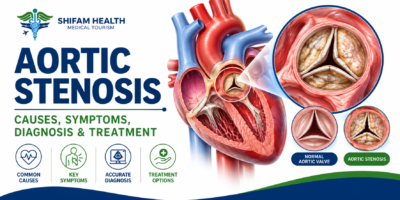
Aortic stenosis means the aortic valve, the one-way door between the heart’s main pumping chamber (the left ventricle) and the aorta, no longer opens fully. Blood has to squeeze through a smaller opening, so the heart works harder to push enough blood to the body.
A healthy aortic valve opens wide with each heartbeat and closes tightly between beats. When the leaflets thicken, stiffen or fuse, the opening narrows. To compensate, the left ventricle generates higher pressure, and over time its muscle wall thickens (left ventricular hypertrophy). Early on this compensation works well, which is why many people feel normal. In severe disease the heart can struggle to keep up, causing symptoms and, if untreated, heart failure.
Three things guide treatment: how narrow the valve is, whether symptoms are present, and how well the heart is coping. A single number never decides it.
What Causes Aortic Stenosis?
The three main causes are age-related calcium build-up on the valve, a bicuspid valve present from birth, and rheumatic heart disease. Which one applies often depends on a patient’s age and background, and less common causes exist.
| Cause | What Happens | Who It Typically Affects |
|---|---|---|
| Calcific (degenerative) aortic stenosis | Calcium deposits and tissue changes gradually stiffen the leaflets | Mostly older adults, though severity varies widely |
| Congenital bicuspid aortic valve | The valve is born with two leaflets instead of three and may wear out earlier | Often diagnosed in younger or middle-aged adults |
| Rheumatic heart disease | Past rheumatic fever scars and fuses the leaflets, often alongside other valves | More common where rheumatic fever has been common; not the main cause everywhere |
| Other causes | Previous chest radiation and some other conditions can damage the valve | Less common |
Calcific disease is not simply “normal aging”. Many older adults have little or no valve narrowing, and others progress faster. A bicuspid valve can also be associated with enlargement of the aorta in some people, so the aorta is usually assessed as well. In rheumatic disease, more than one valve is often involved, which changes treatment planning.
Risk factors include older age, a bicuspid valve, previous rheumatic fever, and cardiovascular risk factors linked to calcific valve disease such as high blood pressure and raised cholesterol. Chronic kidney disease is also associated with faster valve calcification. A risk factor does not mean you have or will develop aortic stenosis, and lifestyle changes alone cannot reverse established valve narrowing.
What Are the Symptoms of Aortic Stenosis?
Mild or even moderate aortic stenosis often causes no symptoms. As the valve narrows further, typical symptoms are breathlessness on exertion, chest pain or tightness, dizziness, and fainting or near-fainting. Symptoms can be gradual, so people often reduce activity without noticing.
- Breathlessness, especially when climbing stairs, walking uphill or carrying shopping
- Reduced stamina and fatigue
- Chest discomfort (angina) during effort
- Light-headedness or fainting, particularly during or after exertion
- Signs of heart failure in some patients: breathlessness lying flat, ankle swelling
These symptoms overlap with lung disease, anaemia, coronary disease and simply being unfit, so only a clinical assessment can tell. Do not deliberately exercise harder to “test” whether symptoms appear. A new drop in what you can comfortably do is itself worth reporting.
When is aortic stenosis an emergency?
Get emergency help immediately for: new or severe chest pain; fainting, especially during exertion; sudden or severe breathlessness; breathlessness at rest with distress; or rapidly worsening symptoms. Do not wait for an online opinion, a second opinion or international travel.
For newly developing or worsening breathlessness on exertion, chest discomfort or dizziness, arrange a prompt medical review.
What Are the Severity Levels of Aortic Stenosis?
Aortic stenosis is graded as mild, moderate or severe mainly from echocardiogram measurements, interpreted alongside symptoms and heart function. Severe disease is generally defined by a peak jet velocity of at least 4 m/s, a mean gradient of at least 40 mmHg, and a valve area of about 1.0 cm² or less.
| Severity | Peak Jet Velocity (Vmax) | Mean Pressure Gradient | Aortic Valve Area |
|---|---|---|---|
| Mild | 2.0–2.9 m/s | Under 20 mmHg | Larger than severe range |
| Moderate | 3.0–3.9 m/s | 20–39 mmHg | Larger than severe range |
| Severe | 4.0 m/s or more | 40 mmHg or more | Typically 1.0 cm² or less (indexed 0.6 cm²/m² or less) |
| Very severe | 5.0 m/s or more | 60 mmHg or more | — |
When the numbers disagree. Gradients depend on how much blood is flowing across the valve. A patient can have a severe-range valve area but a gradient below 40 mmHg, called low-gradient aortic stenosis. This happens with weak heart muscle (low-flow, low-gradient with reduced ejection fraction), or even with a normal ejection fraction when the heart chamber is small and stroke volume is low. Sorting out true severe disease from moderate disease can need extra tests such as a CT calcium score or a stress echo performed under specialist guidance.
Severity is also not the same as symptoms. Someone can have severe stenosis with no symptoms, and someone with moderate stenosis can have breathlessness from another cause.
How Is Aortic Stenosis Diagnosed?
Diagnosis starts with a clinical examination and is confirmed by echocardiography (an ultrasound of the heart). A heart murmur may raise suspicion but does not by itself prove aortic stenosis or show how severe it is.
History and examination. The doctor asks about breathlessness, chest pain, dizziness and exercise tolerance, checks blood pressure, listens to the heart, and looks for signs of fluid build-up.
Transthoracic echocardiogram (TTE). This is the main test. It shows how the valve looks and opens and measures peak velocity, mean gradient and valve area, along with left ventricular size and pumping function and any other valve or aortic problems. It is painless and takes under an hour.
Additional tests, only when needed:
| Test | Why It May Be Used |
|---|---|
| ECG | Checks rhythm and signs of heart strain |
| CT scan | Measures valve calcium, aortic anatomy and is central to TAVR planning |
| Cardiac catheterization / coronary angiography | Checks for coronary artery blockages, and occasionally measures pressures directly |
| Stress testing | In carefully selected patients, usually without symptoms, under specialist supervision |
| Blood tests (e.g. BNP) | May help when severity or the cause of symptoms is unclear |
Not everyone needs every test. Exercise testing is generally not appropriate in patients who already have symptoms from severe stenosis unless a specialist decides otherwise.
What do the terms on an echo report mean?
| Term | Plain Meaning |
|---|---|
| Peak aortic jet velocity | The fastest speed of blood through the valve; higher means tighter |
| Mean pressure gradient | The average pressure difference across the valve |
| Aortic valve area (AVA) | The size of the opening; smaller means tighter |
| Ejection fraction (LVEF) | The percentage of blood the left ventricle pumps out each beat |
| Left ventricular hypertrophy | Thickening of the heart muscle from working against resistance |
| Low-flow, low-gradient | Valve area looks severe but the gradient is lower because less blood is flowing |
No single figure should be read in isolation. Image quality, blood pressure, flow, heart function and symptoms all affect interpretation, so ask your cardiologist to walk through your own report.
Can Aortic Stenosis Be Treated With Medication?
Medicines cannot open a severely narrowed aortic valve. They are used to manage related problems: high blood pressure, fluid overload, coronary artery disease and other cardiovascular risk factors. Valve replacement is the definitive treatment when it is indicated.
Because a tight valve limits how much blood the heart can push out, some blood-pressure and heart medicines need careful selection and monitoring in significant aortic stenosis. Never stop or start a medicine on your own because of something you read online.
No supplement, special diet or alternative therapy has been shown to remove valve calcium or replace a needed valve procedure. What does help: treating blood pressure, not smoking, following general heart-health advice, and attending every scheduled follow-up.
How Is Mild or Moderate Aortic Stenosis Managed?
Mild and moderate aortic stenosis are usually monitored with periodic clinic reviews and repeat echocardiograms, plus treatment of blood pressure and other risk factors. There is no procedure that is routinely recommended for these stages unless another heart condition requires surgery.
How often to repeat the echo depends on severity, how fast the valve is changing, and symptoms. As a rough guide from the 2020 ACC/AHA guideline, mild disease is rechecked every few years, moderate disease about every one to two years, and severe disease about every six to twelve months, but your cardiologist individualises this.
Feeling well is not a reason to skip follow-up. Aortic stenosis can progress quietly, and new breathlessness, chest discomfort or dizziness should prompt an earlier review.
When Is Aortic Valve Replacement Needed?
Valve replacement is recommended for appropriately selected patients with severe aortic stenosis, most clearly when symptoms are caused by the valve or when the heart’s pumping function is falling. For some people without symptoms, earlier intervention may be considered. The decision belongs to a cardiology and cardiac-surgery heart team.
Factors that influence timing:
- Symptoms attributable to severe stenosis (the strongest trigger)
- Reduced left ventricular function
- Rapid progression, very severe measurements, or abnormal test results in selected asymptomatic patients
- Other heart or aortic disease that needs surgery anyway
- Anatomy, procedural risk, life expectancy and the patient’s own priorities
What has changed recently. For years the standard approach for severe aortic stenosis without symptoms was “watchful waiting”. Newer trial evidence, including the EARLY TAVR trial, and the 2025 ESC/EACTS guidelines now support considering earlier valve replacement in selected asymptomatic patients with severe high-gradient stenosis and low procedural risk, as an alternative to close surveillance. The 2020 ACC/AHA guideline predates this evidence. This does not mean every asymptomatic patient needs a procedure now. It means the question “wait or treat?” deserves an informed discussion with a heart team.
What Is Surgical Aortic Valve Replacement (SAVR)?
SAVR is open-heart surgery in which the diseased aortic valve is removed and a mechanical or tissue valve is stitched into place. It has the longest track record of any aortic valve treatment and allows the surgeon to treat other problems, such as bypass grafts or an enlarged aorta, during the same operation.
Surgery may be favoured in younger patients, those with a bicuspid valve or anatomy unsuitable for a catheter valve, an enlarged aorta, coronary disease needing bypass, or when a longer-lasting valve is a priority. It requires general anaesthesia, a heart-lung machine, and a recovery measured in weeks. The surgical team chooses the access (full breastbone incision or smaller approaches).
What Is TAVR (TAVI)?
TAVR, also called TAVI, is a catheter-based procedure that places a folded replacement valve inside the diseased valve, usually through an artery in the groin, without stopping the heart or opening the chest. It suits many, but not all, patients and is not automatically better simply because it is less invasive.
Before TAVR, a CT scan maps the valve, the aorta and the blood vessels to see whether a catheter valve will fit and which access route works. After the procedure, many patients stay in hospital for a short time, though some need longer monitoring, for example if the heart’s electrical system is affected and a pacemaker is needed. Limitations include a risk of leakage around the valve, conduction problems, and less long-term durability data than surgery, particularly for younger patients.
In 2025 the ESC/EACTS guidelines lowered the age at which TAVI is recommended for suitable anatomy to 70, citing improved evidence. Older ACC/AHA 2020 guidance uses different age bands (for example favouring surgery under 65 and TAVR over 80, with shared decision-making between). Guidelines differ, and individual decisions can differ from both.
SAVR vs TAVR: What Is the Difference?
The choice between SAVR and TAVR is made by a heart team after weighing age, life expectancy, valve anatomy, other heart or aortic conditions, procedural risk and patient preference. Neither is better for everyone.
| Factor | SAVR (Open Surgery) | TAVR / TAVI (Catheter) |
|---|---|---|
| How the valve is placed | Chest opened, heart-lung machine, valve sewn in | Catheter, usually via groin artery, valve expanded inside the old valve |
| Recovery | Longer hospital stay and recovery, typically weeks | Often shorter stay and faster early recovery |
| Often favoured when | Younger patients, bicuspid valve, aortic enlargement, need for bypass or other surgery, unsuitable access | Older patients, higher surgical risk, suitable anatomy on CT |
| Anatomy | Less limited by vessel size or valve shape | Needs suitable vessels, valve and coronary anatomy |
| Long-term evidence | Longest follow-up | Strong and growing, less follow-up in younger patients |
| Specific risks | Bleeding, atrial fibrillation, wound problems, stroke | Vascular complications, pacemaker need, leakage around valve, stroke |
| Future procedures | Possible valve-in-valve TAVR later | Lifetime planning matters; redo options depend on valve and anatomy |
The most useful question to ask is not “which is better” but “which is better for me, and why?”
What Are the Types of Replacement Valves?
There are two main types. Mechanical valves are very durable but require lifelong blood-thinning with warfarin-type medicine. Biological (tissue) valves usually avoid lifelong warfarin but can wear out over time, so a repeat procedure may eventually be needed.
| Mechanical Valve | Biological (Tissue) Valve | |
|---|---|---|
| Durability | Very long lasting | Variable; can degenerate over years |
| Blood thinners | Lifelong vitamin K antagonist (warfarin type) with regular INR monitoring | Often not lifelong because of the valve, but medicines may still be needed for other reasons or after the procedure |
| Suits | Younger patients who accept anticoagulation and monitoring | Older patients, those who cannot take warfarin, and all TAVR valves |
| Considerations | Bleeding risk, pregnancy planning, ability to monitor | Possible future valve-in-valve procedure |
No valve has a fixed lifespan that can be promised. Durability depends on the valve, age, kidney function and other factors, and studies keep updating the picture.
What Are the Risks of Aortic Valve Replacement?
All valve replacement carries risk. Shared risks include bleeding, infection, stroke, kidney injury, rhythm problems and the possibility of needing further procedures. Procedure-specific risks differ, and individual risk depends on your health, anatomy and the chosen procedure.
- Both procedures: bleeding, infection, stroke, kidney injury, abnormal heart rhythm, valve dysfunction, bleeding or clots linked to blood-thinning medicines
- SAVR: wound and breastbone healing issues, post-operative atrial fibrillation, longer recovery
- TAVR: vascular access complications, conduction problems that may need a pacemaker, leakage around the valve
Your team uses risk scores and frailty assessment to estimate your personal risk. Be wary of anyone who quotes a single success rate as if it applied to everyone.
What Is Recovery Like After Valve Replacement?
Recovery depends on the procedure, your age and your other health conditions. TAVR patients often go home after a short stay, while SAVR patients typically stay longer and need weeks to months to regain full strength. Your team will give you a personal timeline.
Typical recovery elements include monitoring in hospital, wound or access-site care, gradual activity, cardiac rehabilitation where offered, taking prescribed medicines, a follow-up echocardiogram, and knowing which warning signs need urgent attention (fever, chest pain, fainting, new breathlessness, bleeding, wound changes). Replacement valves still need lifelong follow-up and dental care to reduce the risk of valve infection.
If you are travelling: do not fly until your treating team confirms you are medically fit, and plan enough time near the hospital for early follow-up before heading home.
Can Aortic Stenosis Be Prevented or Reversed?
Established calcific narrowing cannot be reversed by lifestyle changes or medicines, and there is no proven way to prevent most cases. What you can do is protect your overall heart health, monitor a known diagnosis, treat blood pressure, avoid smoking and seek review if symptoms appear.
What Should You Ask a Cardiologist or Heart-Valve Team?
Good questions turn a frightening diagnosis into a plan. Bring your echo reports, imaging, medication list and heart history.
- How severe is my aortic stenosis, and which measurements show that?
- Are my symptoms likely caused by the valve?
- Is my heart function affected?
- Do I need further tests, such as a CT?
- Is monitoring appropriate, and how often should I be reviewed?
- What findings would lead to valve replacement?
- Am I a candidate for SAVR, TAVR, or both, and why?
- What are the benefits, risks and alternatives in my case?
- Which type of valve is recommended, and will I need blood thinners?
- What follow-up and rehabilitation will I need?
- When will I be fit to travel after treatment?
Aortic Stenosis Treatment in India for International Patients
Patients from abroad can seek specialist opinions and treatment for aortic stenosis in India. A sound approach is to share records, get a clear explanation of severity and treatment indication, compare options and obtain an itemised written estimate. Patients with acute symptoms must get emergency care where they are first.
A practical checklist:
- Gather echo reports, original imaging files, recent blood tests, medication list and any prior cardiac procedures.
- Ask the heart team to explain severity, whether symptoms fit the valve, and why a particular procedure is advised.
- Request an itemised estimate: consultations, tests (including CT), procedure fees, the valve or device, hospital and ICU stay, medicines, rehabilitation and follow-up.
- Clarify who handles complications, follow-up after you return home, and how records will be sent to your home cardiologist.
- Confirm with the treating team when you can safely fly home.
What does it cost? Published figures vary widely and should be treated as indicative only. Hospital and aggregator pages list TAVR in India at roughly INR 14–25 lakh in domestic pricing, and some sources quote about USD 23,000 to about USD 50,000 depending on the valve brand and hospital. International patients are often quoted more than domestic list prices. The valve itself is typically the largest cost component. SAVR is usually priced differently depending on valve type and hospital stay.
Myths vs Facts
| Myth | Fact |
|---|---|
| Everyone with aortic stenosis has symptoms. | Many people have none for years, even with significant narrowing. Regular follow-up still matters. |
| Medicine can reverse severe valve narrowing. | Medicines manage related conditions but cannot open a severely narrowed valve. |
| Every severe case gets the same procedure. | SAVR, TAVR or monitoring is chosen individually by a heart team. |
| TAVR is always better than surgery. | TAVR suits many patients, but surgery is often preferred in some, for example younger patients, aortic enlargement or need for bypass. |
| Valve replacement always means open-heart surgery. | TAVR places a valve through a catheter in suitable patients. |
| An echo number can be read without context. | Measurements must be interpreted with flow, heart function, symptoms and image quality. |
| A tissue valve never needs blood thinners. | Many do not need lifelong warfarin for the valve, but some patients need antithrombotic medicines for other reasons or after the procedure. |
| Replacement valve means no more follow-up. | Replacement valves need lifelong monitoring. |
| Wait until symptoms are severe before seeing a doctor. | New or worsening symptoms need prompt assessment, and severe symptoms need emergency care. |
Frequently Asked Questions About Aortic Stenosis
What is aortic stenosis?
A condition in which the aortic valve narrows, restricting blood flow from the heart to the body.
What causes aortic stenosis?
Common causes include age-related calcium buildup, a congenital bicuspid valve, and rheumatic heart disease.
What are the symptoms?
Symptoms include breathlessness, chest pain, dizziness, fainting, and reduced exercise tolerance.
Is aortic stenosis serious?
Mild cases may remain stable for years, but severe disease can become life-threatening, particularly when symptoms develop.
How is aortic stenosis diagnosed?
Echocardiography is the main test. CT scans, ECG, and other investigations may help assess severity and treatment suitability.
Can medication cure aortic stenosis?
No. Medication can manage associated conditions but cannot reverse established valve narrowing.
When is valve replacement necessary?
It is generally recommended for severe aortic stenosis with symptoms or impaired heart function. Selected asymptomatic patients may also benefit from earlier intervention.
What is the difference between SAVR and TAVR?
SAVR replaces the valve through open-heart surgery. TAVR replaces it using a catheter, usually inserted through an artery in the groin.
Is TAVR better than open-heart surgery?
Neither is universally better. The choice depends on age, anatomy, surgical risk, other conditions, and patient preferences.
How long does a replacement valve last?
Durability varies by valve type and individual factors. Mechanical valves are generally highly durable, while tissue valves may deteriorate over time.
Will I need blood thinners after valve replacement?
Mechanical valves require lifelong warfarin. Tissue valves may require temporary or longer-term medication depending on individual circumstances.
Talk to Shifam Health About Your Cardiac Options
If you or a family member has been told you have aortic stenosis and you are weighing a second opinion, Shifam Health can help you organise the process. As a healthcare coordination service, we can help gather and organize your cardiac records, arrange specialist opinions, explore hospital options in India, request and explain itemized treatment estimates, and support international patient arrangements such as visa documents, travel and accommodation.
We do not diagnose or treat, and we cannot say that one procedure is right for everyone.
If you have severe symptoms right now, such as chest pain, fainting or severe breathlessness, seek emergency care where you are. Do not wait to plan international treatment.

Popular Posts From Last Week
- October 12, 2026
- shifamhealth
Normally, the lower end of the spinal cord floats freely inside the spinal canal and glides a little…
- October 12, 2026
- shifamhealth
The word “stenosis” can sound frightening, and a diagnosis of severe aortic stenosis often arrives with the words…


