




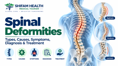
Spinal Deformities: Types, Causes, Symptoms, Diagnosis & Treatment
Filters & Insights
Written By :
Medically Reviewed By :

Shifam Health Medical Team
Shifam Health - Medical Reviews Department
10+ Years Of Experience
Spinal deformity covers scoliosis, kyphosis, lordosis, and adult sagittal imbalance. Learn the real differences, causes, and when treatment is needed.
📅
Last updated: October 2, 2026

Spinal deformity is a broad category, not one disease — it describes abnormalities of spinal curvature, alignment, rotation, or vertebral structure, including scoliosis, kyphosis, lordosis, and combinations of these. Severity and treatment vary enormously by cause, age, and pattern, and not every visible curve represents a disease requiring treatment.
Spinal deformity is genuinely three-dimensional — it can involve:
- Coronal plane abnormalities (side-to-side curvature, as in scoliosis)
- Sagittal plane abnormalities (front-to-back curvature, as in kyphosis and lordosis)
- Axial rotation (twisting of the vertebrae, a component of scoliosis specifically)
- Overall spinal-pelvic alignment, particularly relevant in adult deformity, discussed later
Major categories worth distinguishing clearly from the start:
| Term | What It Describes |
|---|---|
| Scoliosis | Sideways curvature combined with vertebral rotation |
| Kyphosis | Abnormal exaggeration of the spine’s normal forward (front-to-back) curve, usually in the upper back |
| Lordosis | Abnormal exaggeration of the spine’s normal backward curve, usually in the lower back or neck |
| Kyphoscoliosis | Combined coronal (scoliosis) and sagittal (kyphosis) deformity occurring together |
| Sagittal imbalance | A broader alignment problem where the trunk shifts forward relative to the pelvis, discussed in detail below |
| Flatback deformity | Loss of normal lumbar lordosis, a specific cause of sagittal imbalance |
| Degenerative spinal deformity | Deformity developing in adulthood from age-related spinal changes |
| Congenital deformity | Deformity present from abnormal vertebral formation before birth |
| Neuromuscular deformity | Deformity related to an underlying neurological or muscular condition |
Not every visible spinal curve is a disease requiring treatment — this is worth stating plainly at the outset, since it’s a genuine source of unnecessary anxiety for many patients and families.
Table of Contents
ToggleNormal Spinal Alignment — Not Every Curve Is Abnormal
Before discussing abnormal curvature, it’s worth understanding that the spine normally contains curves — this isn’t a flaw to be corrected, it’s how a healthy spine is built:
- Cervical lordosis — a normal backward (concave) curve in the neck
- Thoracic kyphosis — a normal forward (convex) curve in the upper back
- Lumbar lordosis — a normal backward curve in the lower back
- Sacral alignment and the pelvis work together with these spinal curves to support upright posture and balance
The key distinction is between:
- Normal physiological curvature — the expected curves described above
- Postural variation — flexible, position-dependent changes that aren’t fixed abnormalities
- Structural deformity — a genuinely fixed, abnormal curvature or alignment pattern
A person who looks slightly rounded when slouching, but straightens with conscious effort, has postural variation — not structural deformity. This distinction, established through physical examination (discussed later), is foundational to everything that follows in this guide.
Types of Spinal Deformity
Scoliosis — lateral (side-to-side) spinal curvature combined with vertebral rotation — is the most well-known and most extensively covered type of spinal deformity. Given its distinct evidence base and management pathway, particularly in adolescents, we cover scoliosis specifically including Cobb angle interpretation, bracing, and pediatric scoliosis surgery — in much greater depth in our dedicated scoliosis guide. Briefly, scoliosis is categorized as:
| Scoliosis Category | Key Feature |
|---|---|
| Idiopathic (infantile, juvenile, adolescent, or adult-onset) | No specific identified cause; the most common overall category |
| Congenital | Caused by vertebral formation abnormalities present from before birth |
| Neuromuscular | Associated with an underlying neurological or muscular condition |
| Syndromic | Part of a broader genetic or connective-tissue syndrome |
| Degenerative | Develops in adulthood from age-related spinal changes |
Kyphosis
Kyphosis is abnormal exaggeration of the spine’s normal forward curve, most often affecting the upper (thoracic) spine, with several distinct underlying forms — postural, Scheuermann’s, congenital, neuromuscular, degenerative, post-traumatic, and postoperative — each with different causes and implications.
| Type | Key Features |
|---|---|
| Postural kyphosis | Flexible, correctable with conscious posture effort; not a structural, fixed deformity |
| Scheuermann kyphosis | A structural cause of excessive thoracic kyphosis in adolescents, characterized by specific vertebral wedging visible on imaging — genuinely different from simple poor posture |
| Congenital kyphosis | From abnormal vertebral formation before birth |
| Neuromuscular kyphosis | Related to an underlying neurological or muscular condition |
| Degenerative kyphosis | Develops in adulthood from age-related spinal changes, including disc degeneration |
| Post-traumatic kyphosis | Following a vertebral fracture that alters normal alignment |
| Postoperative/iatrogenic kyphosis | Can develop after certain spinal procedures, sometimes related to junctional issues discussed later in this guide |
| Deformity associated with infection or tumor | Vertebral destruction from these processes can also alter spinal alignment |
The distinction between postural and Scheuermann kyphosis matters practically: postural kyphosis generally responds to activity, posture awareness, and general conditioning, while Scheuermann kyphosis, being a genuinely structural condition, may require more specific evaluation and, in more significant cases, bracing or surgical consideration — treating the two identically would misrepresent what’s actually going on in each case.
Lordosis
Excessive lordosis — an exaggerated backward curve, most often discussed in the lower back — can be a structural deformity or, more commonly, a compensatory posture related to another underlying issue, such as pelvic alignment, hip pathology, or a nearby spinal condition like spondylolisthesis.
Potential contributing factors:
- An underlying spinal deformity elsewhere along the spine, with lordosis developing as compensation
- Pelvic alignment
- Hip pathology
- Neuromuscular conditions
- Body weight/composition, where relevant
- Spondylolisthesis (one vertebra slipping relative to another)
- Broader compensatory mechanisms, particularly in the context of sagittal balance issues discussed later
It’s worth being clear: lumbar lordosis itself is not inherently pathological — it’s a normal part of spinal anatomy, and the question is whether it’s excessive relative to what’s normal for that individual, and, if so, what’s driving that excess.
Kyphoscoliosis and Sagittal Imbalance
Kyphoscoliosis describes combined coronal (side-to-side) and sagittal (front-to-back) deformity occurring together — essentially scoliosis and kyphosis coexisting in the same patient. Because this combines two planes of deformity, severe kyphoscoliosis can affect mobility, pain, pulmonary function (discussed later), and, in some cases, neurological function more significantly than either deformity alone.
Sagittal imbalance is a broader, particularly adult-relevant concept describing a forward shift of the trunk relative to the pelvis, often related to:
- Loss of normal lumbar lordosis (“flatback” deformity)
- Compensatory pelvic retroversion (the pelvis tilting backward to try to maintain upright posture)
- Compensatory knee or hip flexion as the body attempts to keep the head over the pelvis
- Resulting difficulty standing fully upright, sometimes described by patients as needing to “lean forward” or bend at the knees to stand comfortably
This concept — sagittal imbalance — is genuinely central to understanding adult spinal deformity specifically, and is expanded on in the dedicated section below on adult deformity and sagittal balance parameters.
Causes
Causes of spinal deformity vary by type — idiopathic (no identified cause after appropriate evaluation), congenital (vertebral formation abnormalities), neuromuscular (underlying neurological/muscular disease), degenerative (age-related spinal changes), traumatic, infectious, tumor-related, and iatrogenic (related to previous spinal surgery).
| Cause Category | Key Points |
|---|---|
| Idiopathic | “Idiopathic” means no specific cause has been identified after appropriate evaluation — it does not mean the condition has no underlying biological basis, simply that a single specific cause hasn’t been pinpointed |
| Congenital | Vertebral formation or segmentation defects, including hemivertebrae (partially formed vertebrae), sometimes associated with abnormalities in other organ systems that developed around the same embryonic period |
| Neuromuscular | Conditions like cerebral palsy, muscular dystrophies, and spinal muscular atrophy affect muscle balance and spinal support — different neuromuscular conditions don’t all behave identically, and generalizing across them oversimplifies real differences |
| Degenerative | Disc degeneration, facet joint degeneration, and often asymmetric age-related vertebral changes, contributing to adult degenerative scoliosis and sagittal imbalance |
| Traumatic | Vertebral fractures that heal in abnormal alignment (malunion) can produce post-traumatic deformity |
| Infectious | Spinal infection can cause vertebral destruction and resulting deformity |
| Tumor-related | Vertebral tumors can alter alignment or cause vertebral collapse |
| Iatrogenic/postoperative | Deformity can develop after certain spinal procedures — including adjacent segment problems, postoperative imbalance, or junctional deformity (like flatback) following some spinal surgeries |
On posture as a cause specifically: postural asymmetry is genuinely flexible and position-dependent, and should not be described as causing structural, fixed deformity — this is worth stating plainly given how commonly it’s assumed otherwise.
Symptoms
Symptoms depend heavily on the deformity type, magnitude, location, whether it’s progressing, and any underlying cause — ranging from no symptoms at all to visible asymmetry, pain, neurological symptoms, functional limitations, and, in severe thoracic cases, respiratory effects.
Visible changes can include uneven shoulders, uneven waist, rib prominence, pelvic asymmetry, a rounded upper back, an excessive lumbar curve, forward trunk posture, or generally altered body alignment.
Pain: some patients have little or no pain despite visible deformity; others experience back pain, neck pain, muscular fatigue, mechanical pain (worse with activity or certain positions), or radicular (nerve-related) pain. The degree of curvature does not directly and reliably predict pain severity — a modest-looking curve can be quite symptomatic in one patient, while a more significant curve causes minimal discomfort in another.
Neurological symptoms — genuinely important to recognize and distinct from mechanical pain — can include numbness, tingling, weakness, gait disturbance, radiculopathy (nerve root-related pain/symptoms), myelopathy (spinal cord-related dysfunction), and, in more severe cases, bowel or bladder symptoms.
Functional limitations can include difficulty standing for extended periods, walking limitations, reduced overall endurance, difficulty sitting comfortably, and impaired ability to perform daily activities.
Respiratory symptoms, relevant specifically for severe thoracic deformity, can include reduced exercise tolerance, shortness of breath, and restrictive pulmonary physiology (the chest wall’s ability to expand normally being mechanically limited by the deformity). This is not something that occurs with ordinary, mild scoliosis or kyphosis — it’s specifically relevant to more severe deformity, discussed further in its own section below.
Red Flags — When Urgent Evaluation Is Needed
While most spinal deformity evaluation proceeds on a routine, non-urgent basis, certain symptoms warrant prompt or urgent medical assessment: new or progressive neurological deficits, new bowel or bladder dysfunction, severe or rapidly worsening pain, signs of infection, or significant unexplained systemic symptoms.
Features warranting prompt or urgent evaluation:
- New or progressive limb weakness
- Numbness accompanied by an actual neurological deficit on examination
- Walking difficulty that is genuinely worsening
- New bowel or bladder dysfunction
- Saddle-area (groin/perineal) sensory changes
- Severe or rapidly worsening pain
- Fever combined with severe spinal pain
- Unexplained weight loss alongside spinal symptoms
- A history of cancer with new spinal symptoms
- Significant trauma
- Breathing difficulty specifically associated with severe deformity
Routine specialist assessment, urgent assessment, and emergency care are genuinely different categories — most spinal deformity evaluation is entirely routine, and this list isn’t meant to create unnecessary fear. But the features above represent a meaningful, real distinction worth recognizing, since they can indicate something beyond straightforward deformity requiring more prompt attention.
Diagnosis and Imaging
Physical examination typically assesses posture, shoulder and pelvic symmetry, overall spinal alignment, the Adam’s forward bend test, rib prominence, leg-length discrepancy, gait, flexibility of the curve, neurological status (strength, sensation, reflexes), balance, and hip examination.
This combination helps determine whether a deformity is genuinely structural (fixed) versus postural (flexible), and whether any neurological involvement is present.
Standing X-rays — including AP/PA and lateral views, often of the whole spine for deformity assessment — are the standard initial and follow-up imaging, since they show alignment under normal load and allow measurement of curvature and sagittal alignment.
MRI is not automatically required for every patient, but becomes relevant with neurological abnormalities, atypical curve patterns, congenital deformity, suspected spinal cord abnormality, severe pain, rapidly progressive deformity, or as part of preoperative assessment where surgery is being considered.
CT scanning has a role in assessing complex bony anatomy, congenital vertebral abnormalities in detail, surgical planning, and selected postoperative evaluation — not recommended routinely when it isn’t specifically needed.
Cobb Angle and Spinal Measurements
The Cobb angle, measured on a standing X-ray, is the standard way to quantify the degree of curvature — but it carries real measurement variability and should never be treated as the sole factor determining whether treatment, including surgery, is needed.
The Cobb angle is measured by drawing lines along the most tilted vertebrae at the top and bottom of a curve and measuring the angle between them. Measurement variability is real — the same curve measured on different occasions, or by different observers, can show some variation, meaning small differences between measurements don’t necessarily represent true progression.
No single Cobb-angle threshold should be presented as a universal treatment rule. Clinicians interpret the Cobb angle together with the patient’s age, skeletal maturity, curve pattern and location, symptoms, and overall clinical picture — not as a number that mechanically dictates treatment on its own.
Adult Spinal Deformity and Sagittal Balance Parameters
Adult spinal deformity assessment goes beyond the Cobb angle alone — modern evaluation incorporates sagittal balance parameters like sagittal vertical axis, pelvic incidence, pelvic tilt, and sacral slope, which together describe how well-balanced the spine is over the pelvis, a concept particularly central to adult (as opposed to pediatric) deformity.
This is genuinely one of the more sophisticated, less commonly well-explained areas of spinal deformity care, worth understanding in accessible terms:
| Parameter | What It Describes, in Plain Terms |
|---|---|
| Sagittal vertical axis (SVA) | How far forward the trunk is positioned relative to the pelvis — a larger SVA reflects more forward imbalance |
| Pelvic incidence | A fixed anatomical measurement of an individual’s pelvis shape, essentially unique to each person and unchanged by posture or surgery |
| Pelvic tilt | How much the pelvis is rotated backward (retroverted) to compensate for spinal imbalance — a higher pelvic tilt often reflects the body working harder to compensate |
| Sacral slope | The angle of the top of the sacrum, related to overall pelvic positioning |
| Lumbar lordosis | The degree of the lower back’s normal backward curve |
| Pelvic incidence–lumbar lordosis (PI-LL) mismatch | A key relationship — ideally, lumbar lordosis should roughly match an individual’s fixed pelvic incidence; a significant mismatch is associated with poorer outcomes and can indicate sagittal imbalance |
Natural History and Progression
Whether spinal deformity progresses depends entirely on the type — pediatric scoliosis progression relates to growth and skeletal maturity, while adult deformity progression relates to ongoing degenerative changes and loss of compensatory capacity over time — there’s no single universal progression rate across all spinal deformity types.
For scoliosis specifically (covered in more depth in our dedicated scoliosis guide), progression risk relates to age, skeletal maturity, curve magnitude, curve pattern, remaining growth, and the underlying condition.
For adult deformity, progression relates more to ongoing degenerative changes, gradual loss of the spine’s ability to compensate for imbalance over time, and, in some cases, compensatory mechanisms (like increased pelvic tilt) eventually becoming insufficient to maintain balance, at which point symptoms and functional limitation can increase.
No single universal progression rate applies across these genuinely different processes — pediatric growth-related progression and adult degenerative progression are fundamentally different phenomena that shouldn’t be described with the same generic statistics.
Non-Surgical Treatment
Non-surgical treatment for spinal deformity depends heavily on cause, age, symptoms, severity, and progression — it can include observation, physiotherapy and core/functional conditioning, pain management, activity modification, bracing (where evidence supports it for the specific condition), and management of any underlying disease or osteoporosis.
- Observation — periodic monitoring, appropriate for many milder, lower-risk deformities
- Physiotherapy and exercise — core and functional conditioning can support pain management and function, though physiotherapy should not be presented as something that permanently corrects every structural deformity — its realistic role is supporting function and symptom management, with a more variable and condition-specific effect on the actual curve itself
- Pain management — addressing the specific pain mechanism involved (discussed further below)
- Activity modification
- Bracing — evidence for bracing’s effectiveness varies genuinely by condition; it has an established evidence base specifically for adolescent idiopathic scoliosis (covered in depth in our scoliosis guide) but evidence varies for other deformity types, and it should never be described as permanently straightening every curve — its purpose is generally limiting progression or supporting alignment/symptoms, not guaranteed permanent correction
- Osteoporosis management and treatment of any other underlying disease contributing to the deformity
There is no single universal exercise program appropriate for every patient with spinal deformity — individualized assessment matters more than a generic protocol.
When Is Surgery Considered — and When Might It Not Be Appropriate?
Surgery may be considered for significant progressive deformity, neurological compromise, severe functional limitation, refractory symptoms, or significant imbalance — but there’s no arbitrary universal threshold, and major reconstruction genuinely isn’t appropriate for every patient, particularly when symptom burden, frailty, or medical comorbidities make the risk-benefit balance unfavorable.
Factors informing the decision:
- Deformity type
- Curve magnitude and pattern
- Documented progression
- Age and skeletal maturity (for younger patients)
- Symptoms and functional impact
- Neurological status
- Bone quality
- Pulmonary function
- Overall health and comorbidities
- Patient goals and priorities
Being genuinely honest here: major spinal reconstruction may not be the right choice for every patient, even with significant deformity present. This isn’t a recommendation against surgery generally — it’s a recognition that the decision should weigh symptom burden, functional goals, frailty, medical comorbidities, bone quality, pulmonary function, and neurological status against the real risks of major surgery (discussed next), individualized after specialist evaluation.
For some patients — particularly older or frailer patients with significant deformity but manageable symptoms — the risk-benefit balance may reasonably favor continued nonoperative management over major reconstruction, and a good specialist should present this option honestly rather than defaulting to surgery whenever deformity is significant.
Surgical Approaches
At a high level, without turning this into an operative manual:
- Posterior spinal fusion — the most common overall approach, using instrumentation (rods and screws) placed from the back of the spine to correct and stabilize alignment, with bone graft encouraging fusion
- Anterior approaches — accessing the spine from the front or side, used in selected cases depending on the deformity pattern
- Osteotomies — surgical bone-cutting techniques used to achieve additional correction, particularly relevant in adult deformity with significant sagittal imbalance
- Vertebral column resection — a more extensive technique reserved for rare, severe, complex deformity
- Minimally invasive approaches — used in appropriately selected cases, with the same caution about minimally invasive not automatically meaning “better” that applies across spine surgery generally
- Navigation/robotic assistance — a surgical tool that may aid planning and implant placement accuracy in appropriate contexts, but does not automatically make surgery safer or produce a better outcome — it assists surgical technique, it doesn’t replace surgical judgment
Surgical technique genuinely depends on the specific anatomy and correction goals — there’s no single universally best approach across all spinal deformity types and patients.
Complex Adult Deformity Surgery and Risks
Why adult deformity reconstruction can be complex: it often involves long-segment fusion, restoration of sagittal alignment (using the balance concepts discussed above), osteotomy for additional correction, correction of coronal imbalance where present, neural decompression where nerve or spinal cord compression coexists, and, in more complex cases, staged procedures performed across more than one surgery.
Being honest about the real risks, without sensationalizing them:
- Neurological injury
- Infection
- Significant blood loss
- Implant failure
- Nonunion/pseudarthrosis (failure of bone to properly fuse)
- Proximal junctional kyphosis or failure — a recognized complication where mechanical stress concentrates at the upper edge of a long fusion construct
- Adjacent segment problems over time
- Cardiopulmonary complications, particularly relevant in older or higher-risk patients given the magnitude of this surgery
- Need for reoperation
Risk varies significantly by the extent of surgery, patient age, bone quality, comorbidities, and overall health — this is precisely why the individualized risk-benefit discussion in the previous section matters so much for adult deformity surgery specifically, given how much larger these procedures often are compared to more limited pediatric scoliosis surgery.
Neurological and Respiratory Effects
Neurological complications relevant to spinal deformity include nerve-root compression (radiculopathy) and spinal cord compression (myelopathy), both of which can occur as a consequence of severe or progressive deformity, not only as a surgical complication. Symptoms requiring urgent assessment were covered in the red flags section above — neurological status genuinely influences treatment decisions, sometimes prompting more urgent surgical consideration than would otherwise be the case based on deformity severity alone.
Respiratory effects, relevant specifically for severe thoracic deformity: restrictive chest-wall mechanics can reduce lung volumes, limit exercise tolerance, and, in selected patients, contribute to sleep-related breathing problems. Pulmonary function testing and respiratory evaluation may be part of assessment for significant thoracic deformity, and perioperative respiratory assessment is particularly relevant when major surgery is being planned for patients with significant thoracic curves. Mild scoliosis or kyphosis does not typically cause significant respiratory impairment — this is specifically a consideration for more severe deformity, not a generalized concern for every patient with any degree of spinal curvature.
Bone Health
Bone quality is a major consideration specifically for adult spinal deformity surgery, since implant fixation strength depends on adequate bone density — osteoporosis or osteopenia can increase the risk of implant-related complications and pseudarthrosis, making bone health optimization a genuine pre-surgical consideration for appropriate candidates.
- Osteoporosis and osteopenia both affect how securely surgical implants can be fixed in bone
- Vitamin D and calcium status may be clinically relevant where bone health is a concern
- Fragility fracture risk is a related consideration
- Bone health may genuinely need optimization before complex spinal reconstruction in appropriate patients — this is a real, evidence-based pre-surgical consideration, not an afterthought, particularly relevant given how much reconstruction and fixation is often involved in adult deformity surgery specifically
Rehabilitation and Pain Management
Rehabilitation after treatment — whether nonsurgical or surgical — typically involves physiotherapy, gait training, posture work, strengthening, mobility support, and occupational therapy support for return to daily activities. Recovery varies genuinely by the specific surgery performed, patient age, baseline function, neurological status, comorbidities, and rehabilitation engagement — no single universal recovery timeline applies across the wide range of spinal deformity treatments.
Pain management should be understood through the lens of the actual pain mechanism involved, since different mechanisms respond to different approaches:
- Mechanical pain (related to abnormal loading/alignment)
- Muscular fatigue
- Facet joint-related pain
- Disc-related pain
- Nerve-root pain
- Spinal stenosis-related pain
- Adjacent segment disease-related pain, particularly relevant after prior spinal surgery
Spinal deformity correction should not be presented as a guaranteed cure for chronic pain — while many patients experience meaningful pain improvement with appropriate treatment, pain has multiple potential contributing mechanisms, and correcting alignment doesn’t automatically resolve every source of discomfort a patient may be experiencing.
Prognosis
There is no single prognosis for “spinal deformity” — outcomes depend on the underlying cause, age, deformity severity, progression, neurological involvement, pulmonary status, bone quality, treatment approach, and rehabilitation — and radiographic correction is genuinely not the same thing as clinical success.
A distinction worth holding onto: radiographic correction (how the spine looks on an X-ray after treatment), pain improvement, functional improvement, and quality-of-life improvement are related but genuinely distinct outcomes.
Equating radiographic correction with clinical success overstates what imaging alone can tell you — a technically well-corrected spine on imaging doesn’t automatically guarantee a patient feels or functions dramatically better, and conversely, meaningful functional improvement can occur without perfect radiographic correction. Realistic prognosis discussions should address all of these dimensions, not just the imaging appearance.
Special Populations
| Population | Key Considerations |
|---|---|
| Children | Growth and skeletal maturity are central to both progression risk and treatment timing |
| Adolescents | Idiopathic scoliosis is most common here; psychosocial considerations (body image, bracing adherence) genuinely matter alongside the medical picture |
| Adults | Degenerative deformity and sagittal imbalance predominate, often combining alignment problems with degenerative spinal disease like stenosis |
| Neuromuscular patients | Progression patterns and treatment decisions depend heavily on the specific underlying condition and require coordinated, multidisciplinary care beyond spine specialists alone |
| Congenital deformity patients | May have associated abnormalities in other organ systems given the shared embryonic developmental timing, warranting broader evaluation |
| Older adults | Bone health, frailty, medical comorbidities, and surgical risk weigh more heavily in treatment decision-making |
| Pregnancy | Relevant primarily for patients with pre-existing deformity or previous spinal surgery/fusion, which can affect delivery and anesthetic planning — worth proactive discussion with both obstetric and spine specialist teams |
Spinal Deformity Treatment in India and Cost
India offers relevant infrastructure across pediatric and adult spinal deformity care, including orthopedic spine surgeons, neurosurgeons, deformity-specific imaging, rehabilitation, and pulmonary and bone-health evaluation — though not every hospital provides every service, particularly for complex adult reconstruction, which requires genuinely specific deformity-surgery expertise.
Potentially available services include pediatric and adult spinal deformity specialist consultation, standing/whole-spine imaging, MRI and CT, pulmonary evaluation, bone-health assessment, bracing services, spinal decompression and fusion surgery, osteotomy and complex reconstruction, ICU-level postoperative care where needed, and rehabilitation. We are not claiming every hospital offers every one of these, particularly complex adult deformity reconstruction specifically, which requires demonstrated experience with this exact category of surgery, distinct from general spine surgery capability.
On cost: based on internationally-quoted pricing for spinal fusion correction procedures (consistent with figures verified for scoliosis correction specifically), international-patient-facing costs commonly range from roughly $6,500 to $16,000 for standard fusion correction, with complex adult deformity reconstruction — involving osteotomy, longer-segment fusion, and greater surgical complexity — tending toward the higher end of this range or beyond, given the genuinely greater complexity, longer operative time, and more extensive instrumentation typically involved compared to more limited pediatric scoliosis correction. Cost components include specialist consultation, imaging (X-rays, MRI, CT), pulmonary and bone-health evaluation where relevant, the surgical procedure itself (varying enormously by extent — decompression alone vs. fusion vs. osteotomy vs. complex reconstruction), implants, hospitalization (including ICU where needed), and rehabilitation.
A personalized estimate, based on your specific deformity type and the actual planned procedure, requires direct consultation with the treating hospital — presenting one figure for the vastly different range of procedures this category encompasses would be genuinely misleading.
International Patient Journey and Travel Safety
Practical journey:
- Collect medical records — previous X-rays, standing whole-spine imaging, MRI, CT, operative reports if previously treated, implant details if relevant, neurological records, physiotherapy records, medication list, and bone-density reports where available
- Specialist review, matching the case to the appropriate spinal-deformity specialist
- Imaging review to determine whether existing imaging is adequate or additional evaluation is needed
- Treatment planning — clarifying whether observation, conservative treatment, bracing, decompression, fusion, or more extensive deformity correction is appropriate
- Procedure-specific cost estimate
- Medical visa support, where applicable
- Travel planning, including an honest assessment of medical stability for travel
- Local coordination — airport transfer, accessible accommodation, hospital visits, caregiver support
- Treatment, with coordinated appointments and hospital logistics
- Rehabilitation and follow-up, ensuring continuity of care after returning home
Travel safety: international travel should not be pursued when urgent treatment is genuinely needed locally first. Patients with acute neurological deterioration, suspected spinal cord compression, rapidly progressive weakness, new bowel/bladder dysfunction, severe infection, major trauma, or severe respiratory compromise should seek urgent local medical assessment — international treatment planning is appropriate only once the situation is medically stable for elective evaluation and travel.
Shifam Health is an international healthcare coordination and medical travel support company — not the treating surgeon, radiologist, physiotherapist, hospital, or diagnostic laboratory. Shifam Health may assist with medical record organization, specialist coordination, hospital coordination, appointment scheduling, treatment estimates, medical visa support, travel logistics, accommodation, interpreters, local coordination, and follow-up communication.
They does not independently decide whether surgery is required, and does not guarantee spinal correction, pain relief, neurological recovery, walking improvement, surgical success, hospital acceptance, exact costs, or visa approval.
Myths vs. Facts
| Myth | Fact |
|---|---|
| Poor posture causes all scoliosis | Postural asymmetry is flexible; it does not cause structural idiopathic scoliosis. |
| Every spinal curve needs surgery | Most cases use observation or nonsurgical treatment; surgery has specific indications. |
| Scoliosis always causes severe pain | Many people have little or no pain. |
| A larger curve always means more pain | Curve size does not reliably predict pain severity. |
| Braces permanently straighten every curve | Bracing mainly aims to limit progression during growth; permanent correction is not guaranteed. |
| Exercise makes scoliosis worse | Exercise is generally encouraged, not restricted. |
| People with scoliosis cannot play sports | Most people can participate in sports, including many after surgery. |
| All scoliosis is diagnosed in childhood | Scoliosis can develop or be diagnosed in adulthood. |
| Spinal deformity always worsens with age | Progression varies by deformity type. |
| A normal X-ray rules out spinal problems | Some neurological or degenerative problems require other imaging or assessment. |
| Spinal surgery completely fixes every problem | Surgery aims for correction and stabilization, not guaranteed symptom resolution. |
| Robotic surgery is always safer | Technology may improve precision but does not guarantee safer outcomes. |
| A high Cobb angle automatically means surgery | Age, growth, symptoms, progression, and other factors also guide treatment. |
| Spinal deformity is only cosmetic | It may affect function, pain, neurological status, and, in severe cases, breathing. |
Frequently Asked Questions
What is spinal deformity?
An abnormal curvature, alignment, rotation, or structural change of the spine, including scoliosis, kyphosis, and lordosis.
What is the difference between scoliosis, kyphosis, and lordosis?
Scoliosis involves sideways curvature and rotation, kyphosis excessive forward curvature, and lordosis excessive inward curvature.
What causes spinal deformity?
Causes include idiopathic, congenital, neuromuscular, degenerative, traumatic, infectious, tumor-related, and postsurgical conditions.
What is the Cobb angle?
A standard X-ray measurement used to quantify spinal curvature and help guide assessment and treatment.
How is spinal deformity diagnosed?
Evaluation usually includes physical examination and standing X-rays. MRI or CT may be added when clinically indicated.
Can spinal deformity be treated without surgery?
Yes. Depending on the condition, treatment may include observation, physiotherapy, activity modification, or bracing.
When is spinal deformity surgery needed?
Surgery may be considered for significant progression, neurological problems, severe functional limitations, or persistent symptoms despite appropriate treatment.
What is Scheuermann kyphosis?
A structural form of excessive thoracic kyphosis in adolescents associated with characteristic vertebral changes on imaging.
Can spinal deformity cause breathing problems?
Severe thoracic deformities can restrict chest-wall movement and reduce lung capacity, although this is uncommon with mild deformity.
Does spinal deformity always worsen?
No. Progression depends on the specific condition, age, growth, and degenerative changes.
Is spinal deformity surgery risky?
Yes. Risks can include infection, neurological injury, blood loss, and implant-related complications, depending on the procedure.
How much does spinal deformity surgery cost in India?
Costs vary by diagnosis and surgical complexity. Complex spinal fusion and reconstruction may cost approximately $6,500–$16,000, but a personalized hospital estimate is required.
Conclusion
Spinal deformity is genuinely a broad category, not a single condition — scoliosis, kyphosis, lordosis, and adult sagittal imbalance each involve different mechanisms, different evaluation approaches, and different treatment logic, even though they’re often discussed together under one umbrella term.
Whether the specific situation is a child with a newly noticed curve, an adolescent with structural kyphosis, or an adult with degenerative sagittal imbalance, the most valuable step is accurate, individualized evaluation including, for adults specifically, assessment of sagittal balance parameters beyond the Cobb angle alone rather than assuming a generic “spinal deformity” label predicts a fixed treatment path.
This article is educational and does not replace clinical examination, diagnosis, imaging interpretation, specialist consultation, or individualized treatment planning. Spinal deformity has many causes and clinical patterns, and treatment must be individualized based on your specific evaluation. Neurological emergencies — including new weakness, bowel/bladder dysfunction, or rapidly progressive symptoms — require urgent local medical care.

Popular Posts From Last Week
- September 25, 2026
- shifamhealth
Spina bifida is a group of neural tube defects caused by incomplete closure or development of the neural…
- September 26, 2026
- shifamhealth
Skeletal dysplasia is not one disease — it’s an umbrella term for a large, genetically diverse group of…
- September 30, 2026
- shifamhealth
Medically reviewed content · Published 2026 · Last reviewed September 2026 Rheumatoid arthritis (RA) primarily affects small joints,…
- October 2, 2026
- shifamhealth
Spinal deformity is a broad category, not one disease — it describes abnormalities of spinal curvature, alignment, rotation,…
- September 28, 2026
- shifamhealth
“Slipped disc,” “herniated disc,” “prolapsed disc” — these terms get used almost interchangeably by patients, and the imagery…
- October 1, 2026
- shifamhealth
Spinal disc herniation occurs when the soft, gel-like center of an intervertebral disc pushes through a weakened or…
- September 29, 2026
- shifamhealth
A spinal fracture is a break in one or more of the bones (vertebrae) that make up the…
- September 27, 2026
- shifamhealth
Last reviewed: 27-Sept-26 · General information for patients from Eritrea planning treatment in India. Not legal or immigration…


