



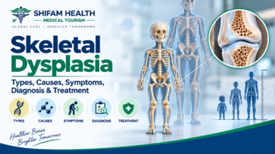
Skeletal Dysplasia (2026): Types, Causes, Symptoms, Diagnosis & Treatment
Filters & Insights
Understand skeletal dysplasia types, causes, diagnosis, and treatment from achondroplasia to osteogenesis imperfecta including care in India

Skeletal dysplasia is not one disease — it’s an umbrella term for a large, genetically diverse group of disorders that affect how bone and cartilage develop, grow, and mineralize. Different skeletal dysplasias have very different features, severity, inheritance patterns, complications, and treatments some are compatible with a normal lifespan and largely orthopedic in impact, while a small number are severe and identified before or shortly after birth.
Diagnosis generally combines clinical examination, growth measurements, skeletal X-rays, and, increasingly, molecular genetic testing, since no single test confirms every type. Treatment is always disease-specific, ranging from monitoring and orthopedic care to, for a small number of conditions, an approved targeted medical therapy.
Table of Contents
ToggleWhy Skeletal Dysplasia Is Not One Disease
Skeletal dysplasia encompasses a large and genuinely heterogeneous group of genetic disorders affecting bone development, cartilage, growth plates, bone mineralization, connective tissue, and skeletal modeling — and, in some conditions, other organ systems as well.
This heterogeneity is the single most important thing to understand about this topic: clinical presentation, severity, inheritance pattern, associated complications, and treatment approach vary substantially between individual disorders. A statement that’s accurate for one skeletal dysplasia can be entirely wrong for another, which is why this article focuses heavily on teaching the framework for understanding these differences rather than treating “skeletal dysplasia” as a single condition with one clinical picture.
It’s also important to distinguish skeletal dysplasia from related but different concepts: disproportionate short stature describes a physical finding that skeletal dysplasias often (but not always) produce; constitutional or familial short stature is a normal-variant pattern unrelated to skeletal dysplasia; endocrine causes of short stature (such as growth hormone deficiency) are a distinct category with different mechanisms; nutritional causes and metabolic bone disease (such as nutritional rickets) can sometimes resemble skeletal dysplasia on examination but have different underlying causes; and acquired skeletal abnormalities (from injury or illness rather than a genetic cause present from birth) are different again.
Not every person with short stature has a skeletal dysplasia — this is a common and understandable point of confusion that proper clinical evaluation resolves.
Types and Classification
Skeletal dysplasias are classified using an internationally recognized nosology that groups conditions by shared genetic and molecular mechanisms, and this classification is periodically revised as new disorders and genes are identified. Given that several hundred distinct skeletal dysplasias are currently recognized, this article does not attempt to list every entity — instead, it explains the classification logic and highlights clinically important, representative examples from major groups:
| Group | General Mechanism | Representative Examples |
|---|---|---|
| FGFR3-related dysplasias | Altered fibroblast growth factor receptor 3 signaling affecting cartilage growth | Achondroplasia, hypochondroplasia, thanatophoric dysplasia |
| Collagen-related disorders | Abnormalities in collagen structure or production affecting bone/connective tissue | Osteogenesis imperfecta (several types), some spondyloepiphyseal dysplasias |
| Growth-plate/epiphyseal disorders | Abnormal endochondral ossification at the growth plate | Multiple epiphyseal dysplasia |
| Spondyloepiphyseal dysplasias | Combined spine and joint (epiphyseal) involvement | Spondyloepiphyseal dysplasia congenita and related conditions |
| Metaphyseal dysplasias | Abnormalities at the metaphysis (the growing end of long bones) | A heterogeneous group of distinct conditions, not one diagnosis |
| Mineralization disorders | Impaired bone mineralization | Hypophosphatasia and related conditions |
| Ciliopathies | Defects in cellular cilia function affecting skeletal and other organ development | Certain short-rib thoracic dysplasias |
| Lysosomal/storage-related skeletal disorders | Metabolic storage abnormalities with skeletal manifestations | Mucopolysaccharidoses (which also present with skeletal findings) |
| Severe/lethal neonatal skeletal dysplasias | Severe disruption of skeletal development, often identified prenatally | Thanatophoric dysplasia, certain forms of osteogenesis imperfecta |
These categories are not always mutually exclusive, and some conditions share features across groups — this table is a teaching framework, not a rigid, non-overlapping taxonomy.
Common and Clinically Important Examples
Achondroplasia
The most common skeletal dysplasia causing disproportionate short stature, caused by a specific pathogenic variant in the FGFR3 gene affecting cartilage growth. Features include rhizomelic limb shortening (the upper arm/thigh segments are shortened more than the forearm/lower leg), macrocephaly with frontal bossing, midface hypoplasia, and a characteristic body proportion pattern.
Important complications to monitor include foramen magnum narrowing (which can affect the brainstem and upper spinal cord, particularly in infancy), sleep-disordered breathing, spinal stenosis (which can develop later in life), thoracolumbar kyphosis, and limb alignment issues. Achondroplasia is also the skeletal dysplasia with the most developed disease-specific medical therapy landscape (discussed below).
Hypochondroplasia
Related to achondroplasia (and also linked to FGFR3, though typically a different specific variant) but generally producing a milder phenotype. Distinguishing it clinically from mild achondroplasia or other causes of short stature can be genuinely difficult, and molecular genetic confirmation is often needed to establish the diagnosis with confidence.
Thanatophoric Dysplasia
A severe skeletal dysplasia, also FGFR3-related, typically identified prenatally or at birth, associated with significant respiratory compromise related to a very small chest cavity and other skeletal abnormalities. It should not be understood simply as “a more severe form of achondroplasia” — while related genetically, it is a clinically and prognostically distinct condition, and its identification prenatally or at birth is important for appropriate counseling and care planning.
Osteogenesis Imperfecta (OI)
A group of genetically heterogeneous disorders (multiple distinct types exist, most commonly related to collagen abnormalities) primarily characterized by bone fragility and recurrent fractures, sometimes with limited height reduction. Associated features can include dentinogenesis imperfecta (abnormal tooth development), hearing loss, scoliosis, and bone deformity from repeated fractures.
OI is clinically distinct from dysplasias primarily characterized by disproportionate short stature — its defining feature is fragility, not necessarily short stature, though severity and features vary across its several recognized types. Bisphosphonate therapy is used in appropriate cases to help reduce fracture frequency, alongside orthopedic and rehabilitation management.
Multiple Epiphyseal Dysplasia (MED)
Characterized by abnormalities at the epiphyses (the rounded ends of long bones near joints), presenting with joint pain, early-onset osteoarthritis, and growth considerations. It is genetically heterogeneous (multiple different genes can cause this pattern), which is part of why molecular diagnosis can be genuinely useful for confirming the specific subtype and informing prognosis.
Spondyloepiphyseal Dysplasia (SED)
Involves both the spine (spondylo-) and the epiphyses, producing a characteristic short-trunk pattern (where the trunk is disproportionately shortened relative to the limbs, the reverse pattern from achondroplasia’s limb-predominant shortening). Spine involvement, hip disease, and, in some forms, myopia or retinal complications and hearing issues are relevant associated features, along with specific cervical spine considerations that matter for both monitoring and anesthesia planning.
Metaphyseal Dysplasias
This is explicitly a heterogeneous group rather than a single diagnosis — multiple distinct, genetically unrelated conditions can all produce abnormalities at the metaphysis (the growing region of long bones), and lumping them together as “metaphyseal dysplasia” without further specification doesn’t provide a complete clinical picture.
Genetic Heterogeneity and Inheritance
This is one of the central themes running throughout skeletal dysplasia, and understanding it helps make sense of why diagnosis and counseling can be complex. Skeletal dysplasias can arise from pathogenic genetic variants following different inheritance patterns: autosomal dominant (a single altered copy of a gene is sufficient to cause the condition, as in achondroplasia), autosomal recessive (two altered copies, one from each parent, are needed), and X-linked patterns (related to genes on the X chromosome), depending on the specific disorder.
A particularly important point for families: many skeletal dysplasias, including the majority of achondroplasia cases, arise from a de novo pathogenic variant — meaning the genetic change occurred newly in the affected individual rather than being inherited from either parent. This means a child can be the first and only affected person in a family with no relevant family history whatsoever, which surprises many parents. Rarely, a parent may carry the variant in only some of their cells (mosaicism) without being clinically affected themselves, which is relevant to recurrence risk for future pregnancies and is something genetic counseling specifically addresses.
Other genetic concepts relevant to skeletal dysplasia include variable expressivity (the same genetic variant can produce different degrees of severity in different individuals, even within the same family) and, for some conditions, incomplete penetrance. Genetic counseling can be valuable even when there is no previous family history — precisely because of the frequency of de novo variants in this group of conditions.
Causes: The Underlying Biology
Rather than a circular explanation like “the cause of skeletal dysplasia is short stature,” it’s worth understanding what’s actually happening biologically. Skeletal dysplasias result from pathogenic genetic variants that disrupt normal skeletal development through various mechanisms: altered cell-signaling pathways (as in FGFR3-related conditions, where signaling that normally regulates cartilage growth is abnormally activated), abnormal cartilage development, disrupted endochondral ossification (the normal process by which cartilage is converted into bone during growth), abnormal bone formation or remodeling, impaired bone mineralization, and abnormalities in the extracellular matrix or collagen that provides bone and connective tissue with structural integrity.
Important Mimics to Distinguish
Several conditions can resemble skeletal dysplasia clinically but have different underlying causes and require different management: nutritional rickets (from vitamin D or calcium deficiency), hypophosphatemic disorders (a distinct group of conditions affecting phosphate regulation), other forms of metabolic bone disease, mucopolysaccharidoses and other storage disorders (which can have skeletal manifestations alongside other organ involvement), constitutional/familial short stature (a normal growth variant), growth hormone deficiency, and various other genetic syndromes with skeletal features.
Not every condition on this differential list is itself a skeletal dysplasia — proper evaluation distinguishes between these possibilities rather than assuming skeletal dysplasia by default whenever growth or skeletal findings are unusual.
Symptoms and Clinical Features
Symptoms depend heavily on the specific skeletal dysplasia — this section organizes possible findings by category, understanding that no individual patient will have every feature listed, and the specific combination present helps point toward (though rarely single-handedly confirms) a specific diagnosis.
Growth and Body Proportions
Findings can include disproportionate short stature, shortening that predominantly affects the limbs (as in achondroplasia) or predominantly affects the trunk (as in spondyloepiphyseal dysplasia), abnormal sitting-height-to-height ratios, altered growth velocity over time, and, in infants, abnormal head circumference growth. Height alone does not diagnose skeletal dysplasia — the pattern of growth and proportion, combined with other findings, is what matters clinically.
Skeletal Findings
Possible findings include bowing of the limbs, joint deformity, scoliosis, kyphosis, lordosis, chest-wall abnormalities, abnormal gait, hip abnormalities, joint laxity or stiffness (depending on the specific condition), recurrent fractures (particularly relevant to osteogenesis imperfecta), bone pain, and early-onset osteoarthritis.
Craniofacial Features
Where relevant to the specific condition: macrocephaly, frontal bossing, midface hypoplasia, craniovertebral abnormalities, and facial asymmetry.
Neurological Symptoms
Possible findings, when relevant to the specific dysplasia and its complications, include weakness, numbness, gait deterioration, developmental concerns in some conditions, headaches, and, in more serious cases, signs of spinal cord or foramen magnum compression, including bowel or bladder dysfunction in advanced cases. These symptoms do not occur in every skeletal dysplasia — they’re most relevant to conditions with known spinal or craniovertebral involvement, such as achondroplasia and several forms of spondyloepiphyseal dysplasia.
Respiratory and Sleep-Related Symptoms
Particularly relevant to conditions with small chest cavities or craniofacial features affecting the airway: obstructive sleep apnea, less commonly central sleep apnea, snoring, recurrent respiratory infections, restrictive chest-wall disease, and, in severe cases, hypoventilation.
Hearing and Vision
Disorder-specific risks can include conductive or sensorineural hearing loss, refractive errors, myopia, and, in some conditions, retinal complications.
Musculoskeletal Function
Broader functional considerations include mobility, chronic pain, joint function, activity tolerance, fatigue, independence in daily activities, and occupational considerations as patients grow into adulthood.
Red Flags: When to Seek Urgent Assessment
The exact emergency risks genuinely differ between skeletal dysplasias, but across this group of conditions, the following warrant urgent medical assessment:
- New or progressive limb weakness
- Loss of previously acquired walking ability
- Rapidly worsening gait
- New numbness
- Severe or progressive back/neck pain accompanied by neurological symptoms
- Bowel or bladder dysfunction
- Signs suggesting spinal cord compression
- Severe breathing difficulty
- Pauses in breathing during sleep (particularly concerning in infants)
- Cyanosis (bluish skin discoloration suggesting inadequate oxygenation)
- Unexplained severe lethargy in an infant
- Signs suggesting raised intracranial pressure (in infants: a rapidly enlarging head circumference, bulging fontanelle, persistent vomiting, irritability)
- Acute neurological deterioration of any kind
- Serious fracture or trauma
- Rapidly worsening respiratory status
For an acutely unstable patient, urgent local medical care takes priority over international travel — this applies throughout this article whenever international treatment coordination is discussed.
How Skeletal Dysplasia Is Diagnosed
Diagnosis generally requires integrating several sources of information together: clinical examination, growth history, body-proportion measurements, family history, detailed phenotype assessment, skeletal radiographs, targeted laboratory testing where clinically indicated, molecular genetic testing, genetic counseling, and specialist review. There is no single universal test that confirms every skeletal dysplasia — this is precisely why the diagnostic process typically involves multiple types of assessment working together rather than one definitive test.
Growth and Body-Proportion Assessment
Clinicians often measure more than standing height alone, including sitting height, arm span, and the upper-to-lower body segment ratio — these proportional measurements can reveal patterns (like predominantly limb-based vs. predominantly trunk-based shortening) that point toward specific diagnostic categories. In children, head circumference and growth velocity over time (rather than a single measurement) are also relevant. This article does not provide arbitrary diagnostic cutoff numbers, since interpretation is age- and sex-specific and should be performed by a specialist familiar with the relevant growth references.
Skeletal Survey and Imaging
A skeletal survey (a systematic series of X-rays covering the skull, spine, pelvis, and limbs) is often central to initial evaluation, potentially supplemented by MRI (particularly for assessing the spine, spinal cord, and craniovertebral junction), CT where specifically justified, and ultrasound in selected settings (including prenatally). Imaging can identify epiphyseal abnormalities, metaphyseal changes, vertebral abnormalities including platyspondyly (flattened vertebral bodies, seen in some spondyloepiphyseal dysplasias), long-bone abnormalities, skull-base abnormalities, spinal stenosis, evidence of cord compression, fractures, and deformity.
Imaging findings alone do not always identify the exact molecular diagnosis — characteristic radiographic patterns can strongly suggest a diagnostic category, but molecular confirmation is often needed for a definitive, specific diagnosis.
Molecular Genetic Testing
This is often a major part of modern skeletal dysplasia diagnosis, including targeted single-gene testing (when a specific condition is strongly suspected clinically), multigene skeletal dysplasia panels (testing many relevant genes simultaneously), exome or genome sequencing in more complex or unclear cases, and copy-number analysis when relevant.
Genetic test results should always be interpreted alongside the clinical phenotype and radiographic findings by an appropriately trained specialist — a genetic test result should not be interpreted in isolation without appropriate clinical and genetics expertise. It’s also important to understand that a negative genetic test does not necessarily exclude a genetic disorder — this can reflect testing limitations, a gene not yet included in a given panel, a newly discovered gene not yet well characterized, mosaicism, or a variant that’s genuinely difficult to classify (a “variant of uncertain significance”).
Prenatal and Neonatal Diagnosis
Because some skeletal dysplasias can be identified before birth, prenatal evaluation is an important part of this topic. Findings on prenatal ultrasound that may raise suspicion include abnormal long-bone measurements, reduced thoracic (chest) size, other skeletal abnormalities, and characteristic craniofacial findings, sometimes considered alongside family history. Prenatal molecular testing and, in selected cases, fetal MRI may be used when clinically appropriate, generally alongside genetic counseling and multidisciplinary planning.
Prenatal ultrasound cannot reliably identify every skeletal dysplasia — some conditions are subtle or not detectable until later in pregnancy or after birth. It’s also important to distinguish prenatal suspicion (a possibility raised by imaging findings) from molecular confirmation (a specific genetic diagnosis) and prognostic assessment (understanding what the diagnosis means for the pregnancy and the child) — these are related but distinct steps, and specialist genetic counseling is particularly important when a severe or potentially life-limiting skeletal dysplasia is suspected prenatally.
Differential Diagnosis
| Group | General Mechanism | Representative Examples |
|---|---|---|
| FGFR3-related dysplasias | Altered fibroblast growth factor receptor 3 signaling affecting cartilage growth | Achondroplasia, hypochondroplasia, thanatophoric dysplasia |
| Collagen-related disorders | Abnormalities in collagen structure or production affecting bone/connective tissue | Osteogenesis imperfecta (several types), some spondyloepiphyseal dysplasias |
| Growth-plate/epiphyseal disorders | Abnormal endochondral ossification at the growth plate | Multiple epiphyseal dysplasia |
| Spondyloepiphyseal dysplasias | Combined spine and joint (epiphyseal) involvement | Spondyloepiphyseal dysplasia congenita and related conditions |
| Metaphyseal dysplasias | Abnormalities at the metaphysis (the growing end of long bones) | A heterogeneous group of distinct conditions, not one diagnosis |
| Mineralization disorders | Impaired bone mineralization | Hypophosphatasia and related conditions |
| Ciliopathies | Defects in cellular cilia function affecting skeletal and other organ development | Certain short-rib thoracic dysplasias |
| Lysosomal/storage-related skeletal disorders | Metabolic storage abnormalities with skeletal manifestations | Mucopolysaccharidoses (which also present with skeletal findings) |
| Severe/lethal neonatal skeletal dysplasias | Severe disruption of skeletal development, often identified prenatally | Thanatophoric dysplasia, certain forms of osteogenesis imperfecta |
Distinctions in this table are not always absolute — some conditions have overlapping features, which is exactly why specialist evaluation matters rather than pattern-matching from a table alone.
Complications by System
- Neurological: foramen magnum compression, spinal stenosis, cervical spine instability in specific conditions, spinal cord compression, and, in some conditions, hydrocephalus or raised intracranial pressure.
- Respiratory: obstructive sleep apnea, less commonly central sleep apnea, restrictive lung disease related to a small chest cavity, thoracic insufficiency, and airway complications relevant to anesthesia.
- Orthopedic: limb deformity, scoliosis, kyphosis, hip disease, joint degeneration, fractures, and chronic pain.
- ENT/hearing: recurrent ear disease, conductive hearing loss, and airway obstruction contributing to sleep-disordered breathing.
- Vision: disorder-specific risks in certain conditions (particularly some forms of spondyloepiphyseal dysplasia) rather than a universal feature across all skeletal dysplasias.
- Dental/oral: relevant craniofacial and dental manifestations in specific conditions, such as dentinogenesis imperfecta in some forms of osteogenesis imperfecta.
- Psychosocial and functional: mobility, accessibility, chronic pain, social experiences related to visible physical difference, school or work accommodations, independence, and mental wellbeing. This is not meant to pathologize the lived experience of short stature or physical disability — many people with skeletal dysplasia live full, independent lives, and this section addresses genuine areas where support and accommodation can matter, not a presumption of poor quality of life.
Treatment: There Is No Single “Skeletal Dysplasia Treatment”
This needs to be explicit: treatment depends entirely on the exact diagnosis, the specific genetic cause, patient age, disease severity, complications present, growth stage, functional status, and patient/family goals and preferences. It’s useful to distinguish disease-modifying therapy — treatment that targets the underlying biological mechanism of a specific condition — from supportive or complication-directed care, which addresses specific problems (a particular joint issue, a sleep-breathing problem, a fracture) as they arise, regardless of whether a disease-modifying option exists for that specific diagnosis.
Disease-Specific Therapies
A small number of skeletal dysplasias now have approved, disease-specific targeted therapies, and it’s essential not to describe a therapy approved for one skeletal dysplasia as a treatment for skeletal dysplasia generally.
Achondroplasia currently has the most developed targeted-therapy landscape: vosoritide (Voxzogo) is an FDA-approved medication indicated for achondroplasia in patients aged 2 years and older whose growth plates have not yet closed, working by counteracting the abnormal FGFR3 signaling that drives the condition. Regulatory status and specific approved age ranges should always be verified directly at the time of any treatment decision, since this is an evolving area with additional therapies in development or newly approved.
This therapy is specific to achondroplasia and its particular underlying genetic mechanism — it is not a treatment for skeletal dysplasia as a category, and would not be expected to help conditions with different underlying biology, such as osteogenesis imperfecta or most metaphyseal dysplasias.
For osteogenesis imperfecta, bisphosphonate therapy is used in appropriate cases to help reduce fracture frequency, representing a different disease-specific approach entirely.
For surgical limb lengthening (discussed separately from medical therapy), this remains an option some patients with achondroplasia and certain other conditions consider, aiming to increase height and limb proportionality and address deformity — though it’s worth noting genuinely mixed opinions exist within the medical and patient communities about limb lengthening, and there is no single internationally accepted standard approach; multiple different surgical techniques and devices exist. Current clinical interest also includes how vosoritide therapy and limb-lengthening surgery might be used together, though this is an evolving area without long-established combined-treatment guidelines.
This article does not describe any of these as “the latest cure” — vosoritide addresses growth-related aspects of achondroplasia specifically; it does not eliminate all achondroplasia-related complications or convert the condition into a different one.
Orthopedic Management
Condition-specific orthopedic management addresses limb deformity (including genu varum/valgum — bowed or knock-kneed alignment), scoliosis, kyphosis, hip disease, fractures, joint degeneration, and gait problems, through options ranging from observation and physiotherapy to orthoses, activity modification, analgesia, corrective osteotomy, and surgery when appropriate.
Surgery should not be recommended simply because an abnormality appears on an X-ray — the decision depends on symptoms, progression, functional impact, neurological status, skeletal maturity, and disease-specific evidence for the particular condition and intervention being considered.
Spine and Neurological Management
Spinal disease is clinically important in several skeletal dysplasias, particularly achondroplasia (where foramen magnum narrowing and later spinal stenosis are recognized concerns) and various spondyloepiphyseal dysplasias (where cervical spine involvement is a specific consideration). Management can involve MRI assessment, regular neurological examination, and, when clinically indicated, surgical decompression and/or stabilization/fusion. Radiographic abnormalities alone do not automatically mean surgery is required — as with other orthopedic decisions, this depends on symptoms, progression, and neurological findings together.
Airway, Sleep, and Respiratory Management
Evaluation of snoring, sleep-disordered breathing, and respiratory function is important in conditions with known risk, through tools including sleep studies (polysomnography), ENT assessment, and respiratory evaluation.
Anesthesia can require special planning in certain skeletal dysplasias because of potential difficult airway anatomy, cervical spine abnormalities, foramen magnum narrowing, restrictive respiratory physiology, and specific positioning risks during surgery — this is a genuinely important consideration for any planned surgical procedure in a patient with a relevant skeletal dysplasia, and should be flagged clearly to the anesthesia team in advance. This article does not provide procedural anesthesia instructions; this planning is the responsibility of the treating anesthesiology team.
Care Across the Lifespan
| Life Stage | Key Focus Areas |
|---|---|
| Infancy/early childhood | Growth monitoring, developmental assessment, hearing and vision screening, early identification of foramen magnum or spinal concerns, sleep evaluation, nutrition |
| School-age children | Continued growth and spinal/neurological monitoring, school support and accommodations, mobility, orthopedic management of emerging deformity, family support |
| Adolescence | Growth-plate-dependent treatment decisions (relevant to some disease-specific therapies and limb surgery timing), continued monitoring, psychosocial support, transition planning toward adult care |
| Adulthood | Spinal stenosis monitoring, chronic pain management, early osteoarthritis, mobility and accessibility, employment considerations, long-term surveillance for condition-specific complications, and, for some conditions, cardiovascular or metabolic considerations |
Children with skeletal dysplasia should not be understood solely through the lens of height measurements — developmental progress, functional independence, and quality of life are equally relevant parts of pediatric care. Adult care requires its own ongoing surveillance rather than assuming monitoring needs end once growth is complete.
Pregnancy, Family Planning, and Genetic Counseling
For individuals or couples affected by or carrying a skeletal dysplasia-related genetic variant, genetic counseling can be valuable for confirming a diagnosis, understanding the specific inheritance pattern involved, estimating recurrence risk for future pregnancies, discussing family testing options, exploring reproductive planning options, and helping interpret genetic test results including areas of genuine uncertainty.
This article does not make deterministic statements about future children without knowing the exact diagnosis and inheritance pattern involved — recurrence risk varies enormously depending on the specific condition and whether it arose de novo or was inherited.
For pregnancy specifically in a parent who has a skeletal dysplasia themselves (rather than carrying an affected fetus), relevant considerations can include maternal skeletal and pelvic anatomy (relevant to delivery planning), and anesthesia considerations similar to those discussed above for any surgical procedure.
This article does not provide personalized reproductive or delivery advice — pregnancy planning for anyone with or connected to a skeletal dysplasia diagnosis should involve appropriate specialists, including maternal-fetal medicine, genetics, and anesthesia as relevant.
Multidisciplinary Care
Given the range of systems potentially involved, skeletal dysplasia care often benefits from a coordinated team, potentially including clinical genetics, pediatrics, orthopedics, spine/neurosurgery, pulmonology, ENT, sleep medicine, neurology, endocrinology/metabolic bone specialists, audiology, ophthalmology, dentistry, physiotherapy, occupational therapy, rehabilitation, and psychological/social support.
Not every patient requires every specialist — the specific team needed depends entirely on the individual diagnosis and which systems are actually affected.
Prognosis and Life Expectancy
This section deserves particular care. There is no single “life expectancy for skeletal dysplasia” that can be honestly stated, because the group encompasses conditions with dramatically different prognoses. Prognosis depends heavily on the exact diagnosis, the specific genetic variant involved, disease severity, whether significant respiratory complications develop, whether significant neurological complications develop, cardiovascular considerations in relevant conditions, and the quality and timeliness of ongoing care.
Broadly, it’s useful to distinguish: lethal or severe neonatal skeletal dysplasias (a smaller subset, often identified prenatally, where survival is significantly affected); severe conditions with significant medical complexity that nonetheless are compatible with survival into childhood and adulthood with appropriate multidisciplinary care; and conditions, including achondroplasia, generally associated with a near-normal lifespan, though with meaningful, manageable complications requiring ongoing monitoring throughout life.
Prognosis for one skeletal dysplasia should never be generalized to another — this is precisely the kind of statement that requires the specific diagnosis to answer responsibly, and a family should seek this information from a specialist familiar with their child’s or their own specific diagnosis rather than general online information.
Surgery: General Principles
Where surgery is relevant to a specific skeletal dysplasia, it’s generally considered for a specific structural or functional problem — a particular joint issue, spinal instability or compression, a significant limb deformity, or, for those choosing it, limb lengthening — rather than as a treatment for the underlying genetic condition itself.
Surgery does not “cure” skeletal dysplasia — it addresses a specific complication or functional goal. Considerations before any surgery include specialist assessment appropriate to the specific procedure, understanding alternatives, the anesthesia considerations discussed above (particularly relevant given airway and cervical spine considerations in several skeletal dysplasias), potential complications, expected rehabilitation, realistic functional goals, and, for some procedures (particularly limb lengthening, which is often staged), the possibility of needing further procedures.
Rehabilitation and Quality of Life
Rehabilitation support can include physiotherapy, occupational therapy, mobility aids and support, pain management, adaptive equipment, condition-appropriate exercise, accessibility accommodations, and school or work accommodations, all aimed at supporting independence and function. Recommendations here must be diagnosis- and patient-specific — a blanket statement like “exercise prevents complications” oversimplifies what’s actually appropriate for any individual patient’s specific condition and current status, and specific activity guidance should come from the treating specialist team familiar with the individual’s diagnosis.
Skeletal Dysplasia Care in India (2026)
Appropriately equipped centers in India can potentially provide clinical genetics evaluation, molecular diagnostic testing, pediatric subspecialty care, orthopedic surgery (including limb-lengthening techniques), spine and neurosurgery, general pediatric care, respiratory/sleep evaluation, and multidisciplinary coordination across these specialties.
India should not be presented as automatically the best destination for every skeletal dysplasia, and not every hospital treats every rare skeletal dysplasia — rare-disease care, almost by definition, requires verifying that a specific center has genuine, relevant experience with the specific diagnosis in question, rather than assuming general “spine surgery” or “orthopedic” capability translates directly to skeletal dysplasia expertise. This article does not claim any specific, unverified hospital capability.
Cost of Skeletal Dysplasia Evaluation and Treatment in India
Because this is not a single procedure, there is no single “skeletal dysplasia treatment cost” — and this article does not manufacture one. Relevant cost components can include genetics consultation, molecular testing (a targeted single-gene test costs meaningfully less than a broad multigene panel or exome sequencing), radiographs, MRI or CT, specialist consultations across relevant disciplines, sleep studies, hearing and vision evaluation, surgery when indicated, hospitalization, medications, any disease-specific therapy, rehabilitation, and long-term follow-up.
Where component pricing could be verified: limb-lengthening surgery in India has published cost estimates ranging broadly from roughly $2,000 to $25,000 USD, with one source specifically citing $6,500–$11,500 USD for standard techniques, varying substantially by the specific technique used (external fixator/Ilizarov method, magnetically controlled internal nail systems like PRECICE, or other approaches), the number of bone segments involved, and whether the femur is included.
Disease-specific medical therapy such as vosoritide is a specialized, likely-imported biologic medication, and reliable current Indian availability and pricing could not be verified through available sources — this should be confirmed directly with a treating center, since availability and cost for such specialized therapies can change and vary significantly by country and hospital.
Never assume one hospital’s quoted estimate represents India-wide pricing — cost varies by diagnosis, specific test or treatment type, hospital, city, disease severity, whether surgery and what type is involved, ICU requirements, medications, rehabilitation needs, and overall duration of care. Given the rarity and individual variability of these conditions, a meaningful cost estimate genuinely requires case-specific consultation rather than a generic published figure.
International Patient Journey
- Collect existing medical records — including previous diagnoses, growth records, X-rays, MRI/CT, genetic reports, laboratory reports, operative reports, and medication history
- Specialist review — coordinating appropriate genetics, orthopedic, pediatric, or other relevant specialists based on the specific case
- Diagnostic confirmation — determining whether the existing diagnosis is sufficient or additional testing is needed
- Treatment planning — clarifying treatment options, monitoring needs, surgery if indicated, rehabilitation, and expected follow-up
- Cost estimation — a treatment-specific estimate based on the actual confirmed plan, not a generic disease-wide figure
- Medical visa/invitation support where applicable
- Travel planning, including an honest assessment of whether the patient is medically stable enough to travel
- Accommodation and caregiver planning — particularly important for children, patients with mobility limitations, or those requiring extended treatment
- Treatment in India — coordinating hospital appointments and logistics
- Follow-up and rehabilitation — with explicit attention to long-term continuity of care after returning home, since genetic conditions require ongoing monitoring rather than being resolved by a single intervention
When International Travel May Not Be Appropriate
Patients experiencing acute respiratory distress, severe neurological deterioration, acute spinal cord compression, serious trauma, unstable medical status, severe infection, or other emergencies should seek urgent local medical care first, rather than pursuing international travel. International treatment coordination is appropriate only when the patient’s condition is medically stable enough for travel.
How Shifam Health Can Help
Shifam Health is an international healthcare coordination and medical travel support company — not a geneticist, surgeon, hospital, laboratory, or prescribing clinician, and does not diagnose skeletal dysplasia or independently select treatment.
Shifam Health may assist with medical record collection, specialist coordination, hospital coordination, appointment scheduling, treatment estimate coordination, medical visa support, invitation documentation, airport pickup, accommodation, interpreter/local support, hospital logistics, and follow-up coordination. They cannot guarantee diagnosis, treatment success, cure, outcome, hospital acceptance, visa approval, surgery date, or exact cost.
Families and patients can share medical records, imaging, and genetic reports with Shifam Health to help coordinate an appropriate specialist review and understand potential evaluation and treatment options in India.
When to Seek a Second Opinion
A second opinion can be particularly valuable when the diagnosis is uncertain, when a rare skeletal dysplasia is suspected but not yet confirmed, when genetic testing has been inconclusive, when surgery has been recommended, when imaging and clinical findings seem to disagree, when a disease-specific therapy is being considered, or when a child presents with a complex, not-easily-categorized phenotype.
Useful records to collect for a second opinion include growth charts, previous diagnoses, genetic reports, radiographs, MRI/CT, laboratory reports, current medication list, operative reports, pathology reports where relevant, and detailed family history.
Myths vs. Facts
| Myth | Fact |
|---|---|
| All skeletal dysplasias are the same. | They include hundreds of distinct disorders with different features, severity, and treatments. |
| Skeletal dysplasia simply means being short. | Some mainly cause short stature, while others, such as osteogenesis imperfecta, primarily cause bone fragility. |
| Genetic testing is always required. | Some types can be diagnosed from characteristic clinical and X-ray findings; genetic testing may confirm uncertain cases. |
| A negative genetic test rules it out. | Testing limitations mean a negative result does not always exclude skeletal dysplasia. |
| All skeletal dysplasias are inherited. | Many, including most achondroplasia cases, result from new genetic variants. |
| Skeletal dysplasia causes intellectual disability. | Most types do not affect intellectual development. |
| It always shortens lifespan. | Many types are compatible with a near-normal lifespan with appropriate care. |
| Everyone needs surgery. | Many people need only monitoring, supportive care, and treatment of specific complications. |
| Growth hormone treats all types. | Its effectiveness and role vary by the specific disorder. |
| Targeted drugs treat all skeletal dysplasias. | Treatments such as vosoritide are specific to particular conditions, such as achondroplasia. |
| No pain means no complications. | Some complications can develop with few early symptoms, making regular monitoring important. |
| Surgery cures skeletal dysplasia. | Surgery addresses specific structural problems but does not cure the underlying genetic condition. |
| Children should avoid physical activity. | Appropriate, condition-specific activity is generally encouraged and should be individualized. |
Frequently Asked Questions
What is skeletal dysplasia?
A group of genetic disorders affecting bone and cartilage development, with different types varying in severity and complications.
What causes skeletal dysplasia?
Pathogenic genetic variants affecting bone formation, cartilage development, or mineralization.
Can skeletal dysplasia occur without a family history?
Yes. Many cases, including most achondroplasia cases, result from new genetic variants.
What are the symptoms?
Depending on the type, symptoms may include disproportionate growth, short stature, skeletal deformities, joint problems, and neurological or respiratory complications.
How is skeletal dysplasia diagnosed?
Diagnosis combines clinical examination, growth and body-proportion measurements, skeletal X-rays, and often genetic testing.
What is achondroplasia?
A common skeletal dysplasia causing disproportionate short stature, usually associated with an FGFR3 gene variant.
What is osteogenesis imperfecta?
A group of genetic disorders characterized mainly by fragile bones and recurrent fractures.
Does skeletal dysplasia affect intelligence?
Most skeletal dysplasias do not directly affect intellectual development.
What complications can occur?
Possible complications include spinal compression, breathing or sleep problems, joint problems, and hearing or vision issues.
Is there a cure for skeletal dysplasia?
There is no universal cure, but supportive care, surgery, and disease-specific treatments can manage complications.
What treatments are available for achondroplasia?
Treatment may include monitoring, orthopedic care, and targeted therapy such as vosoritide for eligible patients.
Can skeletal dysplasia be prevented?
Genetic causes generally cannot be prevented, but genetic counseling can help assess inheritance and recurrence risks.
Can skeletal dysplasia be treated in India?
Genetic evaluation, imaging, orthopedic care, surgery, and rehabilitation are available at appropriately equipped centers.
Conclusion
Skeletal dysplasia is not one disease but a large, genetically diverse group of disorders affecting bone and cartilage development, with dramatically different features, severity, inheritance patterns, complications, and treatments across its hundreds of recognized types. Understanding a specific diagnosis — not the umbrella category — is what actually matters for a family or patient navigating this condition: what caused it, how it’s inherited, what complications need monitoring, and what treatment options genuinely exist for that specific disorder.
Diagnosis typically integrates clinical assessment, growth and proportion measurements, skeletal imaging, and, increasingly, molecular genetic testing, since no single test confirms every type. New or progressive neurological symptoms, breathing problems, or other red flags described in this article deserve prompt medical evaluation.
Treatment is always individualized, ranging from monitoring and supportive orthopedic care to, for a small number of specific conditions, an approved targeted medical therapy — and even where such therapies exist, they address one condition’s specific biology, not skeletal dysplasia broadly.
Families and patients can share medical records, imaging, and genetic reports with Shifam Health to help coordinate an appropriate specialist review and understand potential evaluation and treatment options in India.

Popular Posts From Last Week
- September 21, 2026
- shifamhealth
Hearing “neurofibromatosis” attached to your own or your child’s diagnosis often raises a specific kind of confusion, because…
- September 20, 2026
- shifamhealth
Medically reviewed content · Published 2026 · Last reviewed September 2026 A limb deformity in a child is…
- September 24, 2026
- shifamhealth
Osteopenia means bone mineral density lower than the young-adult reference range but not low enough to meet the…
- September 25, 2026
- shifamhealth
Spina bifida is a group of neural tube defects caused by incomplete closure or development of the neural…
- September 26, 2026
- shifamhealth
Skeletal dysplasia is not one disease — it’s an umbrella term for a large, genetically diverse group of…
- September 21, 2026
- shifamhealth
Medically reviewed content · Published 2026 · Last reviewed September 2026 Hip dysplasia describes a hip joint where…
- September 20, 2026
- shifamhealth
Still having pain after spine surgery — whether it never fully went away, or came back after a…
- September 22, 2026
- shifamhealth
Medically reviewed content · Published 2026 · Last reviewed September 2026 The meniscus is a C-shaped piece of…
- September 24, 2026
- shifamhealth
Osteoarthritis is a whole-joint disorder not simply cartilage “wearing out” involving changes to cartilage, the underlying bone, the…