



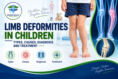
Limb Deformities in Children: Types, Causes, Diagnosis and Treatment (2026 Guide)
Filters & Insights
Learn about limb deformities in children, including types, causes, symptoms, diagnosis, treatment options, and when surgery may be needed.

Medically reviewed content · Published 2026 · Last reviewed September 2026
A limb deformity in a child is an abnormal alignment, shape, rotation, or length of an arm or leg. The most important thing for parents to understand is that a child’s limbs change shape substantially as they grow — bow legs in a toddler and knock knees in a young child are, in the great majority of cases, normal developmental stages, not disease, and resolve on their own without treatment.
A smaller number of children have a true structural (pathological) deformity caused by a congenital condition, a growth-plate problem, an old fracture that healed out of alignment, a metabolic condition like rickets, or a neuromuscular disorder that genuinely does need evaluation and, sometimes, treatment. This guide exists mainly to help you tell the difference, since that distinction not the appearance of the limb alone — is what actually determines whether your child needs anything done at all.
A visible difference in how a child’s legs or arms look is not, by itself, a reason to assume something is wrong. This guide explains normal limb development, the types and causes of true pediatric limb deformity, when evaluation and imaging are actually needed, and current treatment options — from simple observation through guided growth and, in selected cases, surgical correction — along with what care for a genuine limb deformity looks like for international families considering treatment in India.
This is general medical education for parents and caregivers, not a diagnosis, and it is not a substitute for a pediatric orthopedic assessment of your specific child.
Table of Contents
ToggleNormal Development vs. Pathological Deformity
This is the single most important section in this guide, because getting this distinction right is what prevents unnecessary worry, unnecessary imaging, and unnecessary treatment.
Children’s leg alignment follows a well-documented, predictable pattern as they grow, and it is not straight-line development toward “normal” adult alignment — it swings through phases that would look concerning in an adult but are entirely expected in a child:
- Infancy (roughly birth to age 2): Most babies and toddlers have some degree of bow legs (genu varum) — this reflects the folded position of the legs in the womb and is a normal finding, not a deformity, in this age range.
- Early-to-mid childhood (roughly ages 2 to 4): The legs typically swing the other way, and many children develop knock knees (genu valgum), often reaching their most pronounced knock-kneed appearance around age 3 to 4. This, too, is a normal developmental phase in the majority of children.
- Later childhood (roughly age 6 to 7 onward): Alignment gradually straightens further toward the adult pattern, which typically includes a small, normal degree of residual valgus (mild knock-knee alignment) that persists into adulthood for most people.
Because there’s genuine variation in exactly when and how much a given child moves through these phases, precise age cutoffs and angle measurements shouldn’t be treated as rigid diagnostic thresholds — a pediatric clinician assesses the whole picture (the degree of the finding, whether it’s symmetric, whether it’s improving or worsening over time, and the child’s overall growth pattern), not just where a single measurement falls relative to an average.
What tends to distinguish normal developmental variation from something that deserves closer evaluation:
| Finding | More Consistent with Normal Development | More Consistent with a Condition Worth Evaluating |
|---|---|---|
| Symmetry | Roughly symmetric between the two legs | Clearly one-sided or significantly asymmetric |
| Trend over time | Gradually improving as the child grows | Stable, worsening, or appearing/worsening outside the typical age window |
| Associated findings | None — child is otherwise healthy, growing normally, pain-free | Pain, limping, short stature, or other systemic findings present |
| Age pattern | Fits the typical developmental sequence above | Bow legs persisting or worsening well past infancy/toddlerhood, or knock knees that are severe or don’t begin improving by school age |
Persistent, asymmetric, or progressively worsening findings deserve evaluation more than a mild, symmetric, age-appropriate variation does — this single principle does more work in this guide than any other.
Types of Limb Deformities in Children
Angular deformities: bow legs and knock knees
Genu varum (bow legs) describes outward bowing at the knees; genu valgum (knock knees) describes the knees angling inward toward each other. As described above, both are frequently normal developmental stages. When angular deformity is genuinely pathological, it can arise at the femur (thigh bone), tibia (shin bone), or both, and — beyond the knee — similar varus (angled inward) or valgus (angled outward) patterns, or flexion/extension deformities, can occur at other joints.
Rotational deformities: intoeing and out-toeing
These describe how the feet point relative to the direction of walking, rather than how the legs bow side to side. Common causes include femoral anteversion (an inward-rotated thigh bone, a common cause of intoeing, especially in young girls, that very frequently improves on its own through childhood), internal or external tibial torsion (rotation at the level of the shin bone), and variations in foot shape itself. Most rotational differences in young children improve naturally as they grow, and intoeing in a young, otherwise healthy child is not, by itself, a reason to recommend surgery — this is one of the areas where overtreatment (unnecessary bracing or referral for surgical consideration) is a genuine, documented pattern worth actively guarding against.
Limb-length discrepancy
A difference in the length of the two legs (or, less commonly, arms) can be true (an actual difference in bone length) or apparent (the legs are the same length, but something else — like a hip or pelvis alignment issue — makes them look different). Causes include congenital conditions present from birth, a previous fracture or growth-plate injury that affected growth on one side, infection, neurological conditions, hip disorders, and, less commonly, tumors or vascular conditions. How much difference matters, and whether it needs treatment, depends on the current amount, whether it’s expected to grow or shrink as the child continues to grow, and the underlying cause — there’s no single universal threshold above which every discrepancy requires treatment, and small differences are common and usually don’t need any intervention.
Congenital, foot/ankle, upper-limb, and contracture conditions
A smaller group of children have conditions present from birth or that develop through a different mechanism than the common patterns above:
- Congenital limb deficiencies (partial or complete absence or underdevelopment of part of a limb, such as fibular or tibial deficiency, or congenital femoral deficiency) vary enormously in severity and functional impact — some require no intervention beyond monitoring, while others involve complex, staged treatment planning.
- Clubfoot (congenital talipes equinovarus) and other congenital foot conditions like vertical talus are structural conditions generally identified at or shortly after birth, requiring specialist pediatric orthopedic management, most often starting with serial casting.
- Flexible flatfoot — a very common, usually painless finding where the arch is visible when the foot is lifted but flattens with weight-bearing — is a normal variant in most children and does not, on its own, need bracing or surgery; rigid flatfoot, where the arch doesn’t reappear regardless of weight-bearing, is a different, less common finding that does warrant evaluation.
- Upper-limb differences — including cubitus varus or valgus (elbow angulation, sometimes following a fracture that healed with some malalignment), forearm deformity, and congenital hand or arm differences — are managed individually based on cause and functional impact.
- Joint contractures — a fixed limitation in how far a joint can move — can involve the bone itself, the surrounding soft tissue (muscles, tendons, or the joint capsule), or both, and the distinction matters for treatment planning.
Causes of Pathological Limb Deformity
| Cause Category | How It Typically Produces Deformity |
|---|---|
| Normal developmental variation | Not a disease process — reflects the typical phases of childhood alignment described above |
| Congenital conditions and genetic syndromes | Present from birth; severity and pattern vary by the specific condition |
| Growth-plate (physeal) injury | Trauma, infection, or other insult to the growth plate can cause uneven growth on one side, producing progressive angulation over time as the child continues growing |
| Fracture and malunion | A fracture that heals in a less-than-ideal position can leave angulation, rotation, or shortening. This may improve with remaining growth through remodeling, though not always |
| Rickets and metabolic bone disease | Softened, under-mineralized bone, often from vitamin D or calcium/phosphate disorders, can bow under normal weight-bearing forces |
| Neuromuscular disorders, including cerebral palsy | Muscle imbalance, spasticity, and resulting contractures can alter alignment and gait over time |
| Infection | Bone or joint infection near a growth plate can disrupt normal growth |
| Skeletal dysplasia | Genetic conditions affecting bone growth broadly can cause disproportionate limb growth and characteristic deformity patterns |
| Hip conditions (Legg-Calvé-Perthes disease, slipped capital femoral epiphysis) | Disease at the hip can alter gait and produce apparent limb alignment changes. SCFE, most common in adolescents, needs prompt evaluation if suspected |
| Tumors and tumor-like bone conditions | Uncommon cause, relevant to specific clinical presentations rather than routine evaluation |
Symptoms and Signs
How a limb deformity is noticed varies by type. Purely appearance-based findings — bow legs, knock knees, intoeing, out-toeing — are often noticed by parents simply through how a child looks or walks, without the child having any symptoms at all. Others cause functional signs: limping, unequal leg lengths, frequent tripping, difficulty running or keeping up with peers, pain, fatigue with activity, reduced joint range of motion, or difficulty participating in sports. Which of these apply, if any, depends heavily on the specific type and underlying cause — a normal developmental variation typically causes no functional symptoms at all, which is itself a reassuring sign, while pain, limping, or functional limitation point more toward something worth a closer look.
When should parents worry?
Findings that are more concerning, and worth a pediatric or pediatric orthopedic evaluation, include: significant or clearly one-sided deformity, progressive worsening over time rather than gradual improvement, pain, limping, any functional limitation, loss of joint motion, deformity that developed after an injury, or deformity accompanied by other systemic symptoms (poor growth, other health concerns). Persistent asymmetry or progression is generally a more meaningful warning sign than a mild, symmetric variation that a parent simply finds noticeable.
Red flags requiring prompt or urgent evaluation
Seek prompt medical assessment — the same day or urgently, not a routine scheduled visit — for: a sudden inability to walk or bear weight, severe pain following an injury or suspected fracture, acute joint swelling, fever combined with bone or joint pain (a possible sign of infection), a rapidly progressive deformity, new weakness or loss of sensation, or severe hip pain with an inability to bear weight, particularly in an adolescent (which can be a sign of slipped capital femoral epiphysis, a condition that needs prompt diagnosis to protect the hip joint). These situations call for urgent evaluation rather than a wait-and-see approach.
Will My Child Outgrow This?
This is probably the single most common question parents have, and the honest answer depends entirely on which specific finding is present — it is genuinely different for different conditions, which is exactly why an accurate diagnosis matters more than the general question.
Commonly improves on its own: Physiologic (normal developmental) genu varum in infancy and toddlerhood; physiologic genu valgum in early-to-mid childhood; internal tibial torsion in toddlers; and femoral anteversion in young children — all of these typically improve through the child’s own natural growth, usually without any treatment at all.
Does not reliably self-correct, and warrants evaluation rather than assumption: Blount disease (discussed below), deformity from an established growth-plate injury, deformity from rickets or other metabolic bone disease, deformity from a neuromuscular condition, and significant congenital deformities. Assuming any deformity will simply be outgrown without an actual diagnosis is the mirror-image mistake to over-treating normal variation — both stem from skipping the step of finding out which category a specific child’s finding actually falls into.
Blount Disease: An Important Look-Alike
Blount disease is a growth disorder specifically affecting the inner part of the upper shin bone’s growth plate, causing progressive bowing (tibia vara) that — unlike physiologic bow legs — does not improve on its own and can worsen over time. It can appear in younger children (infantile Blount disease, often noticed as bow legs persisting or worsening beyond the age when physiologic bowing should be improving) or in adolescents (adolescent Blount disease, often associated with higher body weight).
Distinguishing Blount disease from ordinary physiologic bow legs matters because the treatment approach is genuinely different, and the two can look quite similar in a younger child before the distinguishing features become clear on examination and imaging — this is not a diagnosis to make from appearance alone, and it’s a good example of why persistent or worsening bow legs, specifically past the age when physiologic bowing typically starts resolving, deserve a specialist look rather than continued reassurance based on appearance.
How Limb Deformities Are Diagnosed
Diagnosis starts with a detailed history — the child’s age, birth history, developmental milestones, family history, any previous trauma or surgery, nutritional history, and the specific symptoms or functional concerns — combined with a thorough physical examination.
Physical examination and measurement
A pediatric orthopedic assessment typically includes observing the child standing (important for accurately assessing lower-limb alignment, since it looks different lying down) and walking or running, along with assessing limb-length differences, hip rotation, knee alignment, tibial torsion, foot progression angle, joint range of motion, muscle strength, and, where relevant, neurological function.
Specific measurements a specialist may reference — such as the mechanical and anatomic axes of the leg, or specific angles at the knee — are clinical tools for precisely describing and tracking a deformity over time, not numbers a parent needs to interpret independently; your child’s specialist will explain what any specific measurement means for your child’s situation.
Imaging
Not every child with a limb difference needs an X-ray, and X-rays are not typically the first step for straightforward, mild, symmetric findings that fit a normal developmental pattern. When imaging is needed, standing (weight-bearing) long-leg X-rays are the standard way to assess alignment, bone length, joint orientation, and growth plates for angular deformities.
CT has a role in planning for complex rotational deformities specifically, with attention paid to minimizing a child’s radiation exposure given how much CT delivers relative to X-ray.
MRI may be used when there’s concern about the growth plate itself, cartilage, soft tissue, an underlying tumor, infection, or complex congenital anatomy that plain X-ray can’t adequately show. None of these advanced imaging tests should be used automatically or as a default step — they’re reserved for situations where the answer will genuinely change what happens next.
Blood tests
Blood tests are not required for every limb deformity evaluation — they’re considered specifically when rickets, another metabolic bone condition, an inflammatory condition, or infection is suspected based on the clinical picture, not as a routine screening step for ordinary developmental variation.
Treatment: Matched to Cause, Severity, and Growth Remaining
Treatment decisions depend on the child’s age and how much growth they have remaining, the underlying cause, severity, location, whether the finding is progressing or improving, symmetry, presence of symptoms, functional impact, and — for conditions caused by an underlying disease like rickets — treatment of that underlying condition. There is no universal treatment appropriate for “limb deformity” as a category; the same visible finding can call for completely different management depending on what’s actually causing it.
Observation
For the large majority of children — those with physiologic bow legs, physiologic knock knees, and many normal rotational variations — observation is the correct and complete treatment, not a placeholder for “doing nothing.” It involves periodic clinical reassessment and growth monitoring, with repeat imaging only if there’s a specific clinical reason to obtain it, not on a fixed schedule regardless of how the child is progressing.
Physiotherapy
Physiotherapy can genuinely help with strength, balance, gait, flexibility, and functional adaptation, and has a real role for children with neuromuscular conditions or those recovering function after treatment. It’s important to be direct about its limits, though: exercises cannot mechanically straighten a structurally deformed growing bone — physiotherapy is not a treatment for genu varum, genu valgum, or a genuine angular or rotational bone deformity in the way it can help with strength or gait, and framing it that way to parents sets an expectation the treatment can’t meet.
Bracing and casting
Bracing has evidence-supported roles in specific situations but is not an effective treatment for physiologic bow legs or normal rotational development — bracing a child for a condition that would have resolved on its own doesn’t speed up that resolution and shouldn’t be recommended for it. Casting, particularly serial casting, has a well-established, different role — most notably as the standard first-line treatment for clubfoot, and for selected other conditions — where it works by gradually, progressively repositioning the limb over a series of cast changes, a genuinely different mechanism and evidence base than bracing for angular deformity.
Guided growth (hemiepiphysiodesis)
Guided growth uses a child’s own remaining growth to gradually correct an angular deformity, rather than surgically cutting and realigning the bone outright. A small plate or screw is placed at the growth plate on the side of the bone growing “too fast” relative to the other side, temporarily slowing growth on that side while the other side continues growing normally — over months, this gradually straightens the limb as the child grows.
This only works while significant growth remains, so it’s not an option for a child near the end of skeletal growth, and it requires ongoing follow-up: undercorrection, overcorrection, and a “rebound” of the deformity after the implant is removed are all recognized possibilities, along with implant-related complications, which is why this isn’t a one-time procedure but one requiring monitoring through to completion.
Osteotomy
An osteotomy is a surgical procedure that directly cuts the bone and realigns it into the corrected position, then stabilizes it with fixation (plates, screws, pins, or a rod) while it heals — used when a deformity is significant, growth remaining is insufficient for guided growth to be effective, or correction is needed more quickly than gradual growth modulation would achieve. This is a more involved procedure than guided growth, with a real recovery period and real surgical risks, and there is no single universal recovery timeline that applies to every osteotomy — it depends on the specific bone, technique, and the individual child’s healing.
Limb-length discrepancy treatment
Options span a genuine range depending on how much difference is present and how much is predicted at skeletal maturity: observation for small differences, a shoe lift for mild ones, epiphysiodesis (deliberately, permanently slowing growth on the longer side to let the shorter side catch up, appropriate only while growth remains), limb shortening procedures on the longer side, and limb lengthening — using external fixation or, in appropriate candidates, an internal lengthening nail — to gradually lengthen the shorter bone through controlled distraction.
Limb lengthening in particular is a demanding treatment, not a simple fix: it carries real risk of infection, joint stiffness, nerve or blood vessel problems, difficulties with the new bone that forms during lengthening (the “regenerate”), malalignment, and device-related complications, and it often involves multiple procedures over an extended period — this should be presented to families honestly as a significant undertaking, not a routine outpatient fix.
When is surgery really necessary?
Surgery — guided growth, osteotomy, or limb lengthening — may be considered for significant or progressive deformity, genuine functional impairment, pain attributable to the deformity, a structural abnormality with little likelihood of spontaneous improvement, meaningful limb-length discrepancy, or complex congenital deformity.
A visible deformity, on its own, is not a reason for surgery — this is worth stating plainly given how much anxiety a “crooked-looking” leg can generate in a parent even when the underlying finding is entirely normal for the child’s age.
Long-Term Consequences and Prognosis
Untreated pathological deformity — meaning a true structural problem, not a normal developmental phase — may, depending on the specific type and severity, contribute over time to abnormal gait, uneven loading across a joint, pain, reduced function, earlier joint wear, and limitations in sports participation. It’s important, though, not to overstate this: not every deformity, treated or untreated, inevitably leads to arthritis or long-term disability — the actual long-term trajectory depends on the specific condition, its severity, and, where treatment is appropriate, how effectively it’s addressed. Prognosis overall depends on the underlying cause, severity, the child’s age and remaining growth, location of the deformity, treatment chosen, timing, and any associated conditions — this guide won’t cite a single success rate or outcome percentage as though it applies universally, since outcomes genuinely vary by these factors.
Prevention
Some contributing factors are modifiable: adequate nutrition and prevention of vitamin D deficiency can prevent nutritional rickets specifically, appropriate injury prevention reduces fracture risk, and appropriately managed fracture care (and prompt attention to any suspected growth-plate injury) reduces the chance of malunion-related deformity. It’s equally important to be direct that many congenital and genetic limb deformities cannot be prevented — they aren’t the result of anything a parent did or didn’t do, and framing prevention as something within every family’s control would be both inaccurate and unfair to families dealing with a congenital condition.
Limb Deformity Treatment in India: Specialists and Cost
Treatment approach and the specialists involved differ substantially depending on whether a child has normal developmental variation needing only reassurance and monitoring, an angular or rotational deformity needing guided growth or osteotomy, a limb-length discrepancy, a congenital deformity, a growth-plate injury, or a deformity related to an underlying neuromuscular or metabolic condition — which is exactly why an accurate diagnosis, not the general topic of “limb deformity,” should drive which specialists and treatment path a family pursues in India or anywhere else.
Cost
Genuinely reliable, current published cost data specific to pediatric guided growth and limb reconstruction procedures in India is more limited than for many of the other topics in this series — this is worth stating directly rather than presenting a cost table that looks more authoritative than the underlying data supports. What can be said with reasonable confidence:
| Component | Approximate Cost (Where Reasonably Established) |
|---|---|
| Pediatric orthopedic consultation | $30–$150 |
| Standing long-leg X-ray | $30–$100 |
| CT or MRI, where needed | $150–$500 |
| Serial casting (e.g., clubfoot treatment, per course) | Variable; needs direct quoting based on the number of cast changes involved |
| Guided growth (hemiepiphysiodesis) surgery | Roughly $1,200–$2,200 based on currently available published figures; confirm directly due to limited independent verification |
| Corrective osteotomy | Roughly $1,400–$2,200 based on currently available published figures, with meaningful variation by complexity and fixation method |
| Limb lengthening | Not reliably established from published sources; complex, often multi-procedure treatment requiring an individualized quote from the treating center |
For any procedure beyond simple diagnostics, request a written, itemized quote based on your child’s specific diagnosis and proposed treatment plan directly from the treating hospital, rather than relying on any published figure, including the ones above — pediatric limb reconstruction pricing varies significantly by complexity, technique, implant type, and length of follow-up required, more than most other topics covered in this content series.
Choosing a center
Look for a genuine pediatric orthopedic team — not adult orthopedic surgeons treating a child as an exception — including a pediatric orthopedic surgeon (ideally with specific limb-reconstruction experience if that’s relevant to your child’s diagnosis), pediatric radiology, pediatric physiotherapy, and, where relevant, genetic or metabolic specialist input. Ask specifically about the center’s experience with your child’s specific diagnosis, not general pediatric orthopedic volume, and verify credentials directly rather than relying on third-party rankings.
The International Patient Journey
- Share medical reports and, where available, photographs and existing X-ray/MRI/CT images
- Pediatric orthopedic specialist reviews the case remotely
- Confirm diagnosis and determine whether the finding is developmental or pathological
- Assess growth remaining and, if needed, develop a treatment plan
- Receive a written cost estimate
- Arrange medical visa and travel documentation
- Travel and attend in-person specialist assessment
- Complete treatment if clinically appropriate
- Begin rehabilitation where relevant
- Receive discharge documentation
- Coordinate follow-up with your child’s home-country pediatric team
Not every child evaluated through this process will be found to need treatment at all, let alone international travel or surgery — a genuine possible (and good) outcome of this process is confirmation that observation alone is appropriate.
Questions for Your Pediatric Orthopedic Specialist
- Is my child’s alignment normal for their age, and is it symmetrical?
- Is it likely to improve naturally, or does it need active treatment?
- What is the underlying cause?
- Is the deformity getting worse, staying the same, or improving over time?
- Does my child actually need an X-ray or other imaging right now?
- How much growth does my child have left, and how does that affect the options?
- Could this be Blount disease or another condition that looks like normal bow legs but isn’t?
- Could there be a metabolic cause, like rickets?
- Should we observe for now, or is treatment appropriate now?
- Would physiotherapy or bracing genuinely help this specific finding?
- Is guided growth an option, or has that window passed?
- What happens if we wait?
- What are the realistic risks of the surgery being proposed?
- What would rehabilitation involve, and could the deformity recur afterward?
How Shifam Health Helps International Families
Shifam Health is a medical tourism facilitator, not a hospital, pediatric orthopedic surgeon, or treating medical provider — we don’t diagnose or treat your child’s limb condition. What we do is help international families navigate the practical side of accessing pediatric orthopedic care in India: collecting and sharing medical records with appropriate specialists, coordinating appointments and hospital logistics, helping obtain a written treatment and cost estimate, assisting with medical visa documentation, arranging airport pickup and family accommodation, providing interpreter support where needed, and staying in touch for follow-up communication once you return home.
We don’t guarantee that your child will need surgery, a specific outcome, or any particular treatment path — that determination belongs entirely to the treating pediatric orthopedic specialist reviewing your child’s actual case.
If you have questions about your child’s limb alignment or a specific diagnosis, share your available reports with our team on WhatsApp or through a quick inquiry form — there’s no obligation, and we typically respond within 24 hours.
Frequently Asked Questions
What are limb deformities in children?
Abnormal alignment, shape, rotation, or length of an arm or leg. Some are normal developmental variations, while others need evaluation.
Are bow legs normal in babies?
Yes. Mild bow legs are common in infants and toddlers and usually improve naturally with growth.
When do bow legs become abnormal?
Persistent, worsening, or clearly asymmetric bowing, especially with pain, may require evaluation for conditions such as Blount disease or rickets.
Are knock knees normal in children?
Yes. Mild knock knees are a normal developmental stage, often most noticeable around ages 3–4, and usually improve with growth.
What causes limb deformities in children?
Causes include normal development, growth-plate injuries, fractures, Blount disease, rickets, skeletal dysplasia, and genetic conditions.
Does intoeing correct itself?
Usually. Common causes such as femoral anteversion and internal tibial torsion often improve without surgery.
What is limb-length discrepancy?
A difference in the length of two limbs. It may result from growth differences, previous injury, developmental conditions, or other causes.
Can growth-plate injuries cause deformity?
Yes. Uneven growth after a growth-plate injury can gradually produce angular deformity or limb-length differences.
Can physiotherapy correct bone deformities?
Physiotherapy can improve strength, gait, and function but cannot mechanically straighten a structurally deformed bone.
How much does limb deformity treatment cost in India?
Costs vary according to the diagnosis, procedure, hospital, and complexity. A personalized estimate requires specialist assessment and imaging.
Conclusion
The great majority of limb differences parents notice in young children are normal developmental variation, not disease — and the single most protective thing a parent can do is get an accurate assessment of which category their child’s specific finding falls into, rather than assuming either extreme (that a visible difference must mean something is wrong, or that any difference will simply be outgrown without checking). When a genuine structural deformity is present, modern pediatric orthopedic treatment — ranging from simple observation through guided growth to surgical correction when truly needed — is generally effective, individualized to the child’s age, growth remaining, and specific diagnosis.
If you’re concerned about your child’s limb alignment, a pediatric orthopedic evaluation, not the appearance of the limb alone, is what will actually answer the question.
This article is for general medical education for parents and caregivers and does not replace individualized assessment by a qualified pediatrician or pediatric orthopedic specialist. It is not a diagnosis and should not be used to decide for or against treatment for any individual child.

Popular Posts From Last Week
- September 18, 2026
- shifamhealth
Written by: Shifam Health Editorial Team — Orthopedic Content Division Medically reviewed by: Shifam Health Clinical Advisory Panel…
- September 16, 2026
- shifamhealth
Ankle valgus deformity is an abnormal alignment in which the ankle tilts toward the outside of the body,…
- September 20, 2026
- shifamhealth
Medically reviewed content · Published 2026 · Last reviewed September 2026 A limb deformity in a child is…
- September 15, 2026
- shifamhealth
Medically reviewed content · Published 2026 · Last reviewed September 2026 Bow legs medically called genu varum —…
- September 14, 2026
- shifamhealth
Medically reviewed content · Published 2026 · Last reviewed September 2026 A “broken ankle” is the common term…
- September 15, 2026
- shifamhealth
Medically reviewed content · Published 2026 · Last reviewed August 2026 A burst fracture is a spinal vertebra…
- September 13, 2026
- shifamhealth
If a lumbar MRI report mentions “kissing spinous processes” or “Baastrup’s disease,” it’s worth pausing before assuming this…
- September 14, 2026
- shifamhealth
Scoliosis is a three-dimensional spinal deformity — sideways curvature combined with rotation — and it is genuinely not…
- September 16, 2026
- shifamhealth
Epithelial ovarian cancer is the most common broad category of ovarian cancer, arising from the cells covering the…
- September 17, 2026
- shifamhealth
Hepatoblastoma is a rare malignant liver tumor that develops primarily in young children, most commonly before age 5….
- September 18, 2026
- shifamhealth
Kyphosis means forward curvature of the spine — and some kyphosis is completely normal anatomy in the mid-back…
- September 19, 2026
- shifamhealth
Lumbar radiculopathy is a neurological syndrome caused by irritation, inflammation, or compression of a nerve root in the…