



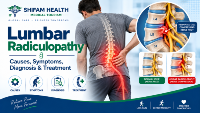
Lumbar Radiculopathy (2026): Causes, Symptoms, Diagnosis & Treatment
Filters & Insights
Learn about lumbar radiculopathy, including its causes, symptoms, diagnosis, and treatment options for nerve-related lower back and leg pain.

Lumbar radiculopathy is a neurological syndrome caused by irritation, inflammation, or compression of a nerve root in the lower back, typically producing leg pain along with numbness, tingling, or weakness in a pattern that follows the affected nerve root. It’s related to, but not identical to, “sciatica,” which describes a pain pattern rather than a confirmed neurological diagnosis.
Most cases particularly those caused by a herniated disc improve with conservative treatment over weeks to a few months. Surgery is generally reserved for cauda equina syndrome, significant or progressive weakness, or persistent disabling symptoms that haven’t responded to appropriate nonoperative care, not for a disc finding on MRI by itself.
Table of Contents
ToggleWhat Is Lumbar Radiculopathy?
Lumbar radiculopathy is a neurological syndrome caused by dysfunction — irritation, inflammation, or compression — of a nerve root in the lumbar (lower back) or lumbosacral spine. This is different from several related but not interchangeable terms:
- Low-back pain: pain localized to the lower back, which may or may not involve a nerve root
- Sciatica: a symptom pattern describing pain along the distribution of the sciatic nerve, into the buttock and leg — this describes where the pain is felt, not a confirmed diagnosis
- Radiculopathy: a neurological syndrome implying actual nerve-root dysfunction, which may include objective sensory loss, weakness, or reflex changes — not just pain
- Radicular pain: pain specifically generated by nerve-root irritation, which can occur with or without the objective neurological findings that define radiculopathy
- Referred pain: pain felt in an area distant from its actual source, without nerve-root involvement
- Lumbar spinal stenosis: narrowing of the spinal canal or nerve pathways, which can cause radiculopathy but is a distinct structural diagnosis
- Peripheral neuropathy: nerve dysfunction from a cause outside the spine (such as diabetes), which can mimic radiculopathy but has an entirely different origin
Sciatica describes a pattern of pain along the sciatic distribution, while radiculopathy implies neurological dysfunction that may include sensory loss, weakness, or reflex changes. Not every case of sciatica turns out to be radiculopathy with objective neurological findings, and not every lumbar radiculopathy presents with the classic sciatic pain pattern — this distinction matters for both diagnosis and expectations.
Lumbar Spine and Nerve-Root Anatomy
The lumbar spine consists of five vertebrae (L1–L5), cushioned by intervertebral discs, with a spinal canal running through the center. In adults, the spinal cord itself ends higher up (around the L1–L2 level), and below that point the canal contains a bundle of nerve roots called the cauda equina (Latin for “horse’s tail”). Individual nerve roots exit the spine at each level through small openings called neural foramina, then travel downward and combine to form major nerve trunks — most notably the sciatic nerve, which is actually formed from the combination of several lumbosacral nerve roots (primarily L4 through S3) rather than being a single nerve root itself.
This anatomical detail matters clinically: because the cauda equina is a bundle of multiple nerve roots rather than a single spinal cord, injury or compression at this level can affect bladder, bowel, and saddle-area function in addition to leg symptoms — which is central to recognizing the cauda equina syndrome emergency discussed later in this article.
Nerve Roots and Dermatomes: L3, L4, L5, and S1 Patterns
| Nerve Root | Typical Pain/Sensory Area | Possible Weakness | Reflex Findings |
|---|---|---|---|
| L3 | Front of thigh, sometimes to the knee | Hip flexion, knee extension (quadriceps) | Knee (patellar) reflex may be reduced |
| L4 | Front/inner thigh to inner shin | Knee extension (quadriceps), sometimes hip adduction | Knee (patellar) reflex may be reduced |
| L5 | Outer thigh/shin to top of foot and big toe | Ankle and big-toe dorsiflexion (lifting the foot/toe upward); weakness here can contribute to foot drop | No reliable single reflex associated with L5 |
| S1 | Back of thigh/calf to outer foot and little toes | Ankle plantar flexion (pushing the foot downward, as in standing on tiptoes) | Ankle (Achilles) reflex may be reduced |
These patterns are general clinical guides, not absolute rules. Real patients frequently don’t follow the textbook pattern exactly, due to anatomical variation, overlapping nerve supply, and the fact that more than one level can be affected simultaneously. Dermatomes (sensory territories) are not perfectly precise from person to person. This is exactly why a hands-on neurological examination by a clinician — rather than self-diagnosis based on symptom location — is the reliable way to determine which level, if any, is involved.
Causes of Lumbar Radiculopathy
The most common cause by far is a herniated (or extruded/protruding) intervertebral disc compressing or irritating a nearby nerve root. Other significant causes include lumbar spinal stenosis (central canal narrowing), foraminal stenosis and lateral recess stenosis (narrowing of the specific passages nerve roots travel through), degenerative spondylolisthesis (one vertebra slipping relative to another due to degenerative changes) and isthmic spondylolisthesis (a distinct cause related to a defect in a specific part of the vertebra), bony osteophytes (bone spurs) and facet-joint degeneration contributing to narrowing, and, less commonly, a synovial cyst arising from a degenerated facet joint.
Less common but clinically important causes include vertebral fracture, spinal tumors, infection (including epidural abscess), postoperative scar tissue in patients with prior spine surgery, and other iatrogenic or structural causes. These causes are not equally common — disc herniation and degenerative stenosis account for the large majority of cases, while tumor, infection, and fracture are comparatively rare but important not to miss, particularly when red-flag features are present.
Risk Factors
Risk factors include advancing age and disc degeneration, occupational heavy lifting, repetitive bending or twisting, smoking, obesity (where evidence supports an association), a previous disc herniation, general spinal degeneration, previous spine surgery, trauma, and certain congenital or anatomical factors. Some evidence also points to an association with prolonged sitting, though this is less consistently established than the other factors listed. These are risk factors, not direct, guaranteed causes — many people with these risk factors never develop radiculopathy, and some people without obvious risk factors do. There’s no reason to view this as a condition patients caused through personal failing.
Symptoms
Symptoms can include low-back pain, buttock pain, leg pain (often the dominant symptom, sometimes more prominent than the back pain itself), burning pain, electric or shooting pain, tingling, pins-and-needles sensations, numbness, weakness, altered sensation, and reduced reflexes on examination. It may worsen with coughing, sneezing, straining, sitting, standing, walking, or specific spinal positions — though not every patient experiences all of these triggers, and the absence of a “classic” trigger doesn’t rule out radiculopathy.
Radicular Pain vs. Radiculopathy: What’s the Difference?
This is one of the article’s most important distinctions. Radicular pain is pain generated by irritation or inflammation of a nerve root — it can be severe and follow a nerve root’s typical distribution, but by itself doesn’t necessarily involve objective neurological loss. Radiculopathy specifically means a neurological deficit caused by nerve-root dysfunction — potentially including measurable weakness, sensory loss, or reduced reflexes on examination, not just pain. A patient can have significant radicular pain without objective radiculopathy, and, less commonly, can have radiculopathy (weakness found on examination) with relatively modest pain. Understanding which of these you actually have — determined through examination, not by how the pain feels — genuinely affects how the situation should be approached.
Is Sciatica the Same as Lumbar Radiculopathy?
Not exactly. Sciatica is a symptom pattern — pain traveling along the path of the sciatic nerve, typically from the lower back or buttock down the back of the leg. Radiculopathy is a neurological syndrome implying actual nerve-root dysfunction. These overlap substantially in practice, since sciatic-distribution pain often does come from lumbosacral nerve-root irritation, but they are not perfect synonyms: not every patient with sciatica has an objective neurological deficit, and not every lumbar radiculopathy presents with the classic sciatic pain pattern — some present predominantly with weakness or numbness, or with pain in a different distribution depending on which nerve root is affected (for example, L3 or L4 radiculopathy more commonly causes anterior thigh symptoms rather than classic posterior-leg “sciatica”).
When Is Lumbar Radiculopathy an Emergency?
Cauda Equina Syndrome
This is the most important emergency to recognize. Warning signs include new urinary retention or difficulty urinating, loss of bladder control, new bowel dysfunction, saddle anesthesia (numbness in the inner thighs, genital, or perineal area), severe or progressive symptoms affecting both legs, and rapidly worsening neurological deficits. New bladder dysfunction, bowel dysfunction, saddle numbness, or rapidly progressive weakness requires urgent medical assessment and should not be managed as routine sciatica.
Other Emergency or Urgent Scenarios
Rapidly progressive motor weakness, major trauma, suspected spinal fracture, fever combined with severe back pain, immunosuppression with severe spinal pain, suspected epidural abscess, known cancer with new severe back or leg symptoms, and unexplained weight loss combined with concerning spinal symptoms all warrant prompt, thorough evaluation rather than routine, delayed assessment.
International patients should not delay emergency assessment to arrange travel. If any of the above symptoms are present, local emergency care should be sought immediately rather than waiting to travel for treatment elsewhere.
How Is Lumbar Radiculopathy Diagnosed?
History
Clinicians assess onset, duration, and progression of symptoms; the location and radiation pattern of pain; presence of numbness or weakness; walking tolerance; bladder and bowel function; saddle sensation; any trauma; fever; cancer history; infection risk factors; unexplained weight loss; previous similar episodes; previous spine surgery; current medications; and functional limitations.
Physical Examination
Examination typically includes assessment of gait and posture, lumbar range of motion, the straight-leg raise test and crossed straight-leg raise test (maneuvers that can reproduce radicular symptoms), motor strength testing, sensory testing, reflex testing, heel-walking and toe-walking (which test specific nerve root function), and, where relevant, hip examination and vascular assessment to help rule out non-spinal mimics. Neurological examination is particularly important whenever radiculopathy is genuinely suspected, since it’s what distinguishes radiculopathy (with objective findings) from radicular pain or nonspecific back pain alone.
Motor Deficits by Level
Weakness patterns can offer clues, though they should always be assessed objectively by examination rather than self-diagnosed: L4 radiculopathy can be associated with quadriceps (knee extension) weakness; L5 radiculopathy can be associated with weakness in ankle or big-toe dorsiflexion (lifting the foot or toe upward) — significant L5 weakness can contribute to a foot drop; S1 radiculopathy can be associated with plantar-flexion weakness (pushing the foot downward). These patterns vary between individuals, and objective testing by a clinician is the reliable way to establish which level, if any, is affected.
Lumbar radiculopathy is primarily a clinical diagnosis, supported by imaging or electrodiagnostic testing when appropriate — it is not established by imaging alone.
When Do You Need an MRI for Lumbar Radiculopathy?
Immediate MRI is not automatically necessary for uncomplicated acute symptoms without red flags. Many episodes of acute radicular pain improve over the initial weeks with conservative treatment, and imaging at that early stage often doesn’t change management. MRI becomes more appropriate when symptoms persist despite an appropriate trial of conservative management, when there’s a significant or progressive neurological deficit, when cauda equina syndrome is suspected, when infection or tumor is suspected, after severe trauma, for surgical planning, or when the clinical picture genuinely needs clarification before a treatment decision.
A critical principle that deserves emphasis: an abnormal MRI does not automatically mean the abnormality is causing the patient’s symptoms. Disc bulges and degenerative changes are extremely common findings on lumbar MRI in people without any symptoms at all — this is well established in the imaging literature. A treatment decision should never be made from an MRI report in isolation; the finding needs to genuinely correlate with the patient’s specific symptoms and examination findings.
X-Ray
Plain X-rays are limited for assessing nerve-root pathology directly but can help evaluate spinal alignment, fractures, spondylolisthesis, degenerative changes, and, with dynamic (flexion-extension) views, instability in selected cases. X-ray is not equivalent to MRI for assessing nerve compression.
CT
CT can be useful for detailed bony anatomy, fracture assessment, evaluating stenosis in selected circumstances, surgical planning, and situations where MRI is contraindicated or unavailable. Radiation exposure is a relevant consideration when choosing between CT and other imaging.
EMG/Nerve Conduction Studies
EMG/NCS is not automatically required for every patient with suspected lumbar radiculopathy. These electrodiagnostic tests measure nerve and muscle electrical activity and can be genuinely useful when the clinical picture and imaging don’t clearly match, when distinguishing radiculopathy from peripheral neuropathy is important, or when the diagnosis remains uncertain after standard evaluation. Timing matters — findings may not be reliably present in the very acute phase — and results should always be interpreted alongside the clinical picture, not in isolation.
Differential Diagnosis
| Condition | Typical Clues | Key Differences |
|---|---|---|
| Nonspecific low-back pain | Pain localized to the back, without leg radiation or neurological findings | No nerve-root involvement |
| Piriformis/deep gluteal syndrome | Buttock-predominant pain, sometimes with leg symptoms | Different anatomical source; can mimic sciatica but arises outside the spine |
| Hip osteoarthritis | Groin or thigh pain, worse with hip movement | Pain pattern and exam findings differ from radicular distribution |
| Sacroiliac-region pain | Pain localized near the SI joint, specific provocative maneuvers positive | Distinct from nerve-root-pattern radiation |
| Peripheral neuropathy | Often bilateral, stocking-glove distribution, associated with diabetes or other systemic causes | Not confined to a single nerve-root pattern |
| Meralgia paresthetica | Numbness/burning on the outer thigh only | Affects a single peripheral (non-spinal) nerve, not a nerve root |
| Vascular claudication | Leg pain with walking, relieved promptly by rest, without positional/postural pattern | Vascular rather than neurological origin |
| Cauda equina/spinal cord pathology | Bilateral symptoms, bladder/bowel involvement, saddle numbness | Surgical emergency; must be actively excluded |
| Shingles (herpes zoster) | Band-like pain often preceding a rash, in a specific dermatomal pattern | Skin findings and different underlying cause |
| Myofascial pain | Localized muscular tenderness and trigger points | No true neurological findings |
| Hamstring pathology | Pain with specific movements, localized to the hamstring | Musculoskeletal rather than neurological in origin |
Natural History
This deserves genuinely honest treatment rather than a simplistic promise. Many acute disc-related radicular syndromes improve without surgery, often over the course of weeks to a few months. Symptom improvement and imaging findings do not always move together — a patient can feel substantially better while the MRI still shows an abnormality, since inflammation around the nerve root can settle and some extruded disc material can regress over time.
That said, some patients develop persistent pain, and some develop recurrent symptoms even after initial improvement. Natural history genuinely differs by underlying cause — for instance, symptomatic spinal stenosis tends to follow a more variable, sometimes more persistent course than an acute disc herniation. The presence of a significant or progressive neurological deficit changes the picture and generally warrants more prompt evaluation rather than an extended trial of conservative care.
First-Line Treatment for Lumbar Radiculopathy
Evidence-based conservative management generally includes staying reasonably active rather than resorting to prolonged bed rest, activity modification around clearly aggravating movements, physiotherapy, graded exercise, patient education about the condition, symptom-focused pain management, and rehabilitation guided by the individual’s specific findings. Treatment specifics depend on the underlying cause, duration and severity of symptoms, any neurological findings, and the degree of functional limitation — not a single generic protocol applied to everyone.
Should You Stay in Bed With Lumbar Radiculopathy?
Generally, no — prolonged bed rest is not preferred for uncomplicated cases. Reasonable, tolerated activity and pacing tend to be favored over strict rest, with the goal of gradually returning toward normal function while avoiding activities that significantly aggravate symptoms. This doesn’t mean pushing through severe pain or, especially, any progressive neurological symptoms — those warrant medical reassessment, not persistence through the activity that’s worsening them.
Medications
Medication categories commonly considered include NSAIDs, acetaminophen/paracetamol, selected neuropathic pain medications for radicular symptoms, short-term muscle relaxants in some cases, and, in selected circumstances, short courses of oral corticosteroids. Evidence for some commonly used medications in radicular pain specifically is genuinely limited or mixed — this should be acknowledged honestly rather than glossed over. Opioids are not casually recommended given their risk profile and limited evidence for durable benefit in this context. Medication suitability depends on individual factors including kidney function, gastrointestinal disease risk, cardiovascular risk, pregnancy status, use of anticoagulants, age, and other medications — this article does not provide individualized prescribing guidance.
Physiotherapy
Physiotherapy typically involves graded strengthening, mobility work, neural mobilization techniques where appropriate, core/trunk conditioning, functional restoration, and patient education, adapted to the individual’s specific symptoms, any neurological deficits, underlying diagnosis, and functional goals.
No single exercise protocol works identically for everyone — a program appropriate for degenerative stenosis differs from one appropriate for an acute disc herniation.
Epidural Steroid Injections
Transforaminal, interlaminar, and caudal epidural approaches are all used depending on the specific clinical situation and target level. These injections can provide meaningful short-term pain relief for appropriately selected patients by reducing inflammation around the affected nerve root, and may have a role in bridging a patient through a difficult period or, in some cases, potentially influencing the need for surgery. They are not a cure — benefit is often temporary, and injections do not remove or repair a herniated disc or structurally correct stenosis. Procedural risks exist, including rare but serious complications, and this article does not provide procedural instructions — these should be discussed directly with the treating pain specialist or surgeon.
When Does Lumbar Radiculopathy Require Surgery?
Surgery may be considered when there is cauda equina syndrome, a severe or progressive neurological deficit, persistent disabling radicular symptoms despite an appropriate trial of nonoperative treatment, imaging-confirmed nerve compression that genuinely matches the patient’s symptoms, significant functional impairment, or selected structural causes (such as significant instability) where surgical stabilization is clinically indicated.
A disc bulge or herniation on MRI alone is not an indication for surgery. The decision depends on the combination of persistent or worsening symptoms, objective neurological findings, and genuine imaging-clinical correlation — not the imaging report by itself.
Surgical Options
Microdiscectomy
The most common surgical procedure for a symptomatic herniated disc, involving removal of the herniated disc fragment compressing the nerve root through a relatively small incision, aiming to decompress the nerve and relieve radicular pain and, where possible, improve neurological function.
Endoscopic Discectomy
A more minimally invasive variant using an endoscope and smaller incisions to achieve a similar goal in selected cases. Potential advantages include smaller incisions and, in appropriate hands, comparable outcomes to standard microdiscectomy for suitable cases, though it involves a meaningful learning curve for the surgeon and appropriate patient selection matters — it is not automatically the right choice for every disc herniation pattern.
Laminectomy/Decompression
Removal of part of the lamina (bony arch) to relieve pressure on nerve structures, most commonly used for lumbar spinal stenosis rather than an isolated disc herniation.
Foraminotomy
A procedure specifically widening the neural foramen (the passage a nerve root exits through) when foraminal narrowing is the specific cause of compression.
Spinal Fusion
Fusion is generally not required for every disc herniation or case of radiculopathy. It’s reserved for selected indications such as spinal instability, significant spondylolisthesis, deformity, certain cases of recurrent pathology, or specific mechanical/structural problems where evidence supports its use. Fusion is not automatically superior for straightforward disc-related radiculopathy without instability — adding fusion when it’s not indicated adds surgical complexity and risk without a corresponding benefit.
What Discectomy Surgery Actually Does
The realistic goal of discectomy is to remove the offending disc material or otherwise decompress the affected nerve root, with the aim of reducing radicular (leg) pain and improving neurological function where possible. Surgery tends to be more effective at relieving leg pain from clear nerve compression than at resolving nonspecific chronic low-back pain — this distinction matters when setting expectations, since a patient whose dominant complaint is back pain rather than leg pain may see a different degree of benefit than one whose primary problem is radicular leg pain from a clearly compressing disc fragment. Complete elimination of all pain cannot be promised.
Surgery vs. Conservative Treatment
| Factor | Conservative Treatment | Surgery |
|---|---|---|
| Invasiveness | Non-invasive | Invasive, with associated surgical risks |
| Typical role | First-line for most cases without red flags | Reserved for specific indications (see above) |
| Speed of relief | Often gradual, over weeks to months | Can provide faster relief of leg pain in appropriately selected patients |
| Neurological deficit | May be appropriate for mild, non-progressive findings | More strongly considered with significant or progressive deficits |
| Recurrence | N/A in the surgical sense, though symptoms can recur | Possible (recurrent disc herniation), particularly in the following months to years |
| Risks | Generally low, medication- and activity-specific | Surgery-specific risks (see below) |
| When generally preferred | Most uncomplicated cases, especially early on | Cauda equina syndrome, progressive weakness, or persistent disabling symptoms unresponsive to appropriate conservative care |
Surgery can provide faster relief for appropriately selected patients, but this does not mean every patient benefits from — or needs — immediate surgery. Many patients who are candidates for either pathway do well with an initial trial of conservative treatment.
Surgical Risks
Risks associated with lumbar spine surgery for radiculopathy include infection, bleeding, dural tear (a tear in the membrane covering the nerves, sometimes leading to a spinal fluid leak), nerve injury, recurrent disc herniation, persistent pain despite technically successful surgery, incomplete neurological recovery, anesthesia-related complications, blood clots in selected higher-risk patients, spinal instability in selected cases, and the potential need for revision surgery. Risk varies according to patient age, overall health, spinal anatomy, surgical approach, whether there has been previous surgery in the area, and the severity and complexity of the underlying pathology.
Recurrent Disc Herniation and Persistent Symptoms After Surgery
Recurrence can occur — a new disc fragment can herniate at the same level after an initially successful discectomy, and this is a genuinely distinct situation from persistent pain that never fully resolved after the original surgery. Recurrent herniation may sometimes require a repeat discectomy; in selected cases involving significant instability or multiple recurrences, fusion may become a more relevant consideration — but not every recurrent disc requires fusion, and this should be assessed individually.
Persistent symptoms after surgery — sometimes loosely called “failed back surgery” — can have multiple distinct causes: recurrent or residual nerve compression, scar tissue formation around the nerve, pathology at an adjacent spinal level, instability, an incorrectly identified original pain source, neuropathic pain that persists independent of ongoing compression, or entirely non-spinal causes. Persistent pain should not be automatically labeled as “failed surgery” without a genuine reassessment to identify what’s actually driving the ongoing symptoms — this determines whether repeat surgery is even relevant, let alone likely to help.
Minimally Invasive and Robotic Surgery
Minimally invasive decompression, tubular retractor techniques, endoscopic approaches, computer navigation, and robotic assistance are all used in appropriate contexts in modern spine surgery. None of the following claims are accurate, and this article avoids them explicitly: robotic surgery is not automatically safer; it does not guarantee greater precision in every case; minimally invasive surgery does not eliminate complications; these technologies do not guarantee faster recovery for every patient; and no patient “should” automatically choose a robotic or minimally invasive approach without individualized assessment of whether it’s appropriate for their specific anatomy and pathology. Technology is a tool that assists a skilled surgeon — it is not, by itself, a guarantee of a better outcome.
Recovery
Recovery varies meaningfully by treatment pathway. Uncomplicated radiculopathy managed conservatively often shows gradual improvement over weeks to a few months, guided by symptom response rather than a fixed calendar. Epidural injections may provide relief within days for those who respond, though benefit duration varies. Microdiscectomy and endoscopic discectomy typically allow relatively early mobilization, often with same-day or next-day walking and discharge within 24–48 hours for straightforward cases, with progressive return to activity over the following weeks. Decompression procedures for stenosis and fusion procedures, when genuinely indicated, generally involve a longer recovery arc than a straightforward discectomy.
Across pathways, recovery generally includes early guided walking, pain management, wound care where relevant, physiotherapy, and a graduated return to driving, work, lifting, and exercise or sport. No universal recovery timeline can be promised — it depends on the specific procedure, pre-treatment severity and duration of symptoms, age, and individual healing.
Return to Work
Timing genuinely differs by job demands: desk-based work is often resumed sooner than light physical work, which in turn is typically resumed before heavy manual labor requiring significant lifting or prolonged standing. The specific timeline depends on the procedure performed (or lack thereof, for conservative management), degree of neurological recovery, pain levels, rehabilitation progress, and guidance from your surgeon or physiotherapist — this article does not provide a universal number of days for any category.
Exercise After Radiculopathy
Walking is generally well tolerated and often encouraged, including in the early recovery period for most patients. Gym exercise, cycling, and swimming can typically be resumed in a graduated manner once cleared by your treating clinician, with specific timing depending on your situation rather than a fixed universal schedule. Running and higher-impact activities are generally reintroduced later in recovery, again guided individually. Stretching and yoga can often be incorporated, with attention to avoiding positions or movements that clearly reproduce nerve-related symptoms. Exercise should be paused and reassessed, rather than pushed through, if you notice new or worsening weakness or other neurological symptoms — this is different from ordinary muscle soreness or mild discomfort, which is a more routine part of graduated rehabilitation.
Pregnancy
Lumbar radicular symptoms can occur during pregnancy, related to postural changes, weight distribution, and pelvic girdle changes. Medication choices during pregnancy require careful individualized consideration with your obstetric and treating team, since many commonly used medications for radicular pain have specific safety considerations in pregnancy. MRI can generally still be performed when clinically necessary during pregnancy (without gadolinium contrast, which is more selectively used), so pregnancy does not, by itself, make appropriate imaging impossible when there’s a genuine clinical need — such as suspected cauda equina syndrome, which remains a surgical emergency in pregnancy and should not be managed expectantly.
Long-Term Prognosis
Most patients with straightforward disc-related radiculopathy do well with appropriate management, whether conservative or surgical when indicated. That said, some patients experience recurrent symptoms, some experience recurrent disc herniation, some develop chronic pain, and future degenerative changes at the same or adjacent levels remain possible over time — none of these outcomes are universal, but none can be entirely ruled out either.
Nerve recovery may take longer than pain relief, and it’s important to be explicit: severe or prolonged nerve compression can result in incomplete neurological recovery even after successful decompression. This is stated not to frighten patients with typical, briefer episodes, but because it’s a genuine, evidence-supported consideration for those with more significant or longstanding deficits, and it’s part of why earlier evaluation matters when weakness is present and progressing.
When to See a Specialist (and When to Get a Second Opinion)
Specialist evaluation is warranted for persistent radicular symptoms not improving with initial conservative measures, any objective weakness, red-flag symptoms of any kind, or uncertainty about the diagnosis. A second opinion is particularly valuable when major surgery — especially fusion — is recommended, when your symptoms and MRI findings don’t seem to clearly match, when the diagnosis remains uncertain, when a neurological deficit is progressing, when previous surgery hasn’t resolved symptoms, when recurrent disc herniation is present, when symptoms persist despite appropriate treatment, or when multiple different treatment options have been proposed by different clinicians.
Useful records to bring to any consultation or second opinion include MRI images and reports, X-rays, CT if available, EMG/NCS results if performed, a full medication history, records of any previous injections, operative notes if you’ve had prior surgery, discharge summaries, and physiotherapy records.
Lumbar Radiculopathy Treatment in India (2026)
Appropriately equipped centers in India coordinate care across spine specialists (orthopedic or neurosurgical), neurologists, radiologists, pain specialists, and rehabilitation physiotherapists. Depending on the specific case, potential treatment approaches include full clinical and imaging evaluation, structured conservative management and physiotherapy, epidural injections for appropriately selected patients, and, when genuinely indicated, microdiscectomy, endoscopic discectomy, laminectomy/decompression, foraminotomy, or fusion for specific structural indications.
Cost of Lumbar Radiculopathy Treatment in India
Published international-patient cost estimates found for lumbar disc/nerve compression surgery show meaningful variation reflecting procedure type, approach, and hospital tier:
- Microdiscectomy: published estimates ranging roughly from $1,500–$7,000 USD, with some sources citing figures toward the higher end (around $5,500–$6,500) and others noting a broader range depending on hospital and complexity
- Endoscopic discectomy: some sources cite a somewhat lower range, roughly $2,200–$4,200 USD, for minimally invasive/endoscopic variants, though this varies by hospital and technique
- Lumbar decompression/laminectomy: published estimates broadly in the range of roughly $1,900–$9,500 USD depending on hospital tier and complexity
- Spinal fusion (when genuinely indicated): generally higher than discectomy alone, with published estimates roughly in the $3,200–$9,000+ USD range depending on levels involved and implants used
These figures show real variance across sources, reflecting differences in hospital tier, city, surgeon, and what’s specifically included in a quoted package (implants, hospital stay length, physiotherapy, follow-up). Costs are also affected by the number of spinal levels involved, anesthesia, hospital stay duration, and any complications. Treat these as reference ranges rather than a firm quote — an itemized, case-specific estimate from the treating hospital, based on your actual imaging and diagnosis, is necessary for an accurate figure.
Separately from medical treatment costs, international patients should budget for accommodation, local transportation, airport transfers, interpreter or local support where needed, and follow-up visits. India should not be presented as automatically the cheapest or best destination — genuine verification of a specific hospital’s and surgeon’s experience with your specific procedure matters more than general cost comparisons.
Can International Patients Travel to India for Lumbar Radiculopathy Treatment?
International treatment may reasonably be considered when the diagnosis is already established, the patient is medically stable, the situation is elective (not an emergency), relevant records and imaging can be reviewed by a specialist in advance, and appropriate specialist care is confirmed to be available.
However, patients with cauda equina symptoms, rapidly progressive weakness, suspected epidural abscess, acute major trauma, or severe neurological deterioration should seek urgent local medical care rather than delaying treatment for international travel — this is not a situation where medical tourism planning should take priority over immediate local emergency evaluation.
International Patient Journey
- Share medical records and describe your symptoms, including onset and progression
- Share MRI, X-ray, and/or CT images and reports
- Specialist review of your case
- Confirm which nerve root(s), if any, appear affected and correlate imaging with your actual symptoms
- Assess neurological severity
- Discuss conservative, interventional (injection), or surgical options as appropriate
- Receive an individualized treatment plan
- Receive a cost estimate
- Coordinate specialist/hospital appointments
- Medical visa documentation support where applicable
- Travel and accommodation coordination
- In-person assessment on arrival
- Additional investigations if needed
- Treatment, if medically appropriate
- Hospital recovery
- Rehabilitation
- Discharge planning
- Follow-up
- Coordination with your local physician after returning home
How Shifam Health Can Help
Shifam Health is a medical tourism facilitator/coordinator — not the treating physician, surgeon, hospital, or a diagnostic laboratory, and does not replace emergency care. Shifam Health may assist with collecting medical records, coordinating specialist opinions, sharing MRI/CT reports and images with treating hospitals, hospital coordination, appointment scheduling, communicating treatment plans, coordinating cost estimates, medical visa support, accommodation, airport transfers, interpreter/local assistance, hospital logistics, and follow-up coordination. They cannot guarantee surgery success, pain relief, neurological recovery, exact cost, exact recovery time, visa approval, or hospital acceptance.
International patients can share their medical records and spinal imaging with Shifam Health to help coordinate an appropriate specialist review and understand potential treatment options in India.
Myths vs. Facts
Myth: Every sciatica case is radiculopathy. Fact: Sciatica describes a pain pattern; radiculopathy specifically requires objective neurological findings on examination, which not every sciatica patient has.
Myth: Every disc bulge causes nerve compression. Fact: Disc bulges are extremely common on imaging in people without any symptoms at all — the finding must correlate with the clinical picture.
Myth: A slipped disc always requires surgery. Fact: Most herniated discs improve with conservative treatment; surgery is reserved for specific indications like significant/progressive deficits or persistent disabling symptoms.
Myth: An MRI is always necessary immediately. Fact: For uncomplicated acute symptoms without red flags, immediate imaging is often not necessary and doesn’t typically change initial management.
Myth: Bed rest is the best treatment. Fact: Reasonable activity and pacing are generally preferred over prolonged bed rest for uncomplicated cases.
Myth: Exercise will make a disc herniation worse. Fact: Appropriately guided, graded exercise is generally part of recommended treatment, not something to avoid entirely.
Myth: Epidural injections cure the problem permanently. Fact: Injections can provide meaningful but often temporary pain relief; they don’t repair a herniated disc or structurally correct stenosis.
Myth: Surgery guarantees complete recovery. Fact: Surgery aims to decompress the nerve and relieve symptoms, but pain, numbness, or weakness can persist, particularly after prolonged or severe compression.
Myth: Fusion is needed for every recurrent disc. Fact: Many recurrent disc herniations are treated with repeat discectomy alone; fusion is reserved for cases involving genuine instability or specific recurrent patterns.
Myth: Robotic spine surgery is always better. Fact: Robotic assistance is a tool that may help with certain technical aspects but doesn’t guarantee a better outcome for every patient.
Frequently Asked Questions
What is lumbar radiculopathy?
Lumbar radiculopathy occurs when a nerve root in the lower spine becomes irritated or compressed, causing pain, numbness, tingling, or weakness in the leg.
What causes lumbar radiculopathy?
A herniated disc is the most common cause. Spinal stenosis, spondylolisthesis, injury, and less commonly tumors or infection can also cause it.
What are the symptoms of lumbar radiculopathy?
Symptoms may include shooting or burning leg pain, numbness, tingling, muscle weakness, and difficulty lifting the foot.
Is lumbar radiculopathy the same as sciatica?
They are closely related. Sciatica describes pain along the sciatic nerve, while radiculopathy specifically indicates nerve-root dysfunction.
How is lumbar radiculopathy diagnosed?
Diagnosis usually involves a medical history and neurological examination. MRI, CT, or EMG may be recommended when necessary.
Can lumbar radiculopathy heal without surgery?
Yes. Many cases improve with activity modification, medication, physiotherapy, and time, particularly when caused by a herniated disc.
Does physiotherapy help lumbar radiculopathy?
Yes. Targeted physiotherapy can improve mobility, strength, function, and pain while supporting recovery.
When is surgery needed?
Surgery may be considered for progressive weakness, severe persistent symptoms despite conservative treatment, or emergencies such as cauda equina syndrome.
What is a microdiscectomy?
Microdiscectomy removes the portion of a herniated disc pressing on a nerve root to relieve leg pain and nerve compression.
What is cauda equina syndrome?
A serious spinal emergency involving compression of multiple lower spinal nerves, potentially causing bladder or bowel problems, saddle numbness, or severe weakness.
How long does lumbar radiculopathy take to improve?
Many patients improve within several weeks to a few months, although recovery varies depending on the cause and severity.
How much does treatment cost in India?
Costs vary by treatment and complexity. Surgical treatment may range from approximately $1,500–$9,500+ USD, requiring an individualized hospital quote.
Final Takeaway
Lumbar radiculopathy is a neurological syndrome caused by nerve-root dysfunction in the lower back — related to, but not identical to, “sciatica,” and not something an MRI finding alone can diagnose or justify treating. Most cases, particularly those from an acute disc herniation, improve with conservative treatment over weeks to a few months, while cauda equina symptoms (new bladder or bowel dysfunction, saddle numbness) represent a genuine surgical emergency requiring immediate assessment, not delayed or self-managed care.
Surgery, when genuinely indicated, aims to decompress the affected nerve root and relieve leg pain — it can provide meaningful, sometimes faster relief for appropriately selected patients, but pain, numbness, or weakness can persist even after technically successful surgery, particularly following prolonged or severe nerve compression. The right treatment path depends on the specific cause, severity, neurological findings, and how symptoms respond to appropriate initial care — not a one-size-fits-all protocol.
International patients can share their medical records and spinal imaging with Shifam Health to help coordinate an appropriate specialist review and understand potential treatment options in India.
This article is for general educational purposes and does not replace clinical examination, imaging review, diagnosis, emergency assessment, or individualized treatment advice from a qualified spine specialist or neurologist.

Popular Posts From Last Week
- September 18, 2026
- shifamhealth
Written by: Shifam Health Editorial Team — Orthopedic Content Division Medically reviewed by: Shifam Health Clinical Advisory Panel…
- September 16, 2026
- shifamhealth
Ankle valgus deformity is an abnormal alignment in which the ankle tilts toward the outside of the body,…
- September 15, 2026
- shifamhealth
Medically reviewed content · Published 2026 · Last reviewed September 2026 Bow legs medically called genu varum —…
- September 14, 2026
- shifamhealth
Medically reviewed content · Published 2026 · Last reviewed September 2026 A “broken ankle” is the common term…
- September 15, 2026
- shifamhealth
Medically reviewed content · Published 2026 · Last reviewed August 2026 A burst fracture is a spinal vertebra…
- September 13, 2026
- shifamhealth
Carpal tunnel syndrome (CTS) is compression of the median nerve as it passes through the carpal tunnel —…
- September 13, 2026
- shifamhealth
If a lumbar MRI report mentions “kissing spinous processes” or “Baastrup’s disease,” it’s worth pausing before assuming this…
- September 14, 2026
- shifamhealth
Scoliosis is a three-dimensional spinal deformity — sideways curvature combined with rotation — and it is genuinely not…
- September 12, 2026
- shifamhealth
Medically reviewed content · Published 2026 · Last reviewed September 2026 Discitis is inflammation or infection involving an…
- September 16, 2026
- shifamhealth
Epithelial ovarian cancer is the most common broad category of ovarian cancer, arising from the cells covering the…
- September 17, 2026
- shifamhealth
Hepatoblastoma is a rare malignant liver tumor that develops primarily in young children, most commonly before age 5….
- September 19, 2026
- shifamhealth
Lumbar radiculopathy is a neurological syndrome caused by irritation, inflammation, or compression of a nerve root in the…