



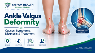
Ankle Valgus Deformity: Causes, Symptoms, Diagnosis & Treatment
Filters & Insights
Ankle valgus deformity has many possible causes — bone, ligament, flatfoot, or arthritis. Learn how it's diagnosed, when surgery is actually needed, and what options exist.

Ankle valgus deformity is an abnormal alignment in which the ankle tilts toward the outside of the body, often because of altered alignment of the tibia, hindfoot, or the structures that support the ankle. It can develop from bone deformity, ligament insufficiency, flatfoot-related mechanics, previous injury, neuromuscular conditions, or arthritis.
A few things worth understanding immediately:
- The ankle joint is formed by the tibia, fibula, and talus working together
- Normal alignment distributes load evenly across the joint; valgus alignment shifts loading toward the lateral (outer) side of the ankle
- The deformity can originate above the ankle (in the tibia), within the ankle joint itself, or below it (in the hindfoot or foot) — and where it originates genuinely changes what treatment makes sense
- The visible appearance alone does not identify the exact cause — a careful clinical examination combined with weight-bearing imaging is generally needed to work that out
- This is not one single condition with one single treatment — that’s the central point this entire guide is built around
Table of Contents
ToggleUnderstanding Normal Ankle Alignment
The ankle isn’t a simple hinge — it’s formed by three bones working together:
- Tibia — the shinbone, forming the top and inner (medial) wall of the ankle joint
- Fibula — the smaller outer leg bone, forming the outer (lateral) wall
- Talus — the bone that sits between the leg bones and the foot, forming the moving part of the joint
Together, the tibia and fibula form what’s called the ankle mortise — a socket that the talus sits within. Stability comes from:
- The deltoid ligament on the inner (medial) side
- The lateral ligament complex on the outer side
- The bony congruence of the mortise itself
- Supporting tendons, including the Achilles tendon and posterior tibial tendon
Below the ankle joint sits the subtalar joint and the rest of the hindfoot, which also contributes meaningfully to overall lower-limb alignment — this is exactly why problems below the ankle (like hindfoot valgus or flatfoot) can influence, or be confused with, the ankle joint itself.
Ankle Valgus vs. Hindfoot Valgus vs. Flatfoot vs. Varus
These terms are related but genuinely not interchangeable. Ankle valgus describes tilt within the ankle joint itself. Hindfoot valgus describes the heel shifting outward relative to the leg, below the ankle joint. Flatfoot (pes planovalgus) is a broader deformity that can include hindfoot valgus alongside arch collapse and forefoot changes. And valgus and varus describe opposite directions of tilt.
| Term | What It Describes |
|---|---|
| Ankle Valgus | The ankle mortise/talus relationship itself tilts outward |
| Hindfoot Valgus | The heel shifts outward relative to the leg, at the subtalar joint below the ankle |
| Pes Planovalgus / Flatfoot | A broader deformity involving medial arch collapse, hindfoot valgus, forefoot abduction, and other structural changes together |
| Alignment | General Direction |
|---|---|
| Valgus | The lower segment deviates outward relative to the segment above it |
| Varus | The lower segment deviates inward relative to the segment above it |
Why this distinction matters practically: flatfoot can genuinely contribute to ankle valgus in some patients, as long-standing hindfoot collapse gradually alters loading on the ankle joint above it — but having a flat foot does not automatically mean a person has ankle valgus, and the two terms should not be used interchangeably. Getting this distinction right is often the first step in understanding your own diagnosis if these terms have come up in a consultation.
Causes of Ankle Valgus Deformity
Ankle valgus can result from a bony malunion after fracture, ligament insufficiency (particularly of the deltoid ligament), flatfoot-related mechanics, growth-related problems in children, neuromuscular conditions, or advanced ankle arthritis — often more than one factor contributes together.
| Cause Category | Examples |
|---|---|
| Post-Traumatic | Malunited ankle fracture, distal tibial deformity from prior injury, syndesmotic (ligament between tibia and fibula) injury, chronic instability after prior ankle surgery |
| Ligament-Related | Deltoid ligament insufficiency, chronic medial ankle instability |
| Flatfoot-Related | Progressive collapsing foot deformity (a current, broader term that has largely replaced the older label “posterior tibial tendon dysfunction,” though you may still encounter both terms), hindfoot valgus, medial column collapse |
| Pediatric/Developmental | Developmental variation, distal tibial growth plate problems, congenital or syndromic conditions |
| Neuromuscular | Conditions like cerebral palsy or Charcot-Marie-Tooth disease, which affect muscle balance around the ankle and foot |
| Arthritic | Advanced ankle osteoarthritis, inflammatory arthritis (like rheumatoid arthritis), post-traumatic arthritis |
This list is not exhaustive, and in many patients more than one factor contributes at once — for example, a prior fracture that healed imperfectly combined with gradual ligament stretching over years. Identifying which factor (or combination) applies to your specific case is exactly what the diagnostic process, discussed below, is designed to work out.
Risk factors that make ankle valgus more likely to develop include previous ankle fracture or surgery, chronic ankle instability, pre-existing flatfoot, tendon or ligament dysfunction, neuromuscular disorders, inflammatory arthritis, and significant prior trauma — though it’s worth being clear that no single one of these factors guarantees the deformity will develop.
Symptoms
Symptoms vary widely depending on the underlying cause, and some patients have little or no pain despite visible deformity. There’s no single symptom pattern that identifies ankle valgus specifically.
Possible symptoms include:
- Ankle pain — which can be medial (inner), lateral (outer), or more generalized around the joint
- Swelling
- A feeling of instability or the ankle “giving way”
- Fatigue or aching during walking
- Difficulty walking on uneven surfaces
- Reduced tolerance for standing or walking for extended periods
- Shoe-fitting problems, sometimes from a widened or misshapen foot
- Progressive visible deformity over time
- Reduced ankle range of motion
Pain location does not by itself establish the underlying cause — medial ankle pain, for example, could relate to deltoid ligament strain, posterior tibial tendon involvement, or several other structures, which is exactly why a full examination matters more than symptom location alone.
What ankle valgus can look like (without this being a basis for self-diagnosis): the ankle may appear tilted, the heel may look shifted outward, the foot may appear flattened, and shoe wear patterns can become uneven over time. Visual appearance alone cannot establish the diagnosis or its cause — it can only prompt the evaluation that does.
When Should You See a Doctor?
Evaluation is generally worthwhile for:
- Progressive or worsening visible deformity
- Persistent ankle pain
- Recurrent ankle sprains or ongoing instability
- Difficulty walking or reduced walking tolerance
- Sudden worsening after an injury
- Significant swelling
- New deformity noticed in a child
- New deformity developing in someone with known inflammatory or neuromuscular disease
More urgent assessment is appropriate after significant trauma, or if you’re unable to bear weight on the ankle at all — these situations shouldn’t wait for a routine appointment.
How Ankle Valgus Is Diagnosed
Diagnosis combines a detailed history and physical examination with weight-bearing X-rays taken under normal standing load — imaging taken without weight-bearing can understate or miss the true deformity, which is why standing films specifically matter here.
General diagnostic pathway:
- Medical history, including any previous injury or surgery
- Standing examination and gait assessment
- Foot and ankle alignment examination
- Range-of-motion assessment
- Ligament stability testing
- Weight-bearing X-rays
- Specialized radiographic alignment measurements
- CT, in selected cases
- MRI, in selected cases
- Blood tests, where inflammatory arthritis is suspected
Diagnosis of Ankle Valgus Deformity
- Physical examination: Checks standing alignment, hindfoot position, arch height, ankle and subtalar motion, ligament stability, tendon function, gait, and neurovascular status.
- Weight-bearing X-rays: Show ankle alignment under normal body weight. AP, mortise, and lateral views, with hindfoot alignment views when needed, help assess deformity and joint alignment.
- CT scan: May be used for complex bone deformities, malunion, bone loss, or detailed surgical planning.
- MRI: Helps evaluate ligaments, tendons, cartilage, and osteochondral lesions when soft-tissue problems are suspected.
- Blood tests: ESR, CRP, rheumatoid factor, or anti-CCP may be ordered when inflammatory arthritis is suspected.
Flexible vs. Rigid Deformity
This distinction meaningfully shapes treatment. A flexible deformity can partially or substantially correct with positioning or when weight is taken off the foot; a rigid deformity remains fixed regardless of positioning, often reflecting structural bone changes, advanced arthritis, or longstanding soft-tissue contracture.
| Type | Characteristics | General Treatment Implications |
|---|---|---|
| Flexible | Alignment improves with positioning or non-weight-bearing testing | Bracing, orthotics, and joint-preserving procedures are more often viable options |
| Rigid | Deformity remains fixed regardless of positioning | Often requires more structural correction, and options may be more limited by the fixed bony or joint changes present |
Severity generally isn’t described using a single universal staging system across all causes of ankle valgus — instead, your specialist will describe severity in terms of your specific symptoms, degree of malalignment, joint congruence, whether arthritis is present, and how much functional limitation you’re experiencing.
Can Ankle Valgus Be Treated Without Surgery?
Some flexible or mild deformities can be managed successfully without surgery, particularly when symptoms are limited — but established, structural (especially rigid) deformity generally cannot be reliably “straightened” through exercises or bracing alone. Nonsurgical treatment is often aimed at reducing symptoms and improving function rather than reversing the underlying structural problem.
Nonsurgical options include:
- Observation, appropriate for mild, asymptomatic deformity
- Activity modification
- Supportive footwear
- Orthotics — custom devices, sometimes with medial or lateral posting, which can support associated flatfoot or help redistribute load, though they cannot permanently straighten established bony deformity
- Bracing — including ankle-foot orthoses or other stabilizing braces, chosen based on whether the deformity is flexible or rigid
- Physical therapy — focusing on strengthening, balance training, gait training, and calf/foot muscle conditioning
- Weight management, where relevant to overall load on the joint
- Pain management, including activity modification and clinician-directed approaches
On exercises specifically: exercise and physical therapy can genuinely improve strength, balance, functional stability, and gait — but exercises do not reliably reverse a fixed, established bony deformity. It’s worth being direct about this distinction, since it’s a common source of unrealistic expectations: therapy helps you function better with the alignment you have; it doesn’t generally restructure the underlying bone or joint.
On injections: corticosteroid or other injections may help symptoms in selected patients, but they do not reliably correct structural valgus alignment, and evidence for regenerative injections (like PRP) specifically for deformity correction should not be overstated — these remain, at most, symptom-management tools rather than deformity-correcting treatments.
When Is Surgery Considered?
Surgery may become a reasonable consideration when:
- Pain persists despite appropriate conservative treatment
- The deformity is progressing over time
- Instability is significant and affecting function
- Walking or daily function is substantially impaired
- The alignment problem threatens the health of the joint cartilage over time
- Arthritis has become advanced
- Structural correction is genuinely needed to address the underlying problem
This is not a checklist where meeting one item means surgery is automatically the right choice — it’s a conversation between you and your specialist weighing your specific symptoms, deformity severity, and goals.
Treatment Must Target the Source of the Deformity
This is arguably the single most important principle in this entire guide, and it’s worth stating as plainly as possible: effective treatment has to address where the deformity is actually coming from, not just where it’s visible.
- If the valgus originates from the tibia, treating only the foot may not correct the underlying problem
- If it’s driven by hindfoot collapse, treatment may need to address the hindfoot and foot specifically, not just the ankle joint
- If deltoid ligament insufficiency is present, ligament stabilization may be a necessary part of any correction
- If advanced arthritis is present, joint replacement or fusion may need to be part of the conversation
No single operation is a universal solution for “ankle valgus” — this is exactly why an accurate diagnosis of the deformity’s actual anatomical source, discussed above, comes before any meaningful treatment planning conversation.
Joint-Preserving Surgery: Osteotomy and Ligament Reconstruction
For appropriate candidates, joint-preserving options aim to correct alignment while keeping the patient’s own ankle joint intact, rather than replacing or fusing it.
Distal tibial osteotomy — a surgical procedure that corrects bony malalignment by deliberately cutting and repositioning the bone into better alignment, then allowing it to heal in the corrected position. This may have a role in selected valgus deformity, particularly where joint-preserving correction is appropriate and the deformity originates significantly from the tibia itself, including in some post-traumatic cases. Not every patient with ankle valgus needs, or is a candidate for, an osteotomy — this depends on where the deformity originates and the condition of the joint itself.
Deltoid ligament reconstruction may be considered when ligament insufficiency is contributing to the deformity, since the deltoid ligament plays a meaningful role in medial ankle stability. Ligament reconstruction alone is not always sufficient — it’s often combined with addressing any associated bony deformity or flatfoot component, since treating the ligament in isolation may not fully correct a deformity that has multiple contributing causes.
Other joint-preserving approaches, selected based on individual anatomy, can include broader realignment procedures, tendon reconstruction, hindfoot correction procedures, and, in selected cases, ankle arthroscopy.
Flatfoot and Progressive Collapsing Foot Deformity
Progressive collapsing foot deformity (PCFD) — the current, broader terminology for what’s sometimes still called flatfoot or, in older literature, posterior tibial tendon dysfunction — can involve hindfoot valgus, medial arch collapse, forefoot abduction, and tendon/ligament dysfunction together, and in advanced cases can contribute to ankle valgus through altered loading over time.
It’s worth being direct: not every flatfoot causes ankle valgus, and equating the two would be inaccurate. But when a longstanding, advanced flatfoot deformity does contribute to ankle-level valgus, effective treatment generally requires addressing the multiple components of the foot/hindfoot problem together, not the ankle in isolation — this is exactly the kind of case where surgical planning may need to correct several structures as part of one coordinated treatment approach, rather than one isolated procedure.
Ankle Valgus With Arthritis: Replacement or Fusion
When ankle valgus is combined with advanced arthritis, treatment may involve alignment correction with either total ankle replacement or ankle fusion.
- Total ankle replacement: Modern implants and surgical techniques have made ankle replacement possible for some patients with valgus deformity. Suitability depends on deformity severity, correctability, bone quality, ligament stability, and surgeon expertise. Additional alignment or ligament-balancing procedures may be needed.
- Ankle fusion: May be preferred for severe deformity, advanced arthritis, poor bone quality, or when replacement is unsuitable. Fusion eliminates ankle motion and may increase stress on nearby joints over time.
The choice between replacement and fusion is individualized based on the deformity, joint condition, activity level, and overall health.
| Feature | Total Ankle Replacement | Ankle Fusion |
|---|---|---|
| Ankle joint motion | Preserved, at least partially | Eliminated |
| Deformity correction | Possible in appropriately selected cases | Also possible |
| Implant involved | Yes | No joint implant (bone-to-bone fusion) |
| Revision surgery | May be required over time | May also be required |
| Effect on adjacent joints | Generally different loading pattern than fusion | Increased stress on adjacent joints is a recognized long-term consideration |
| Best candidate | Highly individualized | Highly individualized |
Neither option is universally “better” — the right choice depends on your specific deformity, bone quality, activity level, arthritis severity, and personal priorities, discussed directly with your surgical team.
Pediatric Ankle Valgus
Children are different from adults because their growth plates are still active, which opens up growth-modulation treatment options that don’t exist once skeletal maturity is reached — but it also means treatment timing has to account for remaining growth, not just current deformity severity.
Possible causes in children include developmental variation, neuromuscular conditions (including cerebral palsy, where muscle imbalance around the foot and ankle can contribute to deformity), distal tibial growth plate disturbances, previous injury, and certain syndromic conditions.
Treatment options specific to the pediatric population:
- Observation, appropriate for many mild or developmental cases, particularly in younger children where some alignment variation can be part of normal development
- Growth-guided correction (sometimes called hemiepiphysiodesis, or temporary guided growth) — a technique that uses a child’s remaining growth to gradually correct alignment over time, appropriate only in selected patients with sufficient growth remaining
- Osteotomy, when deformity is severe or growth-modulation techniques aren’t suitable for the specific case
There is no single universal age at which surgery becomes appropriate for pediatric ankle valgus — this depends on the specific cause, severity, and how much skeletal growth remains, assessed by a pediatric orthopedic specialist.
For children with an underlying neuromuscular condition like cerebral palsy, treatment is generally individualized within the context of that broader condition — orthotics, physical therapy, botulinum toxin injections for muscle imbalance, and surgery can all have a role depending on the full clinical picture, not just the ankle deformity in isolation.
Recovery and Risks
Recovery depends heavily on which specific procedure is performed — there is no single universal timeline that applies across osteotomy, ligament reconstruction, replacement, and fusion.
| Procedure | General Recovery Considerations |
|---|---|
| Osteotomy | Protected weight-bearing during bone healing, followed by gradual rehabilitation |
| Ligament reconstruction | Initial protection, followed by progressive weight-bearing, strengthening, and balance rehabilitation |
| Total ankle replacement | Wound healing, progressive weight-bearing, range-of-motion work, and gait rehabilitation |
| Fusion | Requires full bone healing across the fusion site and adaptation to altered ankle biomechanics afterward |
Possible complications, which vary by specific procedure, can include infection, wound healing problems, nerve injury, blood clots, nonunion or malunion (in osteotomy or fusion), hardware-related problems, recurrent deformity, persistent pain, stiffness, adjacent-joint degeneration over time, and, specifically for replacement, implant loosening or failure requiring revision. These are recognized possibilities to discuss with your surgeon, not predicted outcomes for any individual — and rare complications shouldn’t be treated as likely ones.
Recurrence risk depends on the underlying cause, remaining growth in children, deformity severity, muscle balance, how completely the deformity was surgically corrected, and rehabilitation — there’s no single, reliable recurrence percentage that applies universally across causes and procedures.
What Happens If It’s Left Untreated?
Some mild or flexible deformities can remain stable and manageable for years without surgery, particularly when symptoms are limited. But progressive or more severe deformity, left unaddressed, can potentially lead to:
- Ongoing or worsening pain
- Persistent instability
- Abnormal joint wear over time
- Development or acceleration of arthritis
- Reduced mobility and function
These are possibilities, not inevitabilities — outcomes vary substantially by individual cause and severity, which is exactly why earlier identification of the underlying cause, rather than waiting indefinitely, tends to support better long-term management options.
Prevention
Not every case of ankle valgus can be prevented, particularly congenital, developmental, or neuromuscular causes. That said, some strategies can reduce risk or limit progression:
- Proper treatment and rehabilitation of ankle fractures when they occur
- Thorough rehabilitation after ankle sprains and ligament injuries, rather than returning to activity too early
- Appropriate management of chronic ankle instability rather than letting it persist untreated
- Supportive, well-fitted footwear
- Appropriate treatment of inflammatory arthritis when present
- Early assessment of progressive deformity rather than a prolonged “wait and see” approach
Ankle Valgus Treatment Cost in India
Quick Answer: There is no single “ankle valgus surgery cost” — expenses depend entirely on the underlying cause, deformity severity, and which specific procedure (or combination of procedures) is required.
| Cost Category | What Drives the Cost |
|---|---|
| Diagnosis | Orthopedic consultation, standard and weight-bearing X-rays, CT or MRI where indicated |
| Nonsurgical treatment | Custom orthotics, bracing, physiotherapy course |
| Joint-preserving surgery | Osteotomy, ligament reconstruction, or tendon reconstruction, with associated hardware |
| Flatfoot/hindfoot reconstruction | Complexity varies significantly with how many structures require correction |
| Ankle fusion | Surgical approach, hardware, hospitalization |
| Total ankle replacement | Implant selection, any additional deformity-correcting procedures performed alongside it, hospitalization |
| Rehabilitation and follow-up | Physiotherapy duration, follow-up imaging, and monitoring visits |
| Revision surgery | Costs if a repeat procedure becomes necessary |
Why we’re not quoting one figure: a patient managed with orthotics and physical therapy faces a fundamentally different cost than a patient requiring combined hindfoot reconstruction and total ankle replacement — presenting a single “ankle valgus cost in India” number would genuinely mislead most readers. Cost also varies meaningfully by hospital, city, surgeon, and implant choice where relevant. A personalized estimate, based on your specific diagnosis, deformity source, and recommended procedure, is the only genuinely useful figure, and that requires review of your actual imaging and clinical findings by a specialist.
Ankle Valgus Treatment in India for International Patients
India offers relevant infrastructure for ankle valgus evaluation and treatment at accredited orthopedic centers, including foot-and-ankle surgical subspecialty expertise, deformity-correction experience, pediatric orthopedics where relevant, musculoskeletal imaging, and physiotherapy/rehabilitation services.
Given how specialized deformity correction genuinely is, it’s worth specifically verifying before choosing a center:
- Foot-and-ankle subspecialty expertise specifically — general orthopedic training doesn’t necessarily include deep experience in complex deformity correction
- Genuine deformity-correction experience, including with combined procedures where multiple structures need addressing together
- Pediatric orthopedic expertise, if the patient is a child
- Experience with ankle replacement and fusion in deformed (not just neutral-alignment) ankles specifically, given how much technique matters for outcomes in this scenario
- Adequate weight-bearing imaging capability
- Rehabilitation and physiotherapy support
- Revision surgery capability, should it ever be needed
- Transparent, itemized pricing
This isn’t a claim that India is automatically the right choice for every patient — it depends on the complexity of your specific deformity and practical considerations around travel and follow-up, particularly for procedures requiring extended rehabilitation.
International Patient Journey
- Share medical records, including any prior treatment history
- Share standing (weight-bearing) X-rays — this is genuinely important, as non-weight-bearing images can understate the deformity
- Share CT/MRI, if already available
- Specialist review to help determine the anatomical source of the valgus
- Assessment of flexibility and joint condition
- Evaluation for associated flatfoot, ligament, or tendon problems
- Discussion of nonsurgical options where appropriate
- Determination of whether surgery is genuinely necessary
- Selection of the appropriate corrective procedure, if surgery is indicated
- Transparent cost estimate
- Medical visa assistance
- Travel and accommodation planning
- Treatment in India
- Rehabilitation
- Follow-up and long-term monitoring
This process cannot guarantee complete correction, pain relief, a specific surgical outcome, exact recovery duration, exact cost, or visa approval — no legitimate source can guarantee any of these.
Frequently Asked Questions
What is ankle valgus deformity?
An outward tilt of the ankle that may involve the tibia, ankle joint, hindfoot, or ligaments.
What causes ankle valgus?
Causes include previous fractures, ligament insufficiency, flatfoot, growth problems, neuromuscular conditions, and arthritis.
What are the symptoms?
Pain, swelling, instability, walking difficulty, and visible deformity. Some people have few symptoms.
Is ankle valgus the same as flatfoot?
No. Flatfoot can contribute to valgus alignment, but they are different conditions.
What is hindfoot valgus?
Outward positioning of the heel relative to the leg, usually involving the subtalar region.
What is the difference between valgus and varus?
Valgus means outward angulation, while varus means inward angulation.
Can an old ankle fracture cause valgus?
Yes. A fracture that heals in abnormal alignment can cause post-traumatic valgus.
Can ankle valgus cause arthritis?
Yes. Abnormal joint loading can accelerate wear and contribute to arthritis.
How is ankle valgus diagnosed?
Physical examination and weight-bearing X-rays are usually important. CT or MRI may be needed in selected cases.
Can ankle valgus be treated without surgery?
Mild or flexible cases may benefit from orthotics, bracing, and physical therapy.
Can exercises correct ankle valgus?
Exercise can improve strength and function but cannot reliably correct fixed bony deformity.
When is surgery needed?
Surgery may be considered for persistent pain, instability, progressive deformity, or advanced arthritis despite nonsurgical treatment.
Can ankle replacement be performed?
Yes, carefully selected valgus ankles may undergo replacement when adequate correction is possible.
Is replacement better than fusion?
Neither is universally better. The choice depends on deformity, bone quality, activity, and individual goals.
What does ankle valgus treatment cost in India?
Costs vary by diagnosis and procedure. An accurate estimate requires specialist evaluation and imaging.
Where This Leaves You
Ankle valgus deformity is genuinely not one condition with one treatment — it’s a description of alignment that can come from very different underlying problems, and the right treatment depends entirely on correctly identifying which one applies to you or your child. The single most useful thing you can do is get weight-bearing imaging reviewed by a foot-and-ankle specialist who can identify exactly where your deformity originates, rather than assuming a generic treatment based on the visible appearance alone.
If you’re evaluating treatment options, including treatment in India, sharing your weight-bearing X-rays and any prior imaging or treatment history is the most useful first step toward an accurate, individualized assessment. Shifam Health can help coordinate that record review, specialist and hospital selection, cost estimates, and medical visa guidance — while the actual diagnosis and treatment decisions remain with your orthopedic specialist.




Popular Posts From Last Week
- September 16, 2026
- shifamhealth
Ankle valgus deformity is an abnormal alignment in which the ankle tilts toward the outside of the body,…
- September 10, 2026
- shifamhealth
Medically reviewed content · Published 2026 · Last reviewed August 2026 Blount’s disease, also called tibia vara, is…
- September 15, 2026
- shifamhealth
Medically reviewed content · Published 2026 · Last reviewed September 2026 Bow legs medically called genu varum —…
- September 14, 2026
- shifamhealth
Medically reviewed content · Published 2026 · Last reviewed September 2026 A “broken ankle” is the common term…
- September 15, 2026
- shifamhealth
Medically reviewed content · Published 2026 · Last reviewed August 2026 A burst fracture is a spinal vertebra…
- September 13, 2026
- shifamhealth
Carpal tunnel syndrome (CTS) is compression of the median nerve as it passes through the carpal tunnel —…
- September 13, 2026
- shifamhealth
If a lumbar MRI report mentions “kissing spinous processes” or “Baastrup’s disease,” it’s worth pausing before assuming this…
- September 10, 2026
- shifamhealth
Chronic back pain is one of the most common reasons people seek medical care, and also one of…
- September 11, 2026
- shifamhealth
Myelopathy means dysfunction of the spinal cord itself — not a single disease, but a syndrome that can…
- September 14, 2026
- shifamhealth
Scoliosis is a three-dimensional spinal deformity — sideways curvature combined with rotation — and it is genuinely not…
- September 12, 2026
- shifamhealth
Medically reviewed content · Published 2026 · Last reviewed September 2026 Discitis is inflammation or infection involving an…
- September 12, 2026
- shifamhealth
A femoral hernia occurs when tissue often part of the bowel or fatty tissue — pushes through the…