



Home > Blog > Broken Ankle (Ankle Fracture): Symptoms, Types, Diagnosis, Treatment and Recovery (2026 Guide)
Broken Ankle (Ankle Fracture): Symptoms, Types, Diagnosis, Treatment and Recovery (2026 Guide)
Filters & Insights
Learn about broken ankle (ankle fracture) symptoms, types, diagnosis, treatment options, surgery, healing time, and recovery.

Medically reviewed content · Published 2026 · Last reviewed September 2026
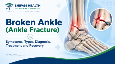
A “broken ankle” is the common term for an ankle fracture — a break in one or more of the bones that form the ankle joint: the distal tibia (shinbone, forming the medial malleolus, the bony bump on the inside of the ankle), the distal fibula (forming the lateral malleolus, the bump on the outside), and, less commonly, the talus (the bone in the foot that sits directly beneath the tibia and fibula).
A broken ankle is not one single injury — fractures range from a small, stable crack in a single bone that may need only a boot, to complex, unstable fractures involving multiple bones and ligaments that require surgery. The bones can fracture together with the ligaments that hold the joint together, and a genuinely important early question — whether the injury is a fracture, a sprain, or both — often can’t be answered by symptoms alone.
This guide explains the different types of ankle fracture, how they’re diagnosed, when surgery is and isn’t needed, what recovery actually looks like, and what treatment and its cost look like for international patients considering care in India. This is general medical education, not a diagnosis, and does not replace emergency or in-person orthopedic evaluation.
Table of Contents
ToggleAnkle Anatomy, Briefly
The ankle joint is formed by three bones fitting together: the tibia (shinbone), whose lower end forms the medial malleolus on the inside of the ankle and the flat weight-bearing surface called the tibial plafond; the fibula, the thinner outer leg bone, whose lower end forms the lateral malleolus on the outside; and the talus, a foot bone that sits in the socket formed by the tibia and fibula together — this socket arrangement is called the ankle mortise. Maintaining the precise shape of this mortise matters enormously for how the joint moves and bears weight, which is why even a fracture that looks small on an X-ray can meaningfully disrupt joint mechanics if it shifts the mortise out of shape.
The syndesmosis — a set of ligaments connecting the lower tibia and fibula just above the ankle joint — holds these two bones together; injury to the syndesmosis can make an otherwise straightforward-looking fracture considerably less stable than it appears.
Types of Ankle Fractures
| Type | What’s Involved | Key Clinical Point |
|---|---|---|
| Lateral Malleolus Fracture | Distal fibula only | The most common single-bone ankle fracture; stability depends heavily on whether the syndesmosis and medial structures are also injured |
| Medial Malleolus Fracture | Distal tibia only | Can occur alone or alongside other malleolar fractures |
| Bimalleolar Fracture | Both the lateral and medial malleolus | Generally less stable than a single-malleolus fracture, since two of the three points holding the mortise together are disrupted |
| Trimalleolar Fracture | Lateral, medial, and posterior malleolus (the back of the tibia) together | Generally the least stable pattern of the malleolar fractures, more often needing surgery |
| Posterior Malleolus Fracture | The back rim of the distal tibia | Often occurs alongside other malleolar fractures; whether it needs separate fixation depends on fragment size, displacement, and joint surface involvement, not fragment size alone |
| Maisonneuve Fracture | A fracture near the top of the fibula (near the knee), with syndesmotic and often medial ankle injury | The ankle X-ray can look deceptively unremarkable while the actual fracture is up near the knee — missing this pattern is a recognized pitfall |
| Pilon (Tibial Plafond) Fracture | The weight-bearing surface of the distal tibia itself | A higher-energy, more severe injury pattern with major soft-tissue involvement, genuinely different in severity from typical malleolar fractures |
| Ankle Fracture-Dislocation | A fracture combined with the joint being knocked out of its normal position | More urgent — dislocation threatens skin, blood supply, and nerves and often needs prompt reduction |
| Stress Fracture | A gradual, overuse-related crack rather than a single traumatic injury | Different mechanism and often different presentation than an acute traumatic fracture |
Maisonneuve fracture: a pattern worth knowing about specifically
This deserves particular attention because it’s a genuine, recognized diagnostic pitfall: a Maisonneuve fracture involves a break near the top of the fibula, close to the knee, combined with disruption of the syndesmosis and often the medial ankle structures — meaning the ankle itself can look nearly normal on an ankle X-ray while the actual fracture and the real source of instability sits much higher up the leg. Because of this, a clinician evaluating a significant ankle sprain-type injury with tenderness or swelling near the ankle syndesmosis will often specifically examine and, if indicated, X-ray the proximal fibula and knee area as well — missing this pattern and treating only the ankle can leave the underlying instability uncorrected.
Pilon fractures: a genuinely different severity class
A pilon fracture involves the weight-bearing surface of the tibia itself, typically from high-energy mechanisms like a fall from height or a motor vehicle collision. These carry major soft-tissue swelling and damage alongside the bone injury, which is why definitive surgical fixation is sometimes deliberately delayed for days to weeks until swelling has improved enough for surgery to be done safely — sometimes with a staged approach (temporary external stabilization first, definitive fixation later). Pilon fractures are not simply “a bad ankle fracture” — they represent a genuinely different injury severity and treatment complexity than the more common malleolar fracture patterns, and they carry a meaningfully higher long-term risk of post-traumatic arthritis given the joint-surface damage involved.
The Weber Classification
For fibula fractures specifically, the Weber (or Danis-Weber) classification describes where the fracture sits relative to the syndesmosis, since this location has real implications for how likely the syndesmosis is to also be injured:
- Weber A — fracture below the level of the syndesmosis; generally the least likely to involve syndesmotic injury
- Weber B — fracture at the level of the syndesmosis; syndesmotic involvement is possible and needs specific assessment
- Weber C — fracture above the level of the syndesmosis; syndesmotic injury is more likely
It’s important to understand that the Weber classification alone doesn’t determine treatment — clinicians also weigh medial-side injury, whether the syndesmosis is actually unstable (not just anatomically “at risk” based on fracture level), displacement, joint congruity, and the overall fracture pattern together. A related concept, the Lauge-Hansen classification, describes injury based on the mechanism of the ankle at the moment of injury (for example, the foot rotating outward while turned inward, or vice versa) — this is mainly useful to clinicians for predicting which associated structures might be injured based on how the injury happened, rather than being something patients need to interpret themselves.
Causes and Risk Factors
Ankle fractures result from twisting injuries (a very common mechanism, especially in sports and simple missteps), falls, sports collisions, road traffic accidents, direct blows, and — particularly in older adults — lower-energy falls onto bone weakened by osteoporosis. High-energy mechanisms (falls from height, vehicle collisions) tend to produce more severe, complex fracture patterns like pilon fractures, while a simple twisting injury more often produces an isolated malleolar fracture. Risk factors include older age, osteoporosis or reduced bone density, previous fractures, participation in high-impact or contact sports, poor balance or neuromuscular conditions affecting stability, certain medications associated with increased fracture risk, previous ankle instability, and occupational hazards involving heights or uneven terrain.
These describe increased population-level risk, not a guarantee — many ankle fractures occur in people with no particular risk factor at all, from an ordinary misstep or fall.
Symptoms
Common symptoms include sudden ankle pain at the time of injury, swelling, bruising, tenderness directly over the injured bone, difficulty walking or an inability to bear weight, visible deformity in more significant injuries, reduced range of motion, pain with movement, and, in less common but more concerning presentations, numbness or tingling. Symptom severity doesn’t always perfectly track fracture severity — some genuinely broken ankles are less dramatically painful or swollen than a severe sprain, which is part of why clinical and radiographic evaluation, not symptoms alone, is what actually confirms a diagnosis.
Can you walk on a broken ankle?
Yes — the ability to bear some weight does not rule out a fracture. Some stable fractures, small or nondisplaced fractures, and stress fractures allow a person to walk, sometimes with significant pain and sometimes with surprisingly little. This is exactly why “I could still walk on it” isn’t reliable evidence against a fracture, and it’s not a good reason to skip evaluation after a significant injury — and it’s not advisable to deliberately test an injured ankle by walking on it to “see how bad it is,” since doing so on an actual unstable fracture can worsen displacement.
Fracture vs. Sprain
| Feature | Ankle Fracture | Ankle Sprain |
|---|---|---|
| What’s Injured | Bone | Ligament (soft tissue) only |
| Swelling | Often significant, though not always | Can also be significant |
| Bruising | Common | Common |
| Ability to Bear Weight | Variable — some fractures allow walking | Variable — severe sprains can also prevent walking |
| Deformity | Present in more significant fractures | Not typically present |
| Point Tenderness | Often directly over bone | Often over the ligament, slightly differently located |
| X-ray | Shows the fracture | Normal (ligament injury doesn’t show on X-ray) |
| Treatment | Ranges from boot/cast to surgery depending on stability | Typically rest, bracing, and rehabilitation |
A severe sprain and a fracture can look and feel remarkably similar clinically — significant swelling, bruising, and difficulty walking can occur with either. This overlap is exactly why X-ray evaluation, guided by clinical judgment about who actually needs one (discussed below), is the standard way to distinguish them rather than relying on how the injury looks or feels.
Red Flags: When to Seek Immediate Care
Seek emergency medical assessment immediately for: visible deformity of the ankle, an open wound over the fracture site or visible bone through the skin, severe uncontrolled pain, a foot that looks pale, blue, or feels cold, absent or markedly reduced pulses in the foot, numbness or new weakness, rapidly increasing swelling, suspected dislocation, or inability to move the foot at all following major trauma. These findings can indicate neurovascular compromise (the blood supply or nerves to the foot being affected), an open fracture (with real infection risk), a joint dislocation, or severe soft-tissue injury — all situations needing prompt emergency evaluation, not a wait-and-see approach or a routine clinic appointment. Never attempt to push a visibly deformed ankle back into position yourself — leave reduction to trained medical personnel.
Immediate first aid while seeking care
Avoid unnecessary weight-bearing on the injured ankle, keep it supported and elevated where possible, apply appropriate cold therapy for swelling if suitable, and remove tight footwear, socks, or jewelry near the ankle early if swelling is developing and this can be done safely without forcing movement. This is supportive first aid on the way to evaluation, not a substitute for it.
How a Broken Ankle Is Diagnosed
Diagnosis combines history and mechanism of injury, physical examination, neurovascular assessment (checking pulses, sensation, and movement in the foot), and imaging.
Physical examination
Beyond assessing the ankle itself — swelling, bruising, tenderness, deformity, skin condition, and range of motion — a thorough examination for a suspected ankle fracture should also check the proximal fibula (near the knee, given the Maisonneuve pattern described above) and specifically test for syndesmotic tenderness, since missing either can mean missing a genuinely unstable injury pattern that an ankle-only exam would overlook.
X-rays and the Ottawa Ankle Rules
Standard views include AP (front-to-back), lateral (side), and mortise (angled to show the joint socket clearly) X-rays, with weight-bearing X-rays sometimes added in selected situations since they can reveal instability that doesn’t show on a non-weight-bearing image. Not every ankle injury needs an X-ray — the Ottawa Ankle Rules, a well-validated clinical decision tool, help determine when imaging is actually likely to be useful, based on factors including specific bone tenderness and the ability to bear weight both immediately after injury and at the time of evaluation. It’s worth understanding that these rules help decide who should get an X-ray — they don’t diagnose a fracture themselves, and they don’t replace clinical judgment in unusual presentations.
CT and MRI
CT is useful for complex fracture patterns, pilon fractures, assessing posterior malleolus involvement and joint-surface extension in detail, and surgical planning — it’s not needed for every straightforward ankle fracture. MRI isn’t routinely required for an acute, clearly-diagnosed fracture; it has a role in selected situations, including suspected stress fractures not visible on X-ray, cartilage or ligament injury assessment, or persistent pain when X-rays are negative and something is still being missed. MRI is not indicated simply because an X-ray already shows a fracture.
Stable vs. Unstable Fractures: The Central Treatment Concept
This distinction, more than the specific fracture type alone, is what actually drives the treatment decision. A stable fracture is one where the ankle mortise remains properly aligned and isn’t expected to shift out of position under normal loading with appropriate treatment. An unstable fracture is one where the fracture pattern, associated ligament damage, or both create a real risk that the joint will lose alignment — this includes fractures with talar shift (the ankle bone visibly displaced within the mortise), significant medial-side injury alongside a fibula fracture, syndesmotic disruption, involvement of multiple malleoli, or any fracture-dislocation. A single-bone, nondisplaced, stable fracture and a bimalleolar fracture with talar shift are treated completely differently, even though both are technically “a broken ankle” — stability, not simply the presence of a fracture, is what treatment planning actually centers on.
Non-Surgical Treatment
For stable, nondisplaced fractures with appropriate joint alignment, conservative (non-operative) management can be entirely appropriate — this may involve a walking boot, a short-leg cast, or a splint, combined with activity modification, a period of protected or limited weight-bearing, and follow-up X-rays to confirm the alignment holds as swelling decreases and healing progresses. There’s no single universal immobilization period that applies to every stable fracture — this is set by the treating clinician based on the specific fracture and how it’s healing.
Cast vs. walking boot
| Feature | Cast | Walking Boot |
|---|---|---|
| Stability Provided | Generally more rigid, less adjustable | Adjustable, generally somewhat less rigid |
| Removability | Not removable by the patient | Removable for hygiene, sleeping, or specific activities as directed |
| Skin/Wound Monitoring | More difficult | Easier |
| Typical Role | Often used earlier in treatment or for fractures needing more rigid immobilization | Often used as swelling decreases, or for injuries needing somewhat less rigid protection |
The choice between them depends on fracture stability, current swelling, stage of healing, and clinician judgment — it isn’t simply a matter of patient preference, though comfort and practicality are real considerations discussed with the treating clinician.
Surgery: When Is It Really Necessary?
Surgery may be considered for displaced fractures, unstable fracture patterns, talar shift, persistent malalignment despite attempted closed (non-surgical) treatment, open fractures, fracture-dislocations, significant syndesmotic instability, and certain fractures extending into the joint surface. Not every broken ankle needs surgery — many stable, nondisplaced fractures heal well with a boot or cast alone, and surgery is specifically reserved for situations where nonoperative treatment is unlikely to maintain proper alignment or joint function.
Open reduction and internal fixation (ORIF)
ORIF is the standard surgical approach: the surgeon directly realigns (“reduces”) the fracture fragments through an incision, then stabilizes them with plates, screws, or, in selected situations, wires — restoring the ankle mortise and joint alignment and holding it in place while the bone heals. Surgery doesn’t make the bone “stronger” than it was before injury; its goal is accurate alignment and stable fixation so the bone can heal in the correct position.
Syndesmotic fixation
When the syndesmosis is confirmed unstable, it needs separate fixation — typically screws or a suture-button device — to hold the tibia and fibula in their correct relationship while the ligament heals. Current evidence comparing these fixation methods continues to evolve, and neither is universally established as superior for every situation; the choice depends on surgeon assessment and the specific injury pattern.
Posterior malleolus: modern decision-making
Older guidance sometimes used a simple fragment-size threshold to decide whether a posterior malleolus fracture needed separate surgical fixation. Current practice considers this more holistically — fragment size alongside displacement, whether the joint surface itself is disrupted (articular step-off), syndesmotic stability, and how the fracture involvement affects the fibular notch — rather than a single size cutoff automatically triggering surgery.
Fracture-dislocations and open fractures
When a fracture is combined with dislocation, urgent reduction (restoring the joint to its normal position) may be needed promptly to protect the skin, blood supply, and nerves — but it’s worth understanding that this emergency reduction is not necessarily the same procedure as the definitive surgical fixation, which may follow at a planned time afterward once swelling allows. Open fractures — where the bone has broken through the skin — carry real infection risk and are managed as a genuine emergency, typically involving prompt antibiotics per clinical protocol, surgical irrigation and debridement (cleaning) of the wound, and stabilization.
Special Situations
Fragility (osteoporotic) fractures in older adults
In older adults, ankle fractures can occur from relatively low-energy falls when bone density is reduced by osteoporosis. Not every older adult’s fracture is automatically osteoporosis-related, but this is a reasonable trigger for bone-density evaluation in the appropriate clinical context, alongside standard fracture treatment.
Children and adolescents
Pediatric ankle fractures are genuinely different from adult fractures because of open growth plates — injuries can involve the growth plate itself, including specific patterns like transitional fractures (Tillaux and triplane fractures), which occur during the period when a growth plate is partially, but not fully, closed. Adult treatment algorithms shouldn’t be applied indiscriminately to a child’s ankle fracture; growth-plate assessment and pediatric-specific evaluation matter, and management is generally best handled by a clinician with pediatric orthopedic experience.
Recovery: What to Realistically Expect
Recovery generally moves through phases, though the pace and specifics vary by fracture type, treatment, and individual healing — this guide won’t promise a specific number of weeks, since that promise wouldn’t be honest across the range of injuries this article covers.
- Protection phase: Immobilization, swelling and pain control, and protecting the healing fracture from excessive load.
- Early movement phase: Once medically permitted, gentle range-of-motion exercises begin, alongside continued swelling management and gradual, guided loading.
- Strengthening phase: Focus shifts to calf and ankle-stabilizer strength, balance, and proprioception (the joint’s sense of its own position) — genuinely important for preventing reinjury, not just optional extra rehabilitation.
- Functional recovery phase: Progression through walking, stairs, work-specific tasks, and, for those returning to sport, running, jumping, and sport-specific drills.
Radiographic healing (bone union visible on X-ray) is not the same as full functional recovery — a bone can be soundly healed while strength, balance, and confidence in the joint still need weeks to months of further rehabilitation to reach their pre-injury level.
Recovery milestones: walking, work, driving, and sport
Return to normal walking depends on fracture stability, treatment type, healing progress, pain, and strength, generally progressing from protected or limited weight-bearing to full weight-bearing under clinician guidance.
- Return to work varies enormously by job demands — desk-based work is often possible sooner than standing or manual labor, and safety-sensitive roles need particular care around when full function and reaction time have returned.
- Return to driving depends on which ankle is injured (the right ankle, for brake control, generally needs more complete recovery for those driving an automatic; either ankle matters for manual transmissions), current weight-bearing restrictions, whether pain medication affecting alertness is still being used, and the ability to perform an emergency stop safely — local legal and insurance considerations may also apply.
- Return to sports depends on demonstrated strength, range of motion, balance, and running or jumping tolerance, ideally assessed through functional testing rather than simply counting weeks since injury. None of these have a single correct timeline that applies to every patient — they depend on the specific injury, treatment, and individual recovery, and are genuinely a discussion with your treating clinician rather than a fixed calendar.
Complications
| Complication | What It Involves | Why It Matters |
|---|---|---|
| Infection | Bacterial infection, more common with open fractures or after surgery | Can require additional treatment, sometimes including further surgery |
| Nonunion / Delayed Union | The bone fails to heal, or heals more slowly than expected | Risk increases with smoking, diabetes, poor blood supply, infection, or more severe initial injury |
| Malunion | The bone heals in a less-than-ideal position | Can affect joint mechanics and function; may need revision surgery in significant cases |
| Post-Traumatic Arthritis | Joint wear developing over time after the injury | More likely with joint-surface fractures, talar shift, or residual joint incongruity, particularly with pilon fractures |
| Hardware Irritation/Failure | Plates or screws causing local discomfort, or, less commonly, breaking | Doesn’t always require removal |
| Nerve or Blood Vessel Injury | Direct injury from the trauma or, rarely, during surgery | Assessed through neurovascular examination at diagnosis and follow-up |
| Blood Clots (DVT/PE) | Clot formation, with immobilization as a contributing risk factor | Risk varies by individual factors |
| Complex Regional Pain Syndrome | A less common chronic pain condition following injury | Can significantly affect recovery and may require specialized management |
None of these complications are inevitable — most ankle fractures heal without them — but a responsible guide shouldn’t only describe the uncomplicated path.
Blood clot warning signs
Seek urgent medical attention for new calf swelling or pain, sudden shortness of breath, chest pain, or coughing up blood — these can indicate a blood clot (deep vein thrombosis) or, more seriously, a clot that has traveled to the lungs (pulmonary embolism), both genuine emergencies.
Hardware removal
Plates and screws used in ORIF do not automatically need to be removed once the fracture has healed — many patients keep their hardware permanently without issue. Removal may be considered specifically for painful or prominent hardware causing symptoms, infection, tendon irritation from hardware position, or specific syndesmotic-screw situations where planned removal is part of the original treatment plan — it’s a situational decision, not a routine second surgery everyone needs.
Factors affecting healing
Smoking is associated with impaired bone healing and higher complication rates after fracture surgery — this is offered as information to support a treatment decision or quit attempt, not as blame. Diabetes can affect infection risk, wound healing, and bone healing, though not every person with diabetes experiences complications — good glucose control around the time of injury and surgery is genuinely protective. Nutrition — adequate protein, calcium, and vitamin D — supports normal bone healing, though there’s no special “miracle food” that accelerates healing beyond adequate overall nutrition, and high-dose supplementation should follow medical guidance rather than self-directed dosing.
Prognosis
Prognosis depends on fracture type, stability, joint-surface involvement, quality of reduction achieved, treatment approach, soft-tissue damage, age, bone quality, other health conditions, and how rehabilitation goes — this guide won’t cite a universal success rate, since outcomes genuinely vary across this range of factors. Most stable, well-treated ankle fractures heal well with good functional recovery; more complex, high-energy, or joint-surface-involving fractures (particularly pilon fractures) carry a meaningfully higher risk of long-term issues including post-traumatic arthritis, which is worth an honest conversation with your treating surgeon specific to your injury pattern rather than a generic reassurance.
When to consider a second opinion
A second orthopedic opinion may be reasonable when surgery has been recommended and you want confirmation, the fracture is complex or a pilon fracture, there’s disagreement about stability between assessments, pain or healing seems unusually delayed, malunion is suspected, or revision surgery is being discussed. This doesn’t mean every case needs a second opinion — for a straightforward, stable fracture with a clear treatment plan, it’s often unnecessary — but it’s a reasonable and available option when genuine uncertainty exists.
Broken Ankle Treatment Cost in India
Cost depends heavily on fracture type and whether surgery is needed. Below is a component breakdown; as with every cost topic in this series, treat these as directional.
| Component | Approximate Cost Range (USD) |
|---|---|
| Emergency/Orthopedic Consultation | $30–$150 |
| X-rays | $20–$80 |
| CT, Where Needed | $100–$300 |
| Cast or Walking Boot | $30–$100 |
| Closed Treatment (Cast/Boot, No Surgery), Full Course Including Follow-Up | $200–$600 |
| ORIF Surgery (Domestic Indian Pricing) | Roughly $1,400–$2,200 based on published domestic figures |
| ORIF Surgery (Published International-Patient Pricing) | Roughly $3,500–$7,000+ |
| Syndesmotic Fixation, Where Needed (Adds to ORIF Cost) | Several hundred dollars additional |
| Physiotherapy Course | $150–$500 |
A clear domestic-vs-international pricing gap is worth flagging directly here, consistent with the pattern seen across other topics in this series: published domestic Indian pricing for ankle ORIF runs roughly ₹1.2–1.8 lakh (about $1,400–$2,200), while sources specifically quoting international-patient pricing put the same procedure at $3,500–$7,000 or more, once consultation, hospital stay, and coordination for a traveling patient are factored in.
If you see a low domestic-sounding figure quoted to you as an international patient, confirm directly whether it reflects what you’ll actually be charged — the gap between these two figures is substantial. As always, request a written, itemized quote based on your specific fracture type and proposed treatment before making any decision.
Ankle Fracture Treatment in India for International Patients
India has substantial orthopedic trauma and foot-and-ankle surgical capability, including centers with dedicated foot-and-ankle specialists, appropriate imaging (X-ray, CT, and MRI where needed), and physiotherapy/rehabilitation services — for international patients, the appeal is typically this combination with costs generally lower than the US, UK, or much of Europe. India isn’t automatically “the best” choice for every patient, and for an acute, displaced, open, or fracture-dislocation injury, international travel should never come before urgent local emergency stabilization — these injuries need immediate care wherever the patient currently is, not a delay for travel planning.
When evaluating a center for ankle fracture care, look for an orthopedic trauma or foot-and-ankle specialist with relevant experience, appropriate imaging capability, physiotherapy and rehabilitation services, and for complex or staged injuries like pilon fractures — genuine multidisciplinary coordination. Verify credentials directly rather than relying on third-party rankings.
The international patient journey
- Share medical records and X-ray/CT images where available
- Orthopedic specialist reviews the case and determines fracture type and stability
- Confirm whether urgent intervention is needed
- Develop a treatment plan and receive a written cost estimate
- Coordinate medical travel documentation, if travel is appropriate for the situation
- Attend in-person assessment and undergo treatment as clinically indicated
- Complete rehabilitation and receive discharge documentation
- Coordinate follow-up with your home-country orthopedic team
No credible provider can guarantee surgical outcome, bone healing on a specific timeline, an exact final cost, or a specific return-to-sport date before reviewing a patient’s actual imaging — be cautious of any provider offering these guarantees upfront.
Questions to Ask Your Orthopedic Surgeon
- What type of ankle fracture do I have, and which bones are involved?
- Is the fracture displaced? Is my ankle stable or unstable?
- Is the syndesmosis injured?
- Do I need a CT scan for more detailed planning?
- Can this heal without surgery, or is surgery recommended — and why?
- What are the realistic risks of waiting or choosing nonoperative treatment?
- If surgery is recommended, what type of fixation would be used?
- How long will I need immobilization, and when can I start putting weight on it?
- When can I start physiotherapy?
- When can I realistically expect to drive, return to work, and return to sport?
- Will the hardware need removal later?
- What is my realistic risk of post-traumatic arthritis given my specific fracture?
How Shifam Health Helps International Patients
Shifam Health is a medical tourism facilitator, not a hospital, orthopedic surgeon, or emergency department — we don’t diagnose or treat ankle fractures. What we do is help international patients navigate the practical side of accessing ankle fracture care in India: coordinating the review of imaging and medical records by relevant specialists, helping identify hospitals with genuine orthopedic trauma and foot-and-ankle experience, obtaining written treatment and cost estimates, assisting with medical visa documentation, arranging airport pickup and accommodation, providing interpreter support where needed, and staying in touch for follow-up communication once you return home.
For an acute injury requiring urgent care, we’d always encourage getting appropriate emergency treatment wherever you currently are rather than delaying for international travel.
If you or someone you’re caring for has a diagnosed or suspected ankle fracture and you’re exploring treatment options in India, share your available reports and imaging with our team on WhatsApp or through a quick inquiry form — there’s no obligation, and we typically respond within 24 hours.
Frequently Asked Questions
What is a broken ankle?
A broken ankle is a fracture of one or more bones forming the ankle, usually the tibia, fibula, or sometimes the talus.
What are the symptoms?
Common symptoms include pain, swelling, bruising, tenderness, difficulty walking, and sometimes visible deformity.
Can you walk on a broken ankle?
Yes. Some fractures allow weight-bearing, so being able to walk does not rule out a fracture.
Can an ankle fracture be mistaken for a sprain?
Yes. Severe sprains and fractures can have similar symptoms. X-rays help distinguish many fractures from sprains.
Does every ankle injury need an X-ray?
No. The Ottawa Ankle Rules can help determine whether X-rays are appropriate based on specific tenderness and weight-bearing ability.
What is a bimalleolar fracture?
It involves fractures of both the medial and lateral malleoli and is often less stable than a single malleolar fracture.
What is a trimalleolar fracture?
It involves the medial, lateral, and posterior malleoli and is generally an unstable fracture pattern.
Can a broken ankle heal without surgery?
Yes. Stable, nondisplaced fractures may heal successfully with a cast or walking boot.
What is ORIF?
Open reduction and internal fixation (ORIF) realigns the broken bones and stabilizes them using plates, screws, or other hardware.
How long does an ankle fracture take to heal?
Healing varies according to fracture type, treatment, age, and overall health. Bone healing and complete functional recovery may take different amounts of time.
Do I need physiotherapy?
Rehabilitation can help restore strength, balance, mobility, and function after an ankle fracture.
Can a broken ankle cause arthritis?
Yes. Fractures involving the joint surface or leaving residual misalignment can increase the risk of post-traumatic arthritis.
Conclusion
A “broken ankle” covers a genuinely wide range of injuries from a small, stable crack needing only a boot, to complex, unstable fracture-dislocations needing urgent surgery and stability, not the fracture label alone, is what actually determines treatment. Getting an accurate diagnosis, understanding whether your specific fracture is stable or unstable, and having a realistic conversation about recovery timeline rather than expecting a fixed number of weeks are the most useful steps toward a good outcome, in India or anywhere else.
This article is for general medical education and does not replace emergency care or individualized advice from a qualified orthopedic surgeon. It is not a diagnosis, treatment recommendation, or guarantee of any outcome. For a suspected acute fracture, seek prompt medical evaluation.



Popular Posts From Last Week
- September 10, 2026
- shifamhealth
Medically reviewed content · Published 2026 · Last reviewed August 2026 Blount’s disease, also called tibia vara, is…
- September 14, 2026
- shifamhealth
Medically reviewed content · Published 2026 · Last reviewed September 2026 A “broken ankle” is the common term…
- September 13, 2026
- shifamhealth
Carpal tunnel syndrome (CTS) is compression of the median nerve as it passes through the carpal tunnel —…
- September 9, 2026
- shifamhealth
A cervical herniated disc occurs when the soft inner material of a disc in the neck pushes through…
- September 10, 2026
- shifamhealth
Chronic back pain is one of the most common reasons people seek medical care, and also one of…
- September 11, 2026
- shifamhealth
Myelopathy means dysfunction of the spinal cord itself — not a single disease, but a syndrome that can…
- September 9, 2026
- shifamhealth
Medically reviewed content · Published 2026 · Last reviewed September 2026 Disc degeneration seen on an MRI does…
- September 12, 2026
- shifamhealth
Medically reviewed content · Published 2026 · Last reviewed September 2026 Discitis is inflammation or infection involving an…
- September 12, 2026
- shifamhealth
A femoral hernia occurs when tissue often part of the bowel or fatty tissue — pushes through the…
- September 11, 2026
- shifamhealth
Kümmell’s disease refers to a delayed collapse of a vertebra — a nonunion or failure to heal properly…
- September 7, 2026
- shifamhealth
Ductal carcinoma in situ (DCIS) is a non-invasive breast condition in which abnormal cells are confined to the…
- September 8, 2026
- shifamhealth
Written by: Shifam Health Editorial Team — Neuro-Oncology Content Division Medically reviewed by: Shifam Health Clinical Advisory Panel…