



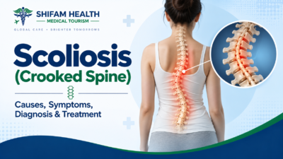
Scoliosis (Crooked Spine): Causes, Symptoms, Diagnosis & Treatment
Filters & Insights
Scoliosis is a 3D spinal curve, not one single condition. Learn the real causes, when bracing helps, when surgery is needed, & what to realistically expect.

Scoliosis is a three-dimensional spinal deformity — sideways curvature combined with rotation — and it is genuinely not one single condition. Its clinical significance depends on the underlying cause, the patient’s age and remaining growth, the size and pattern of the curve, and whether it’s progressing.
Most scoliosis is mild and doesn’t require surgery. Observation, physiotherapy, and bracing (during growth) are appropriate for many patients; surgery becomes a consideration for larger, progressive curves or when there’s significant functional or neurological impact. “Crooked spine” is a fair everyday description, but it undersells how much the underlying cause and pattern actually matter for what happens next.
Table of Contents
ToggleWhat Is Scoliosis?
Scoliosis is a three-dimensional spinal deformity characterized by abnormal sideways (lateral) curvature of the spine combined with rotation of the vertebrae not simply a spine that curves to one side. Whether a given curve matters clinically depends on the patient’s age, the underlying cause, the curve’s size and pattern, remaining growth, and whether it’s progressing.
“Crooked spine” is a genuinely reasonable, patient-friendly way to describe what scoliosis looks like but it’s medically incomplete. Scoliosis isn’t a flat, side-to-side bend; the vertebrae also rotate as the curve develops, which is part of why some patients develop a visible rib prominence (discussed below) even with a modest-looking curve on a simple visual inspection.
This is one of the central points of this entire guide: scoliosis is not one disease. A 12-year-old with an otherwise-unexplained curve, a newborn with a vertebral formation abnormality, a teenager with cerebral palsy and a collapsing spine, and a 68-year-old with degenerative spinal changes can all technically have “scoliosis” — but these are genuinely different clinical situations requiring different evaluation and different treatment logic. Understanding which category applies is the necessary first step before any treatment conversation makes sense.
Types of Scoliosis
Scoliosis is classified primarily by cause: idiopathic (no identified specific cause, the most common type overall), congenital (present from vertebral formation problems before birth), neuromuscular (associated with conditions like cerebral palsy), degenerative/adult (developing later in life from spinal degeneration), and syndromic (associated with certain genetic or connective-tissue disorders).
| Type | Typical Context | Main Cause | Key Features |
|---|---|---|---|
| Idiopathic Scoliosis | Most common overall; subdivided by age of onset (infantile, juvenile, adolescent) | No specific identified cause | “Idiopathic” means the specific cause hasn’t been identified — it does not mean the condition is unexplained biologically or somehow not “real”; it reflects the current limits of what’s been pinpointed as a single cause |
| Congenital Scoliosis | Present from before birth | Abnormal vertebral formation or segmentation during fetal development (including hemivertebrae — partially formed vertebrae) | Can behave differently from idiopathic scoliosis, sometimes progressing in patterns tied to the specific structural abnormality present |
| Neuromuscular Scoliosis | Associated with underlying neurological or muscular conditions | Conditions such as cerebral palsy, muscular dystrophy, or spinal muscular atrophy, where abnormal muscle tone/balance affects spinal support | Management often differs substantially, considering the broader neuromuscular condition, not spinal curvature alone |
| Degenerative/Adult Scoliosis | Develops later in life | Disc degeneration, facet joint arthritis, and other age-related spinal changes | Not simply “childhood scoliosis carried into adulthood” — a genuinely distinct clinical entity, discussed in its own section below |
| Syndromic Scoliosis | Associated with certain genetic or connective-tissue disorders | Part of the broader syndrome’s effects on connective tissue or skeletal development | Management is coordinated within the context of the broader syndrome |
Other secondary causes of scoliosis-like curvature can include spinal tumors, infection, trauma, leg-length discrepancy (which can cause a functional, not structural, curve), and spinal cord abnormalities — these are less common but part of why a thorough diagnostic evaluation, discussed below, matters rather than assuming every curve is idiopathic.
Scoliosis vs. Other Spinal Curvatures
A person appearing visually “crooked” doesn’t automatically mean they have structural scoliosis — poor posture, kyphosis, and functional curvature from leg-length differences can all look somewhat similar on casual observation but are genuinely different conditions with different implications.
| Condition | Main Feature | Structural? | Typical Evaluation |
|---|---|---|---|
| Scoliosis | Sideways curvature plus vertebral rotation | Yes | Standing X-ray, Cobb angle measurement |
| Poor Posture | Slouching, muscular, positional | No — corrects with conscious effort or positioning | Clinical examination generally sufficient |
| Kyphosis | Excessive forward (front-to-back) curvature, typically of the upper back | Can be structural (e.g., Scheuermann kyphosis) or postural | Clinical exam, imaging if structural cause suspected |
| Lordosis | Excessive inward curve, typically of the lower back | Usually postural/functional, occasionally structural | Clinical exam |
| Functional Curvature from Leg-Length Discrepancy | Apparent spinal curve driven by pelvic tilt, not the spine itself | No — the spine itself isn’t structurally deformed | Standing exam with and without a lift to correct leg length; the “curve” typically resolves |
| Scheuermann Kyphosis | A specific structural cause of excessive forward curvature, typically in adolescents | Yes | Standing X-rays showing characteristic vertebral wedging |
The practical point: visual appearance alone — someone looking crooked or uneven — is a reason to seek evaluation, not a diagnosis in itself. These conditions require different assessments and, where relevant, different treatments.
Causes and Risk Factors — Including the Myths
Causes vary by scoliosis type — genetics and family history play a role in idiopathic scoliosis, structural vertebral abnormalities cause congenital scoliosis, and underlying neurological conditions drive neuromuscular scoliosis. Many commonly believed causes — bad posture, school bags, sleeping position — are not established causes of structural scoliosis.
By type:
- Idiopathic scoliosis: genetics and family history appear to play some role, though the complete underlying mechanism isn’t fully identified — hence “idiopathic”
- Congenital scoliosis: abnormal vertebral formation or segmentation during fetal spinal development
- Neuromuscular scoliosis: underlying neurological or muscular conditions affecting spinal muscular support and control
- Degenerative scoliosis: age-related disc degeneration and facet joint arthritis
- Syndromic scoliosis: part of certain genetic or connective-tissue disorders
Myth-busting — this needs to be stated clearly and directly, since these are among the most common misconceptions about scoliosis:
| Commonly Believed Cause | Reality |
|---|---|
| Bad Posture | Not an established cause of structural idiopathic scoliosis |
| Carrying Heavy School Bags | Not an established cause of structural scoliosis |
| Sleeping Position | Not an established cause of structural scoliosis |
| Crossing Legs | Not an established cause of structural scoliosis |
| Exercise or Sports Participation | Not an established cause — and, as discussed later, doesn’t need to be avoided because of scoliosis |
| Normal Childhood Activity | Not an established cause |
If a factor doesn’t cause structural scoliosis, it’s worth saying so plainly rather than hedging — these myths persist widely in popular health content and can lead to unnecessary guilt (particularly in parents) or false confidence in unproven prevention strategies.
Age-Specific Considerations
Scoliosis behaves differently depending on age because remaining growth potential fundamentally changes both progression risk and treatment options — a curve found in a growing child is a genuinely different clinical situation than the same-sized curve found in a skeletally mature adult.
- Infants and young children: early-onset scoliosis (generally before age 10) deserves specialist evaluation given the interaction between spinal deformity and ongoing lung and chest development during this critical growth period
- Adolescents: this is when idiopathic scoliosis is most commonly diagnosed, often around or before puberty; growth remaining (assessed through skeletal maturity markers, discussed below) is central to progression risk assessment and treatment timing, including decisions around menarche timing in girls, which is a recognized clinical marker of remaining growth
- Adults: scoliosis in adults can represent either a curve carried forward from adolescence or newly developing degenerative scoliosis; treatment considerations, discussed in a dedicated section below, are substantially different from pediatric management
- Older adults: degenerative changes can cause new or worsening curvature, sometimes alongside other age-related spinal conditions like spinal stenosis
Not every scoliosis curve worsens over time — this is worth stating clearly, since it’s a common source of anxiety. Progression risk depends on multiple factors (discussed in the “Natural History and Progression” section below), not simply the presence of a curve or a patient’s age alone.
Symptoms
Scoliosis can be completely asymptomatic and discovered incidentally, or it can present with visible body asymmetry — uneven shoulders, an uneven waist, or a rib prominence — and, in some patients, discomfort or, less commonly, neurological symptoms that warrant further evaluation.
Visible signs that may prompt evaluation:
- Uneven shoulders or shoulder blades
- Uneven waist
- Rib prominence on one side (a rib hump, discussed below)
- Uneven hips
- Trunk shift (the torso appearing to lean to one side relative to the pelvis)
- Clothes fitting unevenly
- One side of the back appearing higher than the other, particularly noticeable when bending forward
Rib hump: this refers to a visible prominence of the ribs on one side of the back, most noticeable when bending forward. It reflects the rotational component of scoliosis — as the vertebrae rotate, the attached ribs on one side are pushed backward, creating this visible asymmetry. It’s a genuinely useful clinical sign precisely because it reflects the three-dimensional nature of the deformity, not just the sideways curve visible on a standing X-ray.
On back pain — this needs particular care: scoliosis does not automatically cause back pain, and this claim shouldn’t be made without qualification. Many people with scoliosis, including some with meaningful curves, have no pain at all. When pain is present, it deserves its own evaluation — it may or may not be directly related to the curve itself, and persistent or unusual pain (discussed further under red flags below) can sometimes be a clue toward an underlying cause other than routine idiopathic scoliosis.
Neurological symptoms — including weakness, numbness, tingling, gait changes, balance problems, or bowel/bladder changes — are not typical of uncomplicated idiopathic scoliosis and suggest something beyond routine scoliosis may be present, warranting prompt medical assessment rather than being assumed to simply be “part of” the scoliosis.
Red Flags — When Urgent Evaluation Is Needed
While most scoliosis evaluation can proceed on a routine basis, certain features warrant more urgent assessment: rapidly progressive deformity, significant or persistent pain (especially at night), any neurological symptoms, systemic illness signs, or very early-onset curves.
Features warranting more urgent evaluation:
- Rapidly progressive visible deformity
- Severe or persistent pain
- Significant night pain specifically (pain that wakes someone from sleep or is worse at night can be a particular clue warranting further evaluation)
- Any neurological deficit — weakness, loss of coordination, gait abnormality
- Bowel or bladder dysfunction
- Fever or other signs of systemic illness alongside back symptoms
- Unexplained weight loss alongside spinal symptoms
- New scoliosis diagnosed in an unusual age group, or very early-onset scoliosis
- Significant trauma preceding new spinal symptoms
- Any clinical suspicion of tumor, infection, or spinal cord abnormality
This isn’t meant to create unnecessary fear — the large majority of scoliosis, particularly typical adolescent idiopathic scoliosis, does not involve any of these features. They’re included because a small subset of cases reflect something beyond routine scoliosis, and knowing when to seek more urgent rather than routine evaluation genuinely matters.
How Scoliosis Is Diagnosed
Diagnosis combines a detailed medical history, physical examination (including the Adam’s forward bend test), and standing spinal X-rays to measure the curve — no single test or maneuver establishes the diagnosis on its own.
Medical history typically covers age at onset, growth pattern, family history, presence of pain, any neurological symptoms, menstrual history where relevant (in adolescent girls, as a marker of skeletal maturity), previous treatment, any underlying disease, trauma history, and functional limitations.
Physical examination includes assessment of posture, shoulder and pelvic symmetry, the Adam’s forward bend test, rib prominence, trunk shift, leg-length assessment where relevant, a neurological examination, gait assessment, and, where relevant, skin findings (certain skin markings can be associated with underlying conditions relevant to congenital or syndromic causes).
The Adam’s forward bend test — having the patient bend forward at the waist while the examiner looks along the back for asymmetry or rib prominence — is a useful, simple screening maneuver, but it has real limitations: it can miss milder curves, and a positive finding doesn’t establish the exact curve size or cause on its own — it prompts further evaluation (typically standing X-rays), rather than serving as a complete diagnostic tool by itself.
The Cobb Angle, Explained Properly
The Cobb angle is the standard measurement used to quantify the degree of spinal curvature on a standing X-ray, but it is one piece of clinical information among several, not a number that alone determines treatment.
- The Cobb angle is measured by drawing lines along the most tilted vertebrae at the top and bottom of a curve on a standing spinal X-ray, then measuring the angle formed between them
- It’s the standard, widely used way to quantify curve magnitude and track change over time
- Measurement variability is real — the same curve measured by different observers, or even the same observer on different occasions, can show some variation. This means a small difference between two measurements (a few degrees) does not necessarily represent true clinical progression — it may simply reflect normal measurement variability
- Clinicians interpret the Cobb angle together with growth remaining, skeletal maturity, curve pattern and location, and clinical findings — not as a number that mechanically determines treatment on its own
Commonly discussed general ranges exist in clinical practice and literature (for example, broad categories sometimes described as mild, moderate, or larger curves), but these should not be presented as universal, fixed thresholds that automatically dictate a specific treatment — the actual treatment decision, discussed later in this guide, integrates the Cobb angle with multiple other factors specific to the individual patient.
Imaging: X-Rays, Bending Films, MRI, and CT
Standing (weight-bearing) X-rays — typically front-facing (AP/PA) and side (lateral) views of the full spine — are the standard initial and follow-up imaging for scoliosis, since they show the curve under normal load and allow Cobb angle measurement and tracking over time.
Bending X-rays (images taken while the patient bends sideways) may be used specifically for surgical planning, since they help assess how flexible or rigid a curve is — information that can influence surgical technique decisions.
MRI is not automatically required for every scoliosis patient. It becomes relevant in specific situations, including:
- Neurological abnormalities on examination
- Significant or atypical pain
- Atypical curve patterns (for example, an unusual curve direction or pattern that doesn’t fit typical idiopathic scoliosis)
- Very young age at presentation
- Rapid progression
- Any clinical suspicion of an underlying spinal cord abnormality
In these situations, MRI may look for conditions such as syringomyelia (a fluid-filled cavity within the spinal cord), Chiari malformation (a structural abnormality where brain tissue extends into the spinal canal), tethered cord (abnormal attachment restricting normal spinal cord movement), or other intramedullary (within the spinal cord) lesions. It’s important to be clear that these are not common causes of routine adolescent idiopathic scoliosis — they’re specifically screened for in atypical presentations, not assumed to be present in typical cases.
CT scanning may be used in selected situations, particularly for detailed surgical planning in complex cases, with radiation exposure considerations weighed against the specific clinical need, particularly relevant in children given cumulative radiation exposure considerations over time.
Natural History and Progression
Whether a scoliosis curve progresses depends on multiple interacting factors — curve magnitude, patient age and remaining growth, skeletal maturity, curve location and pattern, and, in adults, ongoing degenerative changes — and no single factor determines outcome with certainty for an individual patient.
Factors associated with progression risk in growing patients include curve magnitude at diagnosis, younger age at diagnosis (more growth remaining generally means more time for potential progression), skeletal immaturity, curve location and pattern, and, in girls, timing relative to menarche and growth velocity.
After skeletal maturity is reached, progression risk generally changes — many curves that were monitored during growth become more stable once growth stops, though larger curves at skeletal maturity can, in some cases, continue to slowly progress even in adulthood, and this is part of why some adults are found to have scoliosis that was present but unaddressed since adolescence.
Adult (degenerative) scoliosis has a different progression pattern altogether, driven by ongoing disc and facet joint degeneration rather than growth-related factors — this is discussed further in its own dedicated section below.
It’s important to be careful with precise progression percentages — published progression risk figures vary across studies and patient populations, and presenting one number as if it applies universally to every patient with a given curve size would overstate the precision of what’s actually known. Your specialist’s assessment of your specific curve, age, and growth status is more meaningful than any generic percentage.
Treatment Decision Framework
Scoliosis treatment follows a logical framework rather than a fixed protocol:
Diagnosis → Cause → Age → Skeletal maturity → Curve magnitude → Progression risk → Symptoms → Neurological status → Overall spinal balance → Treatment decision
This means two patients with an identical Cobb angle can reasonably receive different treatment recommendations if their age, remaining growth, curve pattern, or underlying cause differ — which is exactly why treatment can’t be reduced to “curves above X degrees need surgery” as a simple rule.
Observation
Observation — periodic clinical follow-up with imaging when indicated — is a reasonable, genuinely active management approach (not “doing nothing”) for many milder curves, particularly with lower progression risk based on the factors discussed above.
It involves:
- Periodic clinical follow-up visits
- Follow-up imaging when clinically indicated, timed to balance monitoring needs against imaging frequency
- Growth monitoring in pediatric patients
- Clear guidance on what changes (new symptoms, visible progression, or specific findings) should trigger reassessment sooner than scheduled
This is not a passive “wait and see” approach with no plan — it’s a structured monitoring strategy appropriate for curves judged, based on the full clinical picture, to be at lower risk of significant progression.
Physiotherapy and Scoliosis-Specific Exercise
General physical activity and core strengthening support overall function and fitness; scoliosis-specific exercise approaches, including Schroth-based physiotherapeutic scoliosis-specific exercises (PSSE), may help with posture, symptom management, and, in some patients, curve control — but exercise should not be presented as something that “straightens the spine” in the way surgery or, to a lesser extent, bracing during growth can influence curve magnitude.
- General physical activity, conditioning, and core strengthening are broadly beneficial for overall health and function in people with scoliosis, just as in anyone else
- Physiotherapeutic scoliosis-specific exercises (PSSE), including Schroth-based approaches, are structured exercise programs specifically designed for scoliosis, and evidence suggests they may help with posture, pain, and function, with some studies suggesting a role in supporting curve stability in appropriately selected patients
- Being direct about the evidence: claiming these exercises reliably “correct” or “reverse” an established structural curve overstates what’s currently well-supported — the more accurate framing is that they may support function, symptom management, and, in some patients, may play a supportive role alongside other management, rather than functioning as a standalone alternative to bracing or surgery when those are indicated
What exercise can realistically contribute: improved function, strength, conditioning, pain management, postural control, and, in selected patients, a potential supportive effect on progression — this is meaningfully different from a claim that exercise alone corrects structural scoliosis.
Bracing
Bracing is primarily used during growth, in appropriately selected patients with progressive curves, to reduce the likelihood of further progression during the remaining growth period — it is a curve control strategy, not a permanent correction method, and its effectiveness depends heavily on consistent, adherent use.
- Bracing is generally considered for growing patients (skeletal immaturity matters directly here — bracing works by influencing growth, so it’s far less relevant once growth has essentially stopped)
- Different brace types and wear-time strategies exist (including more full-time and more part-time approaches), and the specific choice is individualized based on curve characteristics and clinical judgment
- Adherence is genuinely central to whether bracing works — a brace that isn’t worn as prescribed can’t realistically achieve its intended effect
Real challenges worth acknowledging honestly, since these genuinely affect real families:
- Brace discomfort, particularly initially
- Skin irritation or pressure areas
- Body image and self-consciousness, particularly in adolescents
- School and social challenges related to visible bracing
- The practical difficulty of consistent long-term adherence
- Psychological considerations that deserve genuine attention, not dismissal
The critical distinction to understand: curve control vs. permanent correction. Bracing aims to reduce the likelihood that a curve will progress to the point of needing surgery during the growth period — it does not claim, and should not be marketed as, permanently straightening every spine. Some curves still progress despite appropriate bracing, and this doesn’t necessarily mean the brace “failed” through any fault of the family or child — it reflects the genuine limits of what bracing can achieve for a given curve’s biology.
Adult Scoliosis Treatment
It most often degenerative scoliosis developing from age-related spinal changes — is a genuinely distinct clinical entity from adolescent idiopathic scoliosis, often presenting with back pain, leg pain from nerve compression, and issues with overall spinal balance rather than the growth-related progression concerns of pediatric scoliosis.
Adult scoliosis can present with:
- Back pain
- Leg pain, sometimes from associated spinal (foraminal) stenosis compressing nerves
- Neurogenic symptoms — numbness, tingling, or weakness related to nerve involvement
- Sagittal imbalance (front-to-back spinal alignment problems) and coronal imbalance (side-to-side), which can affect posture, standing tolerance, and function
Nonoperative care for adult scoliosis can include exercise and physiotherapy, activity modification, structured pain management, and, where relevant, treatment of coexisting osteoporosis (bone density affects both symptoms and surgical planning if surgery is eventually considered).
It needs to be said plainly: surgery for adult spinal deformity can be substantially more complex than adolescent idiopathic scoliosis surgery — adult patients often have additional factors (bone quality, coexisting spinal degeneration at multiple levels, other health conditions) that genuinely increase surgical complexity and risk compared to a relatively healthy adolescent. This isn’t meant to discourage appropriate surgical evaluation when indicated, but the risk-benefit conversation is different and deserves to be treated as such, not glossed over.
When Is Surgery Considered?
Surgery may be considered based on a combination of factors — curve magnitude, documented progression, skeletal maturity, symptoms, functional impairment, spinal balance, any neurological involvement, the underlying cause, and whether appropriate nonoperative treatment has been tried and hasn’t adequately controlled the situation — not a single fixed threshold that applies identically to every patient.
It should never be stated as “surgery is required above X degrees” as an absolute rule — actual thresholds discussed in practice vary according to the patient’s age, the underlying cause of the scoliosis, curve pattern, the specific surgical team’s clinical judgment, and patient-specific goals and circumstances. Two patients with technically similar curve measurements may reasonably receive different recommendations based on these other factors.
This is ultimately an individualized decision made together by the patient (and family, in pediatric cases) and the treating spine team, weighing the specific situation against the realistic risks and benefits of surgical versus continued nonoperative management.
Scoliosis Surgery Options
The most common scoliosis surgery is posterior spinal fusion, which uses rods, screws, and bone graft to correct and stabilize the curve and fuse the treated vertebrae into a solid segment.
- Posterior spinal fusion: The most established approach. The number of vertebrae fused depends on the curve. The goal is meaningful correction and long-term stability, not necessarily a perfectly straight spine.
- Anterior approaches: Access the spine from the front or side and may be appropriate for selected curve patterns.
- Vertebral body tethering (VBT): A motion-preserving, growth-modulation option for carefully selected skeletally immature patients with flexible curves. It can provide meaningful correction, but reoperation is a recognized risk and long-term durability is still being studied.
- Growing rods: Used mainly for early-onset scoliosis in young children, helping control the curve while allowing continued spinal growth.
- Cause-specific surgery: Congenital, neuromuscular, syndromic, or tumor-related scoliosis may require specialized procedures based on the underlying condition.
- Minimally invasive and robotic/navigation-assisted surgery: These techniques may reduce surgical access-related trauma or assist with planning and screw placement. However, minimally invasive does not mean minor surgery, and technology does not replace surgical expertise or judgment.
Surgery Risks and Complications
Being honest about this rather than minimizing it: potential complications of scoliosis surgery can include:
- Infection
- Significant blood loss
- Neurological injury (a serious, though uncommon, risk that spine surgical teams take specific measures to monitor for and minimize, including intraoperative neuromonitoring in many cases)
- Dural injury (injury to the membrane surrounding the spinal cord/nerves)
- Implant-related complications
- Nonunion/pseudarthrosis (failure of the bone to properly fuse)
- Junctional problems, including proximal junctional kyphosis or failure (a recognized complication where stress concentrates at the edge of the fused segment)
- Curve progression despite surgery
- Residual deformity — surgery aims for meaningful correction and stabilization, not necessarily a perfectly straight spine
- Adjacent segment problems over time
- Pulmonary complications, particularly relevant in certain patient populations
- Need for revision surgery
- Persistent pain
- Anesthesia-related complications
- Rare, catastrophic neurological complications
Complication risk varies significantly by patient age, underlying cause, curve severity, surgical complexity (number of levels, anterior vs. posterior approach, revision vs. primary surgery), other health conditions, and bone quality. This is not meant to be sensationalized, but scoliosis surgery like any major spinal surgery carries genuine risks that deserve honest discussion, not minimization, before a decision is made.
Recovery After Surgery
Recovery genuinely varies by procedure complexity, patient age, overall health, and individual healing — there is no single, universally accurate recovery timeline that applies to every patient.
General considerations (not fixed promises):
- Hospital stay duration varies by procedure complexity and individual recovery
- Pain control in the immediate postoperative period
- Progressive mobilization, typically starting relatively soon after surgery
- Gradual return to school or work
- Physiotherapy as part of the recovery process
- Activity restrictions that are gradually lifted over time, following the surgical team’s specific guidance
- Driving is typically resumed once pain control and mobility allow safe driving, per surgeon guidance
- Return to sports is individualized and depends on the specific procedure and healing progress
- Follow-up imaging to monitor fusion maturation over time (fusion is a biological healing process that takes time, not an instant result of the surgery itself)
- Long-term monitoring, particularly relevant given some complications (like junctional problems) can develop over the longer term, not just in the immediate postoperative period
No universal recovery timeline should be promised — your surgical team’s assessment of your specific procedure and recovery progress is what should guide realistic expectations.
Living With Scoliosis: Sports, Pregnancy, Long-Term Outlook
Sports and exercise: Most people with scoliosis, including many who’ve had surgery, can participate in sports and general exercise — blanket restriction from physical activity is generally not appropriate or necessary based on a scoliosis diagnosis alone. Specific activity modifications, if any, are individualized based on the specific situation (including, for surgical patients, guidance from the surgical team on timing of return to specific activities) rather than a general “avoid sports” recommendation.
Pregnancy: Scoliosis generally does not prevent pregnancy. Considerations that may be relevant include pain management during pregnancy, and, for patients with more severe deformity or previous spinal fusion, individualized anesthetic planning may be necessary, particularly regarding epidural placement if the relevant spinal levels have been previously fused — this is worth discussing proactively with both an obstetric team and, where relevant, the original spine surgical team, rather than assuming it will be a routine, unplanned consideration at the time of delivery.
Long-term outlook: Being realistic rather than either alarmist or falsely reassuring — many people with scoliosis live full, active lives, with mild-to-moderate curves often requiring only periodic monitoring rather than ongoing treatment. Some patients develop pain or degenerative changes over time, particularly relevant to larger curves or those with adult degenerative scoliosis. Severe, unaddressed curves can, in select cases, have functional or cardiopulmonary consequences — but this reflects a minority of cases, generally larger or more complex curves, not the typical outlook for most scoliosis patients. Outcomes depend strongly on the underlying cause, curve severity, age, and appropriateness of treatment received — there’s no single universal “scoliosis outlook” that applies to everyone carrying this general diagnosis.
Myths vs. Facts
| Myth | Fact |
|---|---|
| “Scoliosis is caused by bad posture.” | Not an established cause of structural idiopathic scoliosis. |
| “Every scoliosis curve gets worse.” | Progression varies significantly; many curves remain stable, particularly after skeletal maturity. |
| “Everyone with scoliosis needs surgery.” | Most scoliosis is mild and managed with observation or bracing during growth. Surgery is reserved for larger, progressive, or functionally significant curves. |
| “A brace permanently straightens the spine.” | Bracing aims to control progression during growth, not permanently reshape the spine. |
| “Exercise makes scoliosis worse.” | No credible evidence supports this; general activity and scoliosis-specific exercise programs are broadly appropriate. |
| “People with scoliosis cannot play sports.” | Most people with scoliosis, including many post-surgical patients, can participate in sports. |
| “Scoliosis always causes severe pain.” | Many people with scoliosis have no pain at all. |
| “A chiropractor can permanently straighten scoliosis.” | Not supported by credible evidence for correcting established structural curves. |
| “Massage can correct the curve.” | Massage may help muscular discomfort but does not correct structural spinal curvature. |
| “Supplements can reverse scoliosis.” | No credible evidence supports supplements as a scoliosis treatment. |
| “Robotic surgery guarantees a better result.” | Robotic/navigation technology may assist precision but does not guarantee outcomes. |
| “Minimally invasive surgery is always safer.” | Minimally invasive refers to access technique, not automatically lower risk; appropriateness depends on the case. |
| “A large Cobb angle automatically means surgery.” | Cobb angle is one factor alongside age, growth remaining, cause, and symptoms. |
| “Scoliosis surgery makes the spine completely straight.” | Surgery aims for meaningful correction and stabilization, not necessarily perfectly straight alignment. |
| “Scoliosis always disappears after growth stops.” | Curves present at skeletal maturity generally remain, and some can slowly progress in adulthood. |
| “Scoliosis means a person has weak bones.” | Scoliosis is a curvature/alignment condition, not the same as a bone-density disorder. |
Scoliosis Treatment Cost in India
International-patient-facing pricing for scoliosis surgery in India commonly ranges from roughly $6,500 to $16,000 for spinal fusion correction, with total cost depending heavily on curve severity, number of vertebral levels involved, surgical approach, and whether the case is straightforward or complex/revision surgery.
| Cost Category | What Drives the Cost |
|---|---|
| Consultation and Diagnostic Imaging | Specialist consultation, standing X-rays, bending films where needed, MRI/CT where clinically indicated |
| Nonsurgical Treatment | Bracing (custom fabrication), physiotherapy course |
| Surgical Correction | Number of vertebral levels fused, surgical approach (posterior, anterior, or combined), implant/instrumentation requirements |
| Vertebral Body Tethering | Where genuinely available and appropriate; specialized technique and implant considerations distinct from standard fusion |
| Hospitalization | Length of stay, ICU needs where relevant, anesthesia |
| Rehabilitation and Follow-Up | Postoperative physiotherapy, follow-up imaging to monitor fusion over time |
| Revision Surgery | Costs if a repeat procedure becomes necessary |
Distinguishing cost categories clearly: consultation and diagnostic costs are generally modest and separate from the surgical package cost; nonsurgical treatment (bracing, physiotherapy) is far less expensive than surgery; and total surgical cost depends heavily on curve complexity — a straightforward, limited-level fusion costs meaningfully less than an extensive, multi-level or revision correction.
Scoliosis Treatment in India for International Patients
India offers relevant infrastructure for scoliosis evaluation and treatment at accredited spine centers, including pediatric and adult spine specialists, neurosurgeons with spinal deformity expertise, imaging capability, bracing services, scoliosis-specific physiotherapy, and both standard fusion and, at select centers, newer techniques like vertebral body tethering where genuinely available.
International patient journey:
- Share previous medical records and imaging
- Imaging/report review by an appropriate specialist
- Clarification of diagnosis and scoliosis type
- Determination of whether in-person examination is required before a treatment plan can be finalized
- Discussion of treatment options
- Hospital/doctor-based treatment cost estimate
- Understanding expected length of stay
- Medical visa documentation support where applicable
- Travel and accommodation planning
- Specialist consultation on arrival
- Completion of required investigations
- Confirmation of the treatment plan
- Treatment, if recommended and agreed upon
- Rehabilitation
- Follow-up before returning home
- Return-home coordination and longer-term monitoring
It’s important to be direct about this: remote review of records and imaging cannot replace an in-person physical examination where one is clinically necessary — for many scoliosis cases, particularly before finalizing a surgical plan, an in-person evaluation by the treating surgical team is a genuine requirement, not a formality that can be skipped based on remote review alone.
Shifam Health is a medical tourism facilitator/coordinator, not a hospital, surgeon, or treating physician. They may assist with collecting medical records, sharing reports with appropriate specialists and hospitals, coordinating consultations, hospital coordination, treatment estimate coordination, travel planning, medical visa documentation support, airport pickup, accommodation coordination, interpreter and local support, hospital logistics, and follow-up coordination. Shifam Health does not diagnose scoliosis, perform surgery, guarantee outcomes, independently select treatment apart from the treating doctors, or guarantee the “best” surgeon or lowest cost — clinical decisions remain with the treating medical team.
If it would help, you’re welcome to share your imaging and medical reports with Shifam Health for assistance coordinating an appropriate specialist evaluation and understanding realistic next steps for treatment in India.
Frequently Asked Questions
What is scoliosis?
Scoliosis is a three-dimensional spinal deformity involving sideways curvature and vertebral rotation.
What causes scoliosis?
Causes vary by type. Idiopathic scoliosis has no single known cause; other forms may result from congenital, neuromuscular, degenerative, or syndromic conditions.
Is scoliosis caused by bad posture?
No. Poor posture is not an established cause of structural scoliosis.
Can scoliosis go away on its own?
Established structural curves generally do not disappear spontaneously, although mild curves may be monitored, especially in growing children.
Does scoliosis get worse with age?
Not always. Many curves stabilize after growth, but larger curves may progress during adulthood.
What is the Cobb angle?
The Cobb angle measures spinal curvature on an X-ray and helps doctors assess and monitor scoliosis.
Does everyone with scoliosis need surgery?
No. Most patients are managed with observation, exercise, physiotherapy, or bracing, depending on age, curve severity, and progression risk.
Can scoliosis be treated without surgery?
Yes. Observation, scoliosis-specific exercises, physiotherapy, and bracing can help selected patients manage symptoms or limit progression.
Does exercise worsen scoliosis?
Generally, no. Exercise is usually encouraged to maintain strength, mobility, and overall health.
Can scoliosis cause back pain?
It can, but many people have no pain. Persistent or severe pain should be evaluated for other causes.
When is an MRI needed?
MRI may be recommended for neurological symptoms, atypical curves, rapid progression, very young patients, or other concerning findings.
When does scoliosis require surgery?
Surgery depends on curve severity, progression, symptoms, functional impact, and overall health. International-patient surgery in India may cost approximately $6,500–$16,000, depending on complexity and treatment needs.
Conclusion
Scoliosis is genuinely not one condition with one treatment path — a curve found in a growing adolescent, a structural abnormality present from birth, curvature associated with a neuromuscular condition, and degenerative changes in an older adult are different clinical situations requiring different evaluation and management. Most scoliosis is mild and doesn’t require surgery; when treatment beyond observation is appropriate, options range from bracing during growth to various surgical approaches for progressive or more significant curves, each with genuinely different goals, risks, and realistic outcomes worth discussing honestly with a specialist rather than assuming from general information.
If you’re evaluating scoliosis treatment options, including treatment in India, sharing your imaging and medical history with a specialist is the most useful first step toward an accurate, individualized understanding of your specific situation. Shifam Health can help coordinate that record review, specialist and hospital selection, cost estimates, and medical visa guidance while the actual diagnosis and treatment decisions remain with your treating spine specialist.
Relatable Reads:

Popular Posts From Last Week
- September 10, 2026
- shifamhealth
Medically reviewed content · Published 2026 · Last reviewed August 2026 Blount’s disease, also called tibia vara, is…
- September 14, 2026
- shifamhealth
Medically reviewed content · Published 2026 · Last reviewed September 2026 A “broken ankle” is the common term…
- September 13, 2026
- shifamhealth
Carpal tunnel syndrome (CTS) is compression of the median nerve as it passes through the carpal tunnel —…
- September 9, 2026
- shifamhealth
A cervical herniated disc occurs when the soft inner material of a disc in the neck pushes through…
- September 13, 2026
- shifamhealth
If a lumbar MRI report mentions “kissing spinous processes” or “Baastrup’s disease,” it’s worth pausing before assuming this…
- September 10, 2026
- shifamhealth
Chronic back pain is one of the most common reasons people seek medical care, and also one of…
- September 11, 2026
- shifamhealth
Myelopathy means dysfunction of the spinal cord itself — not a single disease, but a syndrome that can…
- September 9, 2026
- shifamhealth
Medically reviewed content · Published 2026 · Last reviewed September 2026 Disc degeneration seen on an MRI does…
- September 14, 2026
- shifamhealth
Scoliosis is a three-dimensional spinal deformity — sideways curvature combined with rotation — and it is genuinely not…
- September 12, 2026
- shifamhealth
Medically reviewed content · Published 2026 · Last reviewed September 2026 Discitis is inflammation or infection involving an…
- September 12, 2026
- shifamhealth
A femoral hernia occurs when tissue often part of the bowel or fatty tissue — pushes through the…
- September 11, 2026
- shifamhealth
Kümmell’s disease refers to a delayed collapse of a vertebra — a nonunion or failure to heal properly…