



Kyphosis (2026): Causes, Symptoms, Diagnosis & Treatment
Filters & Insights
Understand kyphosis causes, symptoms, when treatment is needed, and options from bracing to surgery including costs in India.

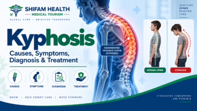
Kyphosis means forward curvature of the spine — and some kyphosis is completely normal anatomy in the mid-back (thoracic spine). Pathological kyphosis refers to an excessive or abnormal curvature, which can result from posture, Scheuermann’s disease, congenital vertebral abnormalities, osteoporosis, trauma, degeneration, infection, tumors, or neuromuscular conditions.
A curved spine on an image does not automatically mean treatment is required — the cause, whether the curve is flexible or fixed, progression, symptoms, and neurological status determine management, which ranges from simple observation to physiotherapy, bracing, or, in selected cases, spinal surgery.
Table of Contents
ToggleWhat Is Kyphosis?
Kyphosis is the term for a forward-curving (convex) curvature of the spine, most often discussed in the thoracic (mid-back) region. This is where the important nuance begins: thoracic kyphosis is a normal part of spinal anatomy — everyone has some degree of forward curve in the upper back. What matters clinically is whether the curve is excessive (beyond the typical range), whether it is structural (fixed, involving actual vertebral changes) or postural (flexible, correctable with position change), and whether it’s associated with pain, progression, or neurological symptoms.
Normal vs. Abnormal Kyphosis
A degree of thoracic kyphosis is present in virtually everyone and serves a genuine biomechanical function, helping the spine absorb load. “Pathological” or excessive kyphosis refers to curvature beyond what’s considered typical, particularly when it’s associated with structural vertebral changes, progression over time, pain, or functional impact.
The presence of a curved spine on an X-ray does not automatically mean treatment is required — a mild, stable, flexible, and asymptomatic curve found incidentally is managed very differently from a progressive, structural, symptomatic one.
Spinal Anatomy and Normal Curves
The spine has a natural series of curves when viewed from the side: cervical lordosis (a backward curve in the neck), thoracic kyphosis (a forward curve in the mid-back), and lumbar lordosis (a backward curve in the lower back), continuing into the sacrum. These alternating curves work together to balance the head over the pelvis and absorb mechanical load efficiently — this overall relationship is referred to as sagittal balance or sagittal alignment.
In more detailed spinal-deformity evaluation, particularly for adults, additional pelvic parameters are considered: pelvic incidence (a fixed anatomical value unique to each person), pelvic tilt, sacral slope, and the sagittal vertical axis (a measure of how far forward the head sits relative to the pelvis). These are introduced here only because they matter for understanding why adult spinal deformity assessment is more than “measuring one curve” — a full explanation of these parameters is a matter for your treating spine specialist, not something to self-interpret from imaging reports.
Types of Kyphosis
| Type | Typical Feature |
|---|---|
| Postural kyphosis | Flexible, correctable with position change; related to posture and muscle imbalance rather than structural vertebral change |
| Scheuermann kyphosis | Structural, involving vertebral wedging and endplate changes, typically presenting in adolescence |
| Congenital kyphosis | Present from birth due to vertebral formation or segmentation defects; can progress early and carries neurological risk |
| Degenerative kyphosis | Related to age-related disc and facet joint changes, typically in adults |
| Osteoporotic kyphosis | Results from vertebral compression fractures and progressive wedging in weakened bone |
| Post-traumatic kyphosis | Follows compression or burst fractures, sometimes with delayed deformity or nonunion |
| Neuromuscular kyphosis | Associated with conditions such as cerebral palsy or other neuromuscular disorders affecting spinal muscle control |
| Iatrogenic / post-surgical kyphosis | Occurs after spinal surgery, including proximal junctional kyphosis near a previous fusion |
| Infectious / destructive kyphosis | Results from spinal tuberculosis, vertebral osteomyelitis, or discitis destroying vertebral structure |
| Tumor-related kyphosis | Results from vertebral destruction by metastatic disease, myeloma, or primary spinal tumors |
These causes are not equally common — postural kyphosis, Scheuermann’s disease, and osteoporotic/degenerative causes make up the large majority of cases seen in practice, while congenital, infectious, and tumor-related causes are comparatively less frequent but carry distinct and important management implications when present.
Kyphosis by Age
Age is one of the most useful organizing lenses for thinking about likely causes and appropriate evaluation.
- Infants and young children: Congenital causes, neuromuscular causes, and structural deformity are the primary considerations, and progression can sometimes be rapid during growth, warranting close specialist monitoring.
- Adolescents: Postural kyphosis and Scheuermann’s disease are the dominant considerations. Growth-related progression is an important factor to track, and symptoms may relate to sports or activity.
- Adults: Degenerative changes, post-traumatic deformity, and the effects of previous spinal surgery become more prominent considerations, alongside chronic structural deformity that may have been present since adolescence.
- Older adults: Osteoporosis and vertebral compression fractures become a leading cause, contributing to progressive sagittal imbalance. Frailty, balance and fall risk, and — in severe deformity — functional and pulmonary effects become relevant considerations.
What Causes Kyphosis?
Causes include poor posture and muscle imbalance (producing flexible, postural kyphosis), Scheuermann’s disease, congenital vertebral abnormalities, osteoporosis with vertebral compression fractures, trauma, spinal infection, tumors, inflammatory spinal disease in some cases, neuromuscular conditions, degenerative spinal disease, previous spinal surgery, and age-related spinal changes generally.
It’s important to distinguish a cause from a risk factor, and to avoid the oversimplification that “bad posture” explains every form of kyphosis — it explains flexible, postural kyphosis reasonably well, but does not explain structural conditions like Scheuermann’s disease, congenital deformity, osteoporotic collapse, or infection- or tumor-related destruction, which involve genuine changes to vertebral structure that posture correction cannot address.
Symptoms
Symptoms depend heavily on the severity and underlying cause, and mild kyphosis may cause no symptoms at all. Possible symptoms include a visibly rounded upper back, back pain, stiffness, fatigue, muscle discomfort, difficulty standing fully upright, reduced physical endurance, activity limitation, neck or shoulder discomfort, low-back pain arising from compensatory changes in spinal alignment, and, in severe deformity, difficulty looking straight ahead.
Neurological symptoms, when present, can include numbness, tingling, weakness, gait disturbance, balance problems, and, in severe spinal cord or cauda equina involvement, bowel or bladder dysfunction — these are discussed further in the red-flag section below.
Red Flags — When Is Kyphosis a Medical Emergency?
Ordinary postural kyphosis is not generally an emergency. However, the following require urgent medical assessment:
- Rapidly progressive deformity
- New, severe pain following trauma
- Inability to stand or walk
- New limb weakness
- Progressive numbness
- Deteriorating gait
- Bowel or bladder dysfunction
- Saddle anesthesia
- Fever combined with severe back pain
- Unexplained weight loss
- Known cancer with new severe spinal pain
- Suspected spinal infection
- An acute vertebral fracture accompanied by neurological symptoms
International patients experiencing any of these symptoms should seek urgent local medical care rather than delaying evaluation to travel — this is not a scenario where international treatment planning should take priority over immediate local emergency assessment.
How Is Kyphosis Diagnosed?
Diagnosis combines history, physical examination, appropriate imaging, and identification of the underlying cause — not a single test in isolation.
History covers age of onset, whether the curve is progressing, presence and character of pain, any trauma history, osteoporosis risk factors or known diagnosis, prior fractures, growth history (in younger patients), any previous spine surgery, infection or cancer risk factors, neurological symptoms, and activity limitations.
Physical examination assesses standing posture and alignment, the contour and flexibility of the curve, range of motion, a forward-bend examination (useful for distinguishing flexible from fixed deformity), a full neurological assessment (gait, strength, sensation, reflexes), and, where relevant, leg-length or pelvic alignment. A curve that corrects with a forward bend or lying down (flexible) is a fundamentally different clinical situation from a curve that remains fixed regardless of position (structural) — this distinction shapes much of what follows in terms of management.
How Kyphosis Is Measured — The Cobb Angle
The Cobb angle is the standard method for measuring spinal curvature on an X-ray, calculated by drawing lines along the endplates of selected vertebrae at the top and bottom of the curve and measuring the angle between them. Measurement technique matters: which vertebrae are selected can affect the result, there is genuine interobserver variability (different examiners can get somewhat different measurements from the same image), and different studies and guidelines have used different numeric thresholds to define “abnormal” kyphosis.
This article deliberately does not present a single universal Cobb-angle number as the definition of pathological kyphosis for every patient and every age — interpretation depends on the patient’s age, the spinal region involved, the imaging technique used, associated symptoms, the presence of structural vertebral changes, and the overall clinical context. A specific angle that would be unremarkable in one context (say, mild, flexible, asymptomatic postural kyphosis in an adult) might be considered more significant in another (progressive Scheuermann’s kyphosis in a growing adolescent). This is a judgment made by your treating specialist, not a number to interpret in isolation from an imaging report.
Imaging
| Imaging / Test | Main Purpose | What It Can Show | When Useful | Limitations |
|---|---|---|---|---|
| Standing X-ray (AP and lateral) | Initial assessment of curvature and alignment | Curve magnitude, vertebral wedging, compression fractures, degenerative changes, congenital abnormalities | First-line for most suspected kyphosis; standing views are often preferred over lying down since they reflect real-world load-bearing alignment | Limited soft-tissue and neural detail |
| Flexion-extension X-rays | Assess flexibility / instability | Whether the curve corrects with position change | Selected cases where flexibility or instability needs clarification | Not needed for every patient |
| MRI | Assess soft tissue and neural structures | Spinal cord, nerve roots, discs, marrow abnormalities, infection, tumors, neural compression | Neurological symptoms, unexplained severe pain, suspected tumor or infection, atypical vertebral collapse, pre-operative planning in selected cases, rapidly progressive deformity | More limited availability / cost in some settings; not needed for uncomplicated, stable, flexible curves |
| CT | Detailed bony assessment | Bone anatomy, fracture detail, fusion status in previously operated spines, complex deformity | Surgical planning, complex bony anatomy, unclear fracture patterns | Involves radiation exposure; limited soft-tissue detail |
Not every patient with kyphosis needs an MRI. MRI becomes particularly useful with neurological symptoms, unexplained severe pain, suspected tumor or infection, atypical or unexplained vertebral collapse, spinal cord compression concerns, congenital anomalies, pre-operative planning in selected cases, or rapidly progressive deformity — not as a default test for a mild, stable, asymptomatic postural curve.
Differential Diagnosis
| Condition | Typical Clues | Imaging Clues | Important Distinction |
|---|---|---|---|
| Postural kyphosis | Flexible, corrects with position, no structural vertebral change | Normal vertebral shape on imaging | Distinguishes from fixed structural deformity requiring different management |
| Scheuermann kyphosis | Adolescent onset, may have back pain, fixed curve | Vertebral wedging (typically ≥3 consecutive vertebrae), endplate irregularities, Schmorl’s nodes | Structural, growth-related; distinct from simple postural kyphosis |
| Osteoporotic / vertebral compression fracture | Older adult, minor or unclear trauma, possible height loss | Wedge-shaped vertebral collapse | Distinguishes age-related fragility fracture from other structural causes |
| Spinal tuberculosis / vertebral osteomyelitis | Constitutional symptoms (though fever can be absent), subacute course | Disc space and adjacent endplate destruction, possible abscess | Requires antimicrobial ± surgical treatment; missing this diagnosis has serious consequences |
| Spinal tumor (metastatic, myeloma, primary) | Known cancer history, unexplained weight loss, night pain | Destructive lesion pattern, possible soft-tissue mass | Requires oncologic evaluation and a fundamentally different treatment pathway |
| Ankylosing spondylitis / inflammatory spine disease | Younger patient, inflammatory back pain pattern, stiffness | Characteristic spinal fusion pattern over time | Requires rheumatologic management alongside spinal considerations |
| Degenerative sagittal imbalance | Older adult, gradual onset, compensatory changes | Disc and facet degeneration, loss of lumbar lordosis | Adult deformity assessment differs from pediatric/adolescent evaluation |
| Scoliosis | Lateral (side-to-side) curvature, sometimes with rotation | Coronal plane deformity on X-ray | A different plane of deformity from kyphosis, though the two can coexist |
Is Kyphosis Just Bad Posture?
Sometimes — but not always, and this distinction matters enormously for what treatment can realistically achieve. Postural kyphosis is flexible: it corrects when the person is asked to stand up straight, and there’s no underlying structural change to the vertebrae themselves. This type genuinely can respond well to posture awareness, strengthening, and general activity.
Structural kyphosis — as seen in Scheuermann’s disease, congenital deformity, or osteoporotic collapse — involves actual changes to vertebral shape, and cannot simply be “stood up straight” or corrected through posture effort alone, because the underlying bone architecture has genuinely changed. Strengthening exercises can still improve function, symptoms, and overall spinal health in structural kyphosis, but they should not be expected to reverse the fixed vertebral deformity itself. There is no reason for shame or blame in either case — this is a matter of anatomy, not effort or willpower.
Scheuermann Kyphosis
Scheuermann’s disease is a structural form of kyphosis typically presenting during adolescence, involving genuine vertebral changes: wedging of multiple consecutive vertebrae, irregularities of the vertebral endplates, and sometimes Schmorl’s nodes (small herniations of disc material into the vertebral body). It most commonly affects the thoracic spine, though a thoracolumbar pattern also occurs. Unlike postural kyphosis, this deformity is genuinely fixed rather than flexible.
Diagnosis relies on the characteristic pattern on X-ray combined with clinical examination. Management depends on curve severity, symptoms, and — importantly — skeletal maturity, since the deformity can progress during the remaining growth period and tends to be more stable once growth is complete. Options range from observation with periodic monitoring, to physiotherapy focused on posture, strength, and flexibility, to bracing in appropriately selected, still-growing patients, to surgical consideration in more severe or progressive cases.
There is no single surgical threshold that applies universally to every patient — the decision depends on curve magnitude, symptoms, growth remaining, and functional impact, assessed individually by a spine specialist.
Kyphosis From Osteoporosis and Vertebral Compression Fractures
Osteoporotic kyphosis develops when vertebral compression fractures cause anterior (front) height loss in one or more vertebrae, producing progressive wedge deformity. Over time, particularly with multiple fractures, this can accumulate into significant increased thoracic curvature, sometimes visibly noticeable as a stooped or “hunched” posture, along with chronic pain and functional decline.
Management centers on both the deformity itself and, critically, the underlying bone disease: osteoporosis evaluation (bone mineral density testing), fracture-risk assessment, appropriate osteoporosis medication, ensuring adequate calcium and vitamin D status, and fall-prevention strategies. Treating the underlying osteoporosis matters as much as addressing the deformity itself — without it, the risk of further vertebral fractures and progressive deformity remains unaddressed regardless of what’s done for the current curve.
Post-Traumatic Kyphosis
Following compression or burst fractures, vertebrae can heal in a malaligned position, or fail to heal properly (nonunion), leading to delayed or progressive deformity. Whether conservative management or surgery is more appropriate depends on the fracture pattern, degree of resulting deformity, spinal stability, and neurological status — conservative management may be reasonable for stable, non-progressive, neurologically intact cases, while surgery becomes more relevant with instability, significant or progressive deformity, or neurological compromise.
Infection- and Tumor-Related Kyphosis
Infection (spinal tuberculosis, vertebral osteomyelitis, discitis/spondylodiscitis) can destroy vertebral structure, leading to collapse and kyphotic deformity, along with pain, constitutional symptoms, and, in more advanced cases, neurological compromise. Fever may be absent, particularly in older adults, so its absence should not be used to confidently rule out infection. Treatment requires addressing the underlying infection with antimicrobial therapy, with surgery reserved for specific circumstances (significant instability, neurological compromise, or need for drainage/debridement) rather than applied routinely to every infection-related case.
Tumors — including metastatic disease, multiple myeloma, and primary spinal tumors — can similarly cause vertebral bone destruction, collapse, deformity, pain, and neurological compromise. Cancer-related vertebral collapse needs to be actively differentiated from osteoporosis-related collapse when clinical features (cancer history, unexplained weight loss, atypical presentation) raise concern, since management is fundamentally different. This is not meant to suggest every kyphotic deformity indicates cancer — most do not — but the possibility should be genuinely considered rather than dismissed, particularly when red-flag features are present.
Natural History and Complications
Not every case of kyphosis progresses, and severity of complications varies enormously by cause. Possible complications of significant or progressive kyphosis include chronic pain, reduced mobility, muscle fatigue, worsening sagittal imbalance, gait abnormalities, increased fall risk, reduced physical function, neurological compression in severe cases, respiratory effects with severe deformity, reduced quality of life, and, in osteoporotic kyphosis specifically, recurrent vertebral fractures. Not every patient develops these complications — this list represents possibilities relevant to more severe or progressive cases, not an inevitable trajectory.
Kyphosis Treatment Overview
Treatment is built around cause, age, severity, flexibility, progression, symptoms, neurological status, bone quality, and functional impact — not a single formula applied to every “kyphosis” diagnosis regardless of cause.
Observation
For mild, flexible, non-progressive, asymptomatic curves — particularly postural kyphosis — observation with periodic reassessment may be entirely appropriate, rather than defaulting to active intervention.
Physiotherapy and Exercise
Physiotherapy focused on posture awareness, core and spinal extensor strengthening, and flexibility work can improve strength, posture, function, and symptoms. It’s important to state clearly: exercise can improve function and symptoms, but it does not reliably reverse a fixed structural spinal deformity. Generic “back exercises” should not be marketed or understood as a universal cure for structural kyphosis, even though they remain genuinely valuable for overall spinal health and symptom management.
Bracing
Bracing may be considered in specific circumstances, most notably in growing adolescents with progressive Scheuermann’s kyphosis, where the goal is to influence the curve during the remaining growth period, and separately, in some adults with osteoporotic vertebral fractures, where the goal is primarily symptomatic support rather than structural correction. Considerations include patient selection, growth/skeletal maturity, adherence, appropriate duration, and potential downsides including muscle deconditioning and skin irritation with prolonged wear. This article does not prescribe a specific brace type or wearing schedule — that decision should be individualized with your treating specialist.
Osteoporosis Treatment
Where osteoporosis is contributing to the deformity, addressing the underlying bone fragility — through bone density assessment, appropriate pharmacotherapy, adequate calcium/vitamin D, and fall prevention — is a core part of management, not a separate consideration from treating the curve itself.
When Is Surgery Needed?
Surgery is not automatically indicated simply because a Cobb angle is elevated. Potential reasons surgery may be considered include severe or clearly progressive deformity, significant functional impairment, persistent disabling pain despite appropriate nonoperative treatment, neurological compromise or spinal cord compression, structural instability, severe post-traumatic deformity, selected cases of congenital deformity, selected cases of more severe Scheuermann’s kyphosis, infection or tumor requiring stabilization or decompression, and severe sagittal imbalance in appropriately selected adults. The exact threshold for surgery varies by cause, age, anatomy, symptoms, and individual surgeon assessment — there is no single universal cutoff that applies to every patient.
Kyphosis Surgery Options
Surgical approaches are not a simple escalation ladder from “smaller” to “bigger” — each addresses a different specific problem:
- Posterior spinal fusion: the most common general approach, using instrumentation (typically pedicle screws and rods) to correct and stabilize the spine, with fusion intended to maintain the correction long-term
- Anterior release/fusion: used in selected cases where anterior spinal structures need to be addressed directly, sometimes combined with a posterior approach
- Osteotomies (bone-cutting procedures that allow the surgeon to reorient the spine): including Smith-Petersen/Ponte-type osteotomies for more moderate correction needs, and more extensive procedures like pedicle subtraction osteotomy or, in exceptional severe rigid deformity, vertebral column resection, for cases requiring substantial correction
More powerful deformity-correction procedures generally carry meaningfully greater surgical complexity, blood loss, and risk — the choice of technique depends on curve flexibility, magnitude, cause, and the specific correction needed, decided individually rather than following a fixed pathway from mild to severe intervention.
Osteotomies in Plain Terms
An osteotomy involves surgically cutting and reorienting a section of bone to change the spine’s alignment, used when the deformity is too rigid to correct through instrumentation and fusion alone. The specific technique chosen depends on how flexible or rigid the deformity is and how much correction is needed. Risks include significant blood loss, neurological injury, nonunion, and implant failure — this article does not provide surgical technical instructions, since the specific approach is a matter of individualized surgical planning.
Adult Sagittal Deformity
Adult spinal deformity is not simply “a large kyphosis angle” — it involves the interplay between thoracic kyphosis, lumbar lordosis, and pelvic compensation. When alignment is significantly off-balance, patients may compensate through knee or hip flexion, leading to fatigue and difficulty standing upright for extended periods. Surgical planning in this context considers global spinal alignment, individual pelvic parameters, spinal flexibility, any previous fusion, bone quality, age, overall frailty and health status, and the patient’s specific functional goals — radiographic correction of a number on an X-ray is not, by itself, the only relevant goal; functional outcome and quality of life matter equally.
Minimally Invasive and Robotic Techniques
Minimally invasive spinal surgery, computer navigation, robotic assistance, and 3D surgical planning may help with aspects such as screw-placement trajectory and accuracy. These technologies should be understood as adjuncts to sound surgical judgment, not substitutes for it — they do not automatically make surgery safer for every deformity, do not guarantee better long-term outcomes, and do not eliminate neurological or implant-related risks. Availability and appropriate use vary by case complexity and surgeon experience with the specific technology.
Surgical Risks
Spinal deformity surgery carries genuine risks that should be discussed transparently: infection, significant blood loss, neurological injury, dural tear, nerve injury, implant failure, screw malposition, nonunion (pseudarthrosis), proximal junctional kyphosis (a specific complication where deformity develops just above a spinal fusion), adjacent-segment problems, rod fracture, deformity recurrence, pulmonary complications, blood clots, anesthesia-related complications, and the potential need for revision surgery. Risk varies considerably according to patient age, the specific procedure, deformity severity, presence of osteoporosis, other medical conditions, previous spine surgery, and overall surgical complexity — these risks should be discussed specifically with your surgical team for your individual case.
Osteoporosis and Spinal Deformity Surgery
Poor bone quality creates specific surgical challenges, including higher risk of screw loosening, fixation failure, new vertebral fractures, proximal junctional failure, and pseudarthrosis. Preoperative bone-health assessment and, where feasible, optimization of osteoporosis treatment before surgery are important considerations, along with fixation strategies adapted for weaker bone (such as cement-augmented screws or extended fixation constructs) where evidence and individual circumstances support them. Treating osteoporosis before surgery does not eliminate surgical risk — it’s a risk-reduction measure, not a guarantee.
Recovery and Rehabilitation
Recovery differs substantially by treatment type and cause. Conservative management of postural or mild Scheuermann’s kyphosis generally involves an ongoing, gradual process rather than a fixed endpoint. Bracing for growing adolescents typically continues over an extended period tied to growth remaining. Vertebral-fracture-related conservative care often shows meaningful pain improvement over weeks to a few months. Minimally invasive fixation generally allows for a shorter hospital stay and faster initial mobilization than open reconstruction, while major deformity correction surgery (osteotomy, extensive fusion) typically involves a longer hospital stay and a more extended, structured rehabilitation course.
Across treatment types, recovery generally includes early guided mobilization, pain management, physiotherapy, and a graduated return to school, work, driving, and exercise or sports. No fixed recovery timeline can be promised — it depends on the specific procedure, deformity severity, age, bone quality, and individual healing.
Exercise and Daily Life
For flexible, postural kyphosis, exercise and posture-focused strengthening genuinely can improve the curve’s appearance and associated symptoms. For fixed, structural kyphosis (Scheuermann’s, congenital, osteoporotic, or post-surgical), exercise supports strength, function, and symptom management, but should not be expected to reverse the established structural deformity.
Walking is generally safe and beneficial across most forms of kyphosis. Yoga and general gym exercise can typically be pursued safely, with attention to avoiding excessive loading in cases of significant osteoporosis or acute vertebral fracture — specific exercise guidance should be individualized with your treating clinician or physiotherapist rather than following a generic program, particularly for children and adolescents with structural conditions, where activity recommendations should come from the treating specialist.
Regarding everyday posture, sitting, and ergonomics: reasonable adjustments (supportive seating, avoiding prolonged static positions, attention to workstation setup) can help with comfort and symptom management, but there is no single “perfect posture” that applies to everyone, and posture correction alone does not treat established structural kyphosis — it’s a supportive measure, not a definitive treatment.
Pregnancy and Kyphosis
Most patients with mild kyphosis, including mild Scheuermann’s kyphosis, can generally expect a normal pregnancy and delivery process, though this should be confirmed with your obstetric team. Patients with more severe spinal deformity or a history of spinal fusion surgery may have additional considerations for pain management and, if relevant, anesthesia planning (previous fusion can occasionally affect the technical approach to neuraxial/epidural anesthesia) — kyphosis does not automatically prevent vaginal delivery or neuraxial anesthesia, but individualized assessment by both obstetric and anesthesia teams is genuinely important for patients with significant deformity or prior spinal surgery.
Prognosis
Outcome depends on the underlying cause, age, severity, curve flexibility, rate of progression, neurological status, bone quality, treatment chosen, engagement with rehabilitation, and other health conditions. Postural kyphosis generally responds well to conservative management. Mild to moderate Scheuermann’s kyphosis often does well with observation or bracing during growth, with more severe or progressive cases sometimes requiring surgical consideration. Osteoporotic kyphosis requires ongoing attention to bone health regardless of how the acute deformity is managed.
Complete correction should not be promised for any cause — the realistic goal is typically pain control, prevention of further progression, and preservation or improvement of function, with the degree of achievable correction varying by individual case.
When to See a Spine Specialist (and When to Get a Second Opinion)
Prompt specialist evaluation is warranted for a rapidly increasing curvature, persistent pain, any neurological symptoms, adolescent structural kyphosis, suspected congenital deformity, a known vertebral fracture, suspected osteoporosis, post-traumatic deformity, kyphosis developing after previous spine surgery, suspected infection or tumor, significant functional impairment, or when surgery is being considered. Which type of specialist is appropriate varies by age and likely cause — pediatric orthopedic or spine specialists for children and adolescents, and adult spine surgeons or deformity specialists for adults, with neurosurgical involvement when neurological compromise is a concern.
A second opinion is particularly valuable when major surgery has been recommended, an osteotomy is proposed, extensive spinal fusion is proposed, the diagnosis remains uncertain, the deformity is progressing despite treatment, neurological symptoms are present, previous surgery has not achieved the expected result, infection or tumor is suspected, different specialists have offered differing recommendations, or severe osteoporosis complicates surgical planning. Useful records to bring include standing X-rays, CT, MRI, radiology reports, bone-density reports, relevant blood tests, previous operative notes, implant records, and rehabilitation records.
Kyphosis Treatment in India (2026)
Appropriately equipped centers in India coordinate care across pediatric or adult spine specialists (depending on age), orthopedic spine surgeons, neurosurgeons, radiologists, osteoporosis specialists where relevant, and rehabilitation physiotherapists. Depending on the specific case, potential treatment approaches include full diagnostic evaluation (X-ray, MRI, CT, bone density testing where indicated), physiotherapy and bracing for appropriate conservative cases, osteoporosis management, and, for selected patients, deformity-correction surgery ranging from posterior fusion to more extensive osteotomy-based reconstruction, supported by structured rehabilitation.
Kyphosis Treatment Cost in India in 2026
Published international-patient cost estimates for kyphosis correction surgery in India show meaningful variation reflecting differences in procedure complexity, hospital tier, and what’s included in the quote:
- Simpler fixation/lower-complexity cases: some sources cite figures starting around $3,500–$5,400 USD
- Standard deformity correction with fusion: commonly cited international-patient range of roughly $8,000–$9,900 USD
- More complex correction involving osteotomy and extended reconstruction: published estimates ranging up to roughly $13,000–$17,000 USD
Osteotomy as a standalone component has been cited separately in the range of roughly $1,200–$2,700 USD, though in practice this is typically bundled into the total surgical cost rather than billed separately, and should be clarified directly with the treating hospital.
Cost varies according to city, hospital, surgeon, the specific procedure and its complexity, implants used, deformity severity, number of spinal levels involved, whether an osteotomy is required, bone quality, ICU requirements, length of hospitalization, and rehabilitation needs. These figures should be treated as reference ranges, not a firm quote — an itemized, case-specific estimate from the treating hospital, based on your actual imaging and diagnosis, is necessary for an accurate figure, particularly given how much complexity (simple fusion vs. multi-level osteotomy) affects the final cost.
Separately from medical treatment costs, international patients should budget for accommodation, airport transfers, local transportation, interpreter or local support where needed, and follow-up visits. India should not be presented as automatically the cheapest or best destination — verifying a specific hospital’s genuine capability and experience with spinal deformity correction, particularly for more complex cases, matters more than general cost comparisons.
International Patient Journey
- Share medical records and describe the history and progression of the curvature
- Share X-rays, CT, and/or MRI images and reports
- Specialist review
- Establish the likely underlying cause
- Determine severity and neurological status
- Discuss treatment options appropriate to the cause and severity
- Receive an individualized treatment plan
- Receive an estimated treatment budget
- Coordinate specialist/hospital appointments
- Medical visa support where applicable
- Travel and accommodation coordination
- In-person assessment on arrival
- Additional investigations if required
- Treatment, if medically appropriate
- Hospital recovery
- Rehabilitation
- Discharge planning
- Follow-up
- Coordination with the patient’s local physician after returning home
Patients with new neurological deficits, bowel/bladder dysfunction, suspected spinal cord compression, severe trauma, or fever with severe spinal pain should seek urgent local medical care rather than delaying treatment for international travel.
How Shifam Health Can Help
Shifam Health is a medical tourism facilitator/coordinator not the treating doctor, surgeon, hospital, or diagnostic provider, and does not make treatment decisions. Shifam Health may assist with medical record collection, specialist and hospital coordination, appointment scheduling, communication of treatment plans, coordination of cost estimates, medical visa support, airport pickup, accommodation coordination, interpreter/local support, hospital logistics, and follow-up coordination. They cannot guarantee cure, correction, pain relief, neurological recovery, surgical success, exact cost, exact recovery time, hospital acceptance, or visa approval.
International patients can share their medical records and spinal imaging with Shifam Health to help coordinate an appropriate specialist review and understand potential treatment options in India.
Myths vs. Facts
Myth: Kyphosis always means bad posture. Fact: Many forms of kyphosis — including Scheuermann’s disease, congenital deformity, and osteoporotic collapse — involve genuine structural vertebral changes unrelated to posture.
Myth: Kyphosis is always permanent. Fact: Postural kyphosis is often fully correctable, and even structural forms can often be improved with appropriate treatment, though not always fully reversed.
Myth: Exercise can completely straighten structural kyphosis. Fact: Exercise improves strength, function, and symptoms but does not reliably reverse an established structural deformity.
Myth: Scheuermann kyphosis is just poor posture. Fact: It’s a structural condition involving genuine vertebral wedging and endplate changes, distinct from flexible postural kyphosis.
Myth: Kyphosis only affects older people. Fact: It occurs across all ages, from congenital forms in infants to postural and Scheuermann’s kyphosis in adolescents to osteoporotic kyphosis in older adults.
Myth: Kyphosis cannot occur in children. Fact: Congenital and neuromuscular kyphosis can present in infancy and early childhood.
Myth: Osteoporosis-related kyphosis is harmless. Fact: It reflects underlying bone fragility with real fracture and complication risk, and warrants active osteoporosis management.
Myth: If there is no pain, kyphosis cannot be serious. Fact: Some structural or progressive kyphosis, including certain congenital and neuromuscular cases, can be clinically significant with minimal early pain.
Myth: Braces permanently straighten every kyphotic spine. Fact: Bracing may help manage progression during specific circumstances (such as growth in adolescents) but does not guarantee permanent structural correction.
Myth: Spinal fusion completely eliminates future problems. Fact: Fusion stabilizes the treated segment but does not eliminate risks like adjacent-segment issues, proximal junctional kyphosis, or the need for future monitoring.
Frequently Asked Questions
What is kyphosis?
An excessive forward curvature of the spine, commonly affecting the upper or mid-back.
What causes kyphosis?
Causes include poor posture, Scheuermann’s disease, congenital abnormalities, osteoporosis, spinal fractures, infection, tumors, and degenerative changes.
Can poor posture cause kyphosis?
Yes, poor posture can cause flexible postural kyphosis, but it does not cause structural conditions such as Scheuermann’s disease.
What are the symptoms of kyphosis?
Back pain, stiffness, fatigue, rounded shoulders, visible spinal curvature, and reduced mobility. Severe cases may cause breathing or neurological problems.
How is kyphosis diagnosed?
Diagnosis typically involves physical examination and standing spinal X-rays. MRI or CT may be recommended for neurological symptoms, complex deformity, infection, tumors, or surgical planning.
Can kyphosis be treated without surgery?
Yes. Many cases improve with observation, physiotherapy, exercise, or bracing, depending on the cause and severity.
Can exercise correct kyphosis?
Exercise can improve posture, strength, flexibility, and symptoms. It may correct flexible postural kyphosis but cannot fully reverse a fixed structural deformity.
What is spinal fusion for kyphosis?
Fusion uses screws, rods, and bone graft to correct and permanently stabilize the affected spinal segments.
Can kyphosis worsen with age?
It can, particularly when caused by osteoporosis, vertebral fractures, or degenerative changes.
Final Takeaway
Kyphosis is not one single condition — it ranges from entirely normal spinal anatomy and flexible postural curves that respond well to simple measures, to structural conditions like Scheuermann’s disease, congenital deformity, osteoporotic collapse, and, less commonly, infection- or tumor-related destruction that require genuinely different evaluation and management.
A curved spine seen on imaging does not automatically mean treatment, let alone surgery, is required — the cause, whether the curve is flexible or fixed, its progression, associated symptoms, and neurological status all factor into the right approach. New neurological symptoms, bowel or bladder dysfunction, fever with severe back pain, or an unexplained cancer-related presentation deserve prompt, thorough evaluation.
Most kyphosis is managed without surgery; when surgery is genuinely appropriate, the specific approach — from standard fusion to more extensive osteotomy-based correction — depends on individual anatomy, cause, and goals, not a fixed escalation ladder.
International patients can share their medical records and spinal imaging with Shifam Health to help coordinate an appropriate specialist review and understand potential treatment options in India.
This article is for general educational purposes and does not replace individualized clinical examination, imaging review, diagnosis, or treatment advice from a qualified spine specialist.

Popular Posts From Last Week
- September 18, 2026
- shifamhealth
Written by: Shifam Health Editorial Team — Orthopedic Content Division Medically reviewed by: Shifam Health Clinical Advisory Panel…
- September 16, 2026
- shifamhealth
Ankle valgus deformity is an abnormal alignment in which the ankle tilts toward the outside of the body,…
- September 15, 2026
- shifamhealth
Medically reviewed content · Published 2026 · Last reviewed September 2026 Bow legs medically called genu varum —…
- September 14, 2026
- shifamhealth
Medically reviewed content · Published 2026 · Last reviewed September 2026 A “broken ankle” is the common term…
- September 15, 2026
- shifamhealth
Medically reviewed content · Published 2026 · Last reviewed August 2026 A burst fracture is a spinal vertebra…
- September 13, 2026
- shifamhealth
If a lumbar MRI report mentions “kissing spinous processes” or “Baastrup’s disease,” it’s worth pausing before assuming this…
- September 14, 2026
- shifamhealth
Scoliosis is a three-dimensional spinal deformity — sideways curvature combined with rotation — and it is genuinely not…
- September 16, 2026
- shifamhealth
Epithelial ovarian cancer is the most common broad category of ovarian cancer, arising from the cells covering the…
- September 17, 2026
- shifamhealth
Hepatoblastoma is a rare malignant liver tumor that develops primarily in young children, most commonly before age 5….
- September 19, 2026
- shifamhealth
Finding a purple or reddish patch on your skin that won’t fade, or hearing “Kaposi sarcoma” mentioned during…
- September 18, 2026
- shifamhealth
Kyphosis means forward curvature of the spine — and some kyphosis is completely normal anatomy in the mid-back…
- September 19, 2026
- shifamhealth
Lumbar radiculopathy is a neurological syndrome caused by irritation, inflammation, or compression of a nerve root in the…