



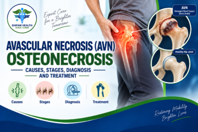
Home > Blog > Avascular Necrosis (AVN) — Osteonecrosis (2026): Causes, Stages, Diagnosis and Treatment
Avascular Necrosis (AVN) — Osteonecrosis (2026): Causes, Stages, Diagnosis and Treatment
Filters & Insights
Complete guide to avascular necrosis (osteonecrosis): causes, staging, MRI diagnosis, core decompression, bone grafting, hip replacement & treatment costs in India

Written by: Shifam Health Editorial Team — Orthopedic Content Division Medically reviewed by: Shifam Health Clinical Advisory Panel | Last updated: September2026
Medical disclaimer: This article is for educational purposes only. It does not constitute medical advice, a diagnosis, or a treatment recommendation for any individual. AVN management requires assessment by a qualified orthopaedic specialist with access to your imaging, full clinical history, and individual risk factors.
Avascular necrosis (AVN), or osteonecrosis, occurs when impaired blood supply causes bone tissue to die. It most commonly affects the femoral head but can involve other bones. Treatment depends heavily on whether the bone has collapsed. Early, pre-collapse disease may be considered for joint-preserving treatment such as core decompression in selected patients, while advanced collapse with painful joint damage may require total joint replacement. No single treatment works for every patient.
What is avascular necrosis (AVN)?
Avascular necrosis (AVN) — also called osteonecrosis — is a condition in which bone tissue dies due to disrupted or insufficient blood supply. Without adequate blood flow, bone cells (osteocytes) cannot survive. The dead bone may initially hold its shape but progressively weakens and, in many cases, collapses. AVN most commonly affects the femoral head (the ball of the hip joint) but can occur at the knee, shoulder, ankle, wrist, and other sites.
Table of Contents
ToggleWhat Is Avascular Necrosis (AVN)?
Avascular necrosis (AVN) also called osteonecrosis — is the death of bone tissue resulting from inadequate blood supply. The bone does not receive enough oxygen and nutrients to sustain cell viability. Over time, the structurally compromised dead bone may collapse under normal loading forces, destroying the joint surface.
The term “avascular” means without blood vessels; “necrosis” means tissue death. These two terms describe what happens biologically: the blood vessels supplying a segment of bone are disrupted, interrupted, or insufficient, and the bone cells dependent on that supply die.
Why AVN Is Distinct
AVN is not simply another form of arthritis or bone disease. Several distinctions matter:
- The primary injury is vascular — a problem with blood supply — not primarily cartilage degradation (as in osteoarthritis) or bone density loss (as in osteoporosis)
- AVN can affect younger patients — sometimes in their twenties, thirties, and forties — an age group rarely affected by primary osteoarthritis
- AVN may be bilateral — particularly in steroid-induced and alcohol-related disease, both hips are often affected simultaneously or sequentially
- Collapse is the key prognostic event — once the femoral head (or other affected bone) collapses, the clinical course changes fundamentally and joint-preserving options become unavailable
The Femoral Head — Why It Is Most Vulnerable
The femoral head (the rounded upper end of the femur that forms the ball of the hip joint) has a blood supply that is anatomically vulnerable. The main vessels supplying the femoral head — branches of the medial and lateral femoral circumflex arteries — travel in close proximity to the femoral neck and can be interrupted by fracture, dislocation, or vascular disease. When these vessels are compromised, the femoral head loses its blood supply and AVN develops.
How AVN Differs from Osteoporosis and Arthritis
AVN, osteoporosis, and osteoarthritis are fundamentally different conditions with different causes, imaging appearances, populations affected, and treatments. Confusing them leads to incorrect management.
Comparison Table
| Feature | AVN / Osteonecrosis | Osteoarthritis | Osteoporosis |
|---|---|---|---|
| Primary problem | Bone tissue death from impaired blood supply | Cartilage breakdown | Reduced bone density and bone microarchitecture |
| Typical age | Any age — often younger adults (20–50s) | Typically older adults (50s+) | Typically older adults; postmenopausal women |
| Common sites | Femoral head, knee, humeral head, talus, lunate | Weight-bearing joints: knee, hip, spine, hands | Spine, hip, wrist, other trabecular-rich sites |
| X-ray finding | Sclerosis, crescent sign, collapse — often normal early | Joint-space narrowing, osteophytes, sclerosis | Reduced bone density; insufficiency fractures |
| MRI appearance | Characteristic double-line sign in early disease | Cartilage thinning, bone marrow changes | Low signal on T1 in fractures; bone marrow oedema |
| Cause | Vascular disruption — trauma, steroids, alcohol, sickle cell, others | Multifactorial — age, load, injury, genetics | Low bone density — oestrogen deficiency, ageing, medications |
| Collapse risk | High in untreated or advanced disease | Does not collapse in the same way | Fragility fractures — different mechanism |
| Primary treatment aim | Preserve joint before collapse; replace if collapsed | Symptom management; joint replacement when advanced | Fracture prevention; pharmacotherapy; fall prevention |
A patient told they have “hip problems” or even “hip arthritis” may actually have AVN particularly if they are younger, have taken corticosteroids, or drink alcohol heavily. Misclassification delays appropriate treatment.
Causes and Risk Factors
AVN has multiple identifiable causes, divided broadly into traumatic and non-traumatic. Non-traumatic AVN is associated with several well-established risk factors — corticosteroid use and alcohol consumption being the most common in most studied populations. In some patients, no cause is identified (idiopathic AVN).
Traumatic Causes
Disruption of the blood supply by direct physical injury:
- Femoral neck fracture: One of the most common causes of femoral head AVN; the fracture disrupts the vessels supplying the femoral head. Risk is higher with displaced fractures and increases with time to surgical fixation.
- Hip dislocation: Posterior hip dislocation, particularly if not reduced promptly, stretches or tears the retinacular vessels supplying the femoral head.
- Other joint fractures: Fractures at other sites with vulnerable end-arterial blood supply (talus fractures, scaphoid fractures, humeral head fractures).
Non-Traumatic Causes and Risk Factors
| Risk Factor / Cause | Notes |
|---|---|
| Corticosteroid use | One of the most common non-traumatic causes. Risk correlates with cumulative dose and duration, not a single course. High-dose or prolonged systemic steroids are the main concern; inhaled steroids carry much lower risk. AVN may be bilateral and multifocal. |
| Alcohol use | Heavy alcohol consumption is a well-established risk factor. Mechanisms include fat emboli, altered lipid metabolism, and possibly direct osteocyte toxicity. |
| Sickle cell disease | Among the highest-risk conditions. Sickling of red blood cells in small bone vessels causes repeated ischaemic episodes. AVN is frequently multifocal. |
| Systemic lupus erythematosus (SLE) | Risk comes from both the disease itself and the high-dose corticosteroids used to treat it; disentangling the two contributions is difficult. |
| Coagulopathies and thrombophilias | Hypercoagulable states such as factor V Leiden, antiphospholipid syndrome, and protein C or S deficiency can cause microvascular thrombosis within bone. |
| Radiation therapy | Radiation to areas containing bone can damage the blood supply — particularly the femoral head after pelvic radiation and the humeral head after shoulder or axillary radiation. |
| Decompression sickness (“the bends”) | Nitrogen bubbles in blood following rapid decompression, such as during scuba diving or caisson work, can block bone vessels. |
| HIV and antiretroviral therapy | AVN risk is elevated in people with HIV. Contributions from antiretroviral drugs, the underlying disease, and associated lifestyle factors are complex. |
| Gaucher’s disease | Lipid-laden macrophages accumulate in bone and can impair blood supply. |
| Organ transplantation | High-dose corticosteroids used for immunosuppression after transplant significantly elevate risk. |
Idiopathic AVN
In a proportion of cases — estimates vary across studies — no specific risk factor is identified. This is called idiopathic osteonecrosis. Its existence does not change the diagnostic or management approach, but patients should not feel that an absence of risk factors makes the diagnosis less likely if imaging is characteristic.
Sites of AVN — Beyond the Hip
While the femoral head is the most common and most studied site for AVN, osteonecrosis can occur at multiple other sites — and the clinical presentation, natural history, and treatment differ by location.
Femoral Head (Hip)
The most common site. Accounts for the majority of clinical cases and nearly all the published research. Disease in the femoral head carries significant risk of collapse, which destroys the hip joint surface.
Knee — Medial Femoral Condyle
The medial femoral condyle (inner aspect of the lower thigh bone at the knee) is the most common site of knee osteonecrosis. Presents with medial knee pain, often in older patients and sometimes confused with meniscal pathology. Two distinct forms:
- Spontaneous osteonecrosis of the knee (SONK): Now understood to often represent subchondral insufficiency fracture rather than true osteonecrosis in the classical sense — distinction matters for management
- Secondary osteonecrosis of the knee: Follows the same aetiology as hip AVN (steroids, alcohol, etc.); more likely to be multifocal
Humeral Head (Shoulder)
The humeral head (ball of the shoulder joint) is the second most commonly affected site in the upper extremity. Often associated with steroid use, trauma, or sickle cell disease. Presents with shoulder pain and restricted movement. If collapse occurs, shoulder replacement may be required.
Talus (Ankle)
The talus — the bone connecting the lower leg to the foot — is vulnerable after talar fractures or dislocations. The talus has a precarious blood supply (approximately 60% is covered by articular cartilage, limiting blood vessel entry points). AVN of the talus presents with ankle pain and difficulty weight-bearing.
Lunate (Wrist) — Kienböck Disease
Kienböck disease is osteonecrosis of the lunate bone in the wrist. It causes wrist pain, swelling, and stiffness and is classified using its own staging system (Lichtman classification). Treatment ranges from immobilisation and activity modification to various surgical procedures depending on stage.
Femoral Condyle and Other Knee Sites
Less common than medial femoral condyle involvement but can occur, particularly in secondary disease.
Multifocal Disease
In steroid-induced and sickle-cell-related AVN, multiple sites may be affected simultaneously. A patient diagnosed with AVN at one site should have the other typical sites assessed — at minimum the contralateral hip and other joints if symptomatic.
Symptoms of AVN
AVN may be completely asymptomatic in its early stages — the disease is sometimes discovered incidentally on MRI performed for another reason. When symptoms develop, the most common presentation is pain in the affected joint, often without a prior history of obvious injury at that time.
Hip AVN Symptoms
- Groin pain: The most common presentation — a deep, aching pain in the groin, sometimes radiating to the buttock or thigh. Groin pain is the typical location for hip pathology, but patients sometimes report it as “hip pain” meaning lateral hip or buttock.
- Pain with weight-bearing: Walking, standing, and climbing stairs are commonly aggravating. In early disease, pain may occur only with activity and resolve with rest.
- Pain at rest or at night: Develops as disease progresses.
- Limp: Antalgic gait to reduce loading on the painful joint.
- Restricted range of motion: Internal rotation of the hip is often the earliest movement to become restricted.
- Progressive limitation: As collapse develops, symptoms typically worsen.
Symptoms at Other Sites
- Knee AVN: Medial knee pain, swelling, tenderness over the medial femoral condyle, pain with stairs or walking.
- Shoulder AVN: Shoulder pain, restricted elevation and rotation, pain with overhead activities.
- Ankle (talar) AVN: Ankle pain, difficulty with weight-bearing, swelling.
- Wrist (Kienböck): Wrist pain, reduced grip strength, swelling, limited wrist motion.
The Asymptomatic Phase
Early AVN — before collapse — may be entirely silent. This means that in patients with significant risk factors (high-dose steroids, heavy alcohol use, sickle cell disease), AVN may be present and progressing without any symptoms. This is why screening MRI is sometimes considered in high-risk groups, though universal screening is not standard practice.
How AVN Is Diagnosed
MRI is the gold standard for early diagnosis of AVN, before changes are visible on X-ray. X-ray is adequate for detecting advanced disease with collapse but misses early stages. CT provides bone detail useful for surgical planning. Bone scan has been largely superseded by MRI but may be used in settings where MRI is unavailable.
Imaging
| Investigation | Role | Sensitivity for Early AVN |
|---|---|---|
| X-ray (weight-bearing) | First-line; detects collapse, sclerosis, and joint-space narrowing — but often normal in early disease | Low in early stages |
| MRI | Gold standard for early detection. The double-line sign on T2-weighted MRI is characteristic of AVN | High — can detect AVN weeks to months before X-ray changes appear |
| CT scan | Provides bone detail, assesses collapse extent, and assists with surgical planning | Intermediate — better than X-ray for bone detail; inferior to MRI for early detection |
| Bone scintigraphy (bone scan) | Can show reduced or increased uptake in the affected area | Moderate — largely superseded by MRI in most settings |
The Double-Line Sign
The double-line sign on MRI — a band of low signal (dark) on T2-weighted images with a parallel inner band of high signal (bright) — represents the interface between living and dead bone. Its presence is considered characteristic (though not pathognomonic) of AVN and helps distinguish AVN from other causes of bone marrow oedema.
Blood Tests
There is no blood test that diagnoses AVN. Blood tests may be ordered to investigate underlying causes (coagulation screens, lipid profiles, haemoglobin electrophoresis for sickle cell, inflammatory markers for SLE) and to assess overall health before treatment decisions are made.
Biopsy
Rarely performed for routine AVN diagnosis. May be done in atypical presentations to exclude other diagnoses.
Staging: How Severe Is the AVN?
AVN is staged to guide treatment decisions, with the most critical distinction being whether the bone has collapsed or not. Several staging systems exist; the ARCO classification (Association Research Circulation Osseous) is among the most widely used internationally.
ARCO Staging
| ARCO Stage | Description | Key Feature |
|---|---|---|
| Stage 0 | Normal imaging; diagnosis suspected from risk factors or biopsy only | Imaging normal |
| Stage 1 | Abnormal on MRI or bone scan; X-ray normal | MRI positive; no X-ray change |
| Stage 2 | Abnormal on X-ray (sclerosis, cysts) but femoral head shape preserved | No collapse |
| Stage 3 | Subchondral collapse — the crescent sign on X-ray; femoral head shape begins to change | Collapse begins |
| Stage 4 | Joint destruction — joint-space narrowing, acetabular changes, established secondary osteoarthritis | Advanced collapse; joint destroyed |
The Crescent Sign
The crescent sign — a thin radiolucent (dark) line beneath the subchondral bone surface on X-ray — represents subchondral fracture: the bone has begun to collapse. Its presence marks the transition from stage 2 to stage 3 and is a critically important clinical landmark because it signals that joint-preserving options have become much less likely to succeed.
Why Staging Drives Everything
The central treatment question in AVN is: has the bone collapsed?
- Pre-collapse (Stages 1–2): Joint-preserving procedures may be considered in appropriate patients
- Early collapse (Stage 3): Prognosis for joint-preserving procedures deteriorates significantly; some procedures may still be attempted in selected patients
- Established collapse with joint destruction (Stage 4): Joint replacement is typically the appropriate surgical option
Staging is not just a number — it is the clinical decision framework.
Treatment Overview — The Collapse Question
No single treatment works for every patient with AVN. Treatment selection depends on the stage of disease, the size of the affected area within the bone, the site of involvement, the patient’s age and activity level, the underlying cause, and the patient’s overall health. The most important question driving treatment decisions is whether the bone has collapsed.
The Treatment Decision Framework
Has the bone collapsed?
         │
    ─────┴─────
    │          │
   NO         YES
    │          │
Pre-collapse   Established collapse
(Stages 1–2)   (Stage 4) or significant
    │          collapse (Stage 3)
    │          │
Consider:      Consider:
- Observation  - Total hip/joint
- Core           replacement
  decompression - Symptom management
- Bone graft   - Address underlying
- Osteotomy      cause
- Address
  underlying
  cause
What “Joint-Preserving” Means
Joint-preserving surgery refers to procedures that aim to treat the AVN without replacing the joint. These procedures — core decompression, bone grafting, osteotomy — are only meaningful options when the joint surface is largely intact. Once collapse has destroyed the articular surface, preserving that surface is no longer the goal.
The Evidence Reality
It is important to be clear: the evidence base for joint-preserving procedures in AVN is imperfect. Many studies are small, short-term, or lack control groups. No procedure has been shown in large randomised controlled trials to reliably halt progression or restore normal bone biology. This does not mean these procedures have no role — it means they should be selected carefully for appropriate patients and discussed with realistic expectations.
Observation and Conservative Management
In selected patients particularly those with small lesions, early-stage disease, asymptomatic findings, or medical reasons precluding intervention observation with symptom management and addressing the underlying cause is a legitimate management approach. Conservative management does not cure AVN but may slow progression or manage symptoms while the natural history is observed.
When Observation May Be Appropriate
- Small, incidentally detected early-stage lesions in asymptomatic patients
- Patients for whom surgery carries unacceptable risk
- Patients who have reached an informed decision to avoid surgical intervention
- While awaiting assessment or specialist review
What Conservative Management Involves
Protected weight-bearing: Crutches or reduced weight-bearing on the affected limb can reduce mechanical load on the compromised bone. Evidence that this consistently slows progression is limited, but it is a reasonable symptomatic measure.
Analgesic management: Appropriate pain control — paracetamol, NSAIDs (where not contraindicated), and step-up analgesia — for symptom relief. This does not treat the underlying disease.
Addressing the underlying cause: If AVN is related to corticosteroid use, discussing with the prescribing physician whether dose reduction is possible (without compromising the condition being treated) may be appropriate. Alcohol cessation should be encouraged in alcohol-related AVN. These measures may reduce progression risk, though reversal of established AVN is not reliably achieved.
Observation imaging: Serial MRI at intervals to monitor for progression.
Core Decompression
Core decompression is a surgical procedure performed for pre-collapse AVN in which a drill or trephine is used to create a channel through the femoral neck into the necrotic zone of the femoral head. The aims are to reduce elevated intraosseous pressure and promote revascularisation of the affected area.
What Core Decompression Does and Does Not Do
Core decompression reduces intraosseous pressure within the femoral head — which is elevated in AVN and may stimulate some vascular ingrowth. Whether it reliably halts necrosis progression is less clear. The procedure is most commonly performed arthroscopically or with fluoroscopic guidance and typically involves a short hospital stay.
When It May Be Considered
- Pre-collapse disease (Stages 1–2) — most evidence supports use before the crescent sign appears
- Symptomatic disease unresponsive to conservative management
- Patient age and anatomy appropriate for the procedure
What the Evidence Shows — Honestly
Studies on core decompression show that it can be helpful for early-stage AVN, particularly Stage 1 and Stage 2, but results vary considerably:
- Some studies show reduced progression to collapse compared with conservative management alone
- Success is substantially lower for Stage 3 (early collapse) and has no established role in Stage 4
- Outcomes are worse for large necrotic lesions than small ones
- The quality of evidence — mostly observational studies and small RCTs — limits confident conclusions about long-term outcomes
A patient considering core decompression should understand it as a procedure that may reduce the risk of collapse in selected early-stage disease, not as a procedure that reliably cures AVN or prevents the eventual need for joint replacement.
Core Decompression with Bone Grafting or Biological Augmentation
Core decompression is sometimes performed with structural bone graft, allograft, or — in investigational settings — with mesenchymal stem cell concentrates. The evidence for augmentation beyond core decompression alone is discussed in Sections 11 and 15.
Bone Grafting Procedures
Bone grafting procedures for AVN involve placing bone — the patient’s own bone (autograft), donor bone (allograft), or bone substitutes — into the necrotic area to provide structural support and a scaffold for potential new bone formation. The most established grafting procedures are non-vascularised and vascularised bone grafts; the latter requires specialised microsurgical expertise.
Non-Vascularised Bone Graft
Bone harvested from elsewhere in the patient’s body (typically the iliac crest) or processed allograft is placed into the necrotic zone, usually through the channel created by core decompression.
Evidence: Modest and largely from observational studies. Most useful for early-stage, small-to-medium lesions. Provides structural support but lacks its own blood supply to revascularise the necrotic area directly.
Vascularised Fibula Graft
A segment of fibula bone with its attached blood vessels is harvested from the patient’s leg and transplanted — with microvascular anastomosis — into the femoral head. The aim is to bring a living bone segment with its own blood supply directly into the necrotic zone.
Evidence: More extensive than for non-vascularised grafts, with some longer-term follow-up data. May be most useful for younger patients with pre-collapse or early-collapse AVN who want to delay or avoid hip replacement. The procedure requires significant surgical expertise (microsurgery) and is performed at specialist centres.
Important context: Vascularised fibula graft can delay the need for total hip replacement in selected patients and has a reasonable track record at experienced centres — but it does not reliably prevent eventual replacement in many patients with progressive disease. Reporting this accurately is important to avoid misrepresenting outcomes.
Structural Allograft and Tantalum Implants
Structural allografts and porous tantalum (trabecular metal) implants are alternatives used in some centres to provide mechanical support after core decompression. Evidence is primarily from case series; long-term data in direct comparison with other approaches are limited.
Osteotomy
Osteotomy — cutting and repositioning bone to redistribute load — may be considered in selected patients with pre-collapse or early-collapse AVN to shift the necrotic segment away from the primary weight-bearing zone, allowing relatively unaffected bone to bear load.
Types of Osteotomy for Hip AVN
- Intertrochanteric osteotomy (varus or valgus): Repositions the femoral head to bring healthier bone into the primary weight-bearing area
- Rotational osteotomy: Rotates the femoral head to move the necrotic segment to a non-weight-bearing position
When It May Be Considered
Selected younger patients with pre-collapse or early-collapse disease where the necrotic segment can realistically be moved out of the main loading zone, and where adequate healthy bone is available to bear load after repositioning.
Evidence and Limitations
Osteotomy for hip AVN is performed at some specialist centres, particularly in Japan where literature is more extensive. Results are variable and technique-dependent. Patient selection is critical — the procedure is not widely performed and outcomes data are more limited than for other approaches. Not all patients are anatomically suitable.
Total Hip Replacement for AVN
Total hip replacement (THR) is the most reliable surgical treatment for advanced, collapsed AVN of the femoral head. It replaces the destroyed femoral head and damaged acetabulum with prosthetic components. For patients with Stage 4 disease — established joint destruction — total hip replacement is the most evidence-supported option for pain relief and functional restoration.
What Total Hip Replacement Achieves
- Removes the collapsed, painful femoral head
- Replaces both the ball (femoral component) and socket (acetabular component) with prosthetic implants
- Provides reliable pain relief in the large majority of appropriately selected patients
- Restores functional mobility and quality of life
Considerations Specific to AVN
Hip replacement for AVN has specific considerations compared with primary osteoarthritis:
Patient age: AVN frequently affects younger patients (30s, 40s, 50s) compared with primary hip osteoarthritis. Younger patients are more active and place higher demands on the implant, which is relevant to implant selection and long-term revision risk.
Bone quality: In steroid-induced AVN, bone quality may be reduced. In sickle cell disease, bone abnormalities from repeated sickling episodes affect the surgical environment. These factors affect fixation of the implant and surgical complexity.
Bilateral disease: When both hips are affected — common in steroid-induced AVN — staged bilateral replacement may be required.
Prior surgery: Patients who have had prior joint-preserving procedures (core decompression, vascularised fibula graft) may have altered bone anatomy that adds complexity to replacement surgery.
Implant Longevity — Honest Framing
Hip replacement implants do not last indefinitely. Revision rates increase over time and are generally higher in younger, more active patients — the same demographic commonly affected by AVN. Stating specific 10-year or 20-year survival percentages for hip replacement in AVN patients specifically would require citing particular implant registry data that varies considerably by implant design, fixation method, and patient population. What is accurate to state is:
- Modern hip replacement in AVN generally provides reliable short-to-medium-term pain relief and function
- Younger patients are more likely to outlive their implants and require revision surgery
- Implant selection (bearing surface, fixation method) is an important consideration in younger AVN patients
- This discussion should happen between the patient and their surgeon based on individual factors
Joint Replacement at Other Sites
When AVN at sites other than the hip progresses to collapse and joint destruction, joint replacement may be considered — applying the same basic principle as hip replacement (replacing the destroyed joint surface). The evidence base is smaller than for hip replacement given the lower prevalence at these sites.
Knee
Knee osteonecrosis with collapse and joint destruction may be treated with:
- Unicompartmental knee replacement (partial knee replacement) — if involvement is limited to one compartment and the other compartments are healthy
- Total knee replacement — for more extensive joint involvement
The choice depends on the extent of involvement, alignment, and patient factors.
Shoulder
Humeral head AVN with collapse may be treated with:
- Hemiarthroplasty (replacing the humeral head only, if the glenoid socket is preserved)
- Total shoulder replacement (replacing both humeral head and glenoid)
The choice depends on the condition of the glenoid socket.
Ankle
Talar AVN with collapse and tibiotalar joint destruction may ultimately require ankle fusion (arthrodesis) — which eliminates painful motion but preserves the foot’s weight-bearing function — or, in selected cases, total ankle replacement. Talar AVN surgery is among the most challenging in foot-and-ankle surgery.
Wrist (Kienböck Disease)
Management of Kienböck disease varies by stage and may include immobilisation, unloading procedures (radial shortening osteotomy or ulnar lengthening), proximal row carpectomy, partial or total wrist fusion, or wrist joint replacement in selected cases.
Regenerative Treatments — What the Evidence Actually Shows
Regenerative treatments including platelet-rich plasma (PRP), mesenchymal stem cell concentrates, and various biological augmentation approaches — are offered for AVN at many clinics. The current evidence does not support these treatments as established, reliably effective options for reversing established AVN or reliably preventing collapse. Patients should approach commercial claims about regenerative therapies with appropriate caution.
This section is included because the gap between marketing claims and clinical evidence for regenerative treatments in AVN is substantial, and patients deserve accurate information.
Stem Cell Therapy in AVN
Several small studies have examined the use of concentrated bone marrow aspirate (containing mesenchymal stem cells) injected into the necrotic zone — often in conjunction with core decompression. Some studies have shown promising short-term results; others have not. The evidence to date is characterised by:
- Small patient numbers
- Short follow-up periods
- Variable techniques and cell preparations (making comparison between studies difficult)
- Absence of large, well-controlled randomised trials demonstrating consistent benefit over standard core decompression alone
- No regulatory approval specifically for AVN from major regulatory bodies (FDA, EMA) at the time of writing
What this means for patients: Stem cell procedures for AVN may eventually prove beneficial for selected patients, and research continues. However, the current evidence does not justify marketing these treatments as cures or as proven alternatives to established management. A patient paying significant sums for commercial stem cell therapy for AVN should understand they are paying for a procedure with limited evidence, not an established standard of care.
Platelet-Rich Plasma (PRP)
PRP involves concentrating growth factors from the patient’s own blood and injecting them into the affected area. Evidence specifically for PRP in AVN is minimal — a handful of small studies, insufficient to draw conclusions about efficacy. PRP should not be represented as a validated treatment for AVN.
Hyperbaric Oxygen Therapy
Some case reports and small studies have examined hyperbaric oxygen therapy in early AVN. The rationale is increasing oxygen delivery to ischaemic bone. Current evidence is insufficient to support hyperbaric oxygen as a standard treatment for AVN.
Extracorporeal Shockwave Therapy (ESWT)
Small studies have examined ESWT — applying focused shockwaves to stimulate biological repair — in early AVN. Results are mixed and evidence insufficient for standard recommendations.
The Honest Clinical Position
The honest clinical position in 2026 is that none of these regenerative approaches has sufficient evidence to be recommended as a standard first-line treatment for AVN. Some — particularly core decompression with bone marrow concentrate — are being actively studied in clinical trials. Patients interested in these approaches are best served by participation in registered trials where outcomes are systematically monitored and reported.
Bisphosphonates and Other Medical Treatments
Bisphosphonates — drugs that inhibit bone resorption, used primarily for osteoporosis — have been studied in AVN based on the rationale that reducing bone resorption might slow collapse. Evidence is limited and bisphosphonates are not established as standard treatment for AVN.
Bisphosphonates in AVN
The theoretical basis: by inhibiting osteoclast activity, bisphosphonates might reduce the resorption of necrotic bone that contributes to structural weakening before collapse. Small studies and case series have reported some benefit; a larger randomised trial (the CORR study) did not demonstrate clear benefit in preventing collapse.
Current guidelines do not recommend bisphosphonates as standard treatment for AVN. Their use in this context should be considered investigational and discussed case-by-case with a specialist.
Lipid-Lowering Agents (Statins)
Some epidemiological and laboratory evidence suggests that statins may have a protective effect in preventing steroid-induced AVN (through effects on lipid metabolism and potentially on osteocyte survival). Evidence for using statins to treat established AVN is minimal. This remains an area of ongoing interest.
Anticoagulants
In patients with coagulopathies contributing to AVN, anticoagulation therapy to address the underlying thrombophilic state has been proposed. Evidence for meaningful clinical benefit specifically in AVN is limited.
Vasodilators
Iloprost (a prostacyclin analogue with vasodilatory properties) has been studied in AVN, particularly in Europe, with some positive results in small trials. It is not widely available and is not a standard treatment.
AVN in Special Populations
Steroid-Induced AVN
Corticosteroid-induced AVN is one of the most common non-traumatic forms and has specific clinical characteristics: it is often bilateral, can affect multiple joints simultaneously, and may occur even after relatively short courses of high-dose steroids. The risk correlates most strongly with high cumulative doses.
Key clinical points:
- Bilateral assessment is essential — both hips (and other typical sites) should be imaged
- Timing: AVN can develop within months of starting steroids or years after discontinuation
- Dose vs duration: High single doses and prolonged use both contribute; the relationship is not strictly linear
- Not every patient on steroids develops AVN — many do not, and predicting individual risk is imperfect
Alcohol-Related AVN
Heavy chronic alcohol consumption is an established risk factor. The mechanism likely involves alcohol-induced fat emboli in bone vessels and altered lipid metabolism. Alcohol cessation should be encouraged — whether it halts progression of established AVN is uncertain, but reducing ongoing insult is reasonable.
Sickle Cell Disease
AVN in sickle cell disease is frequently multifocal, affecting both hips and sometimes other sites. Management is complicated by:
- Haematological complexity and surgical risk
- Younger average age of presentation
- Need for multidisciplinary care including haematology
- Anaesthetic risk requiring careful pre-operative planning
Children and Adolescents (Perthes Disease)
Legg-Calvé-Perthes disease is avascular necrosis of the femoral head in children, typically between ages 4 and 10. It is a distinct condition from adult AVN with its own natural history, staging, and treatment algorithms. Management is focused on containing the femoral head within the acetabulum during the revascularisation process and varies significantly with age and stage. This article does not provide paediatric management detail, which requires specialist paediatric orthopaedic assessment.
Post-Transplant AVN
Organ transplant recipients receive high-dose corticosteroids for immunosuppression, placing them at elevated AVN risk. Bilateral hip AVN in transplant recipients is a recognised complication requiring monitoring and, when symptomatic, appropriate management — complicated by the overall medical complexity of transplant patients.
Prognosis and What Affects Outcomes
The prognosis of AVN is primarily determined by stage at diagnosis, size of the necrotic lesion, whether the underlying cause can be addressed, and the site of involvement. Pre-collapse disease managed appropriately has a more favourable outlook than established collapse with joint destruction, but no treatment reliably prevents progression in all patients.
Factors Associated with Better Outcomes
- Early stage at detection (Stages 1–2)
- Small necrotic lesion — lesions occupying a smaller proportion of the femoral head have better prognosis than large lesions
- Lateral pillar preservation — in the femoral head, lesions in the medial zone progress more favourably than those in the weight-bearing lateral segment
- Addressing the underlying cause — discontinuing or reducing steroids (where clinically appropriate), ceasing alcohol, treating coagulopathy
- Younger age — paradoxically, younger patients are often more appropriate for joint-preserving procedures
Factors Associated with Worse Outcomes
- Advanced stage at diagnosis (Stage 3–4)
- Large necrotic lesion involving a substantial portion of the femoral head
- Lateral zone involvement in the femoral head
- Ongoing causative exposure (continued high-dose steroids, continued alcohol use)
- Bilateral disease — both joints affected, requiring bilateral management
- Sickle cell disease — underlying haematological condition continues to affect bone vasculature
The Natural History Reality
A meaningful proportion of patients with AVN — particularly those with larger lesions — will eventually progress to collapse regardless of non-replacement intervention. This is not a counsel of despair, but it is an honest statement of the biology. The goal of joint-preserving procedures is to delay this progression in appropriate patients, not invariably to prevent it permanently. For patients who progress to collapse, total joint replacement remains available and provides reliable pain relief.
AVN Treatment Cost in India
AVN treatment cost in India varies substantially based on procedure type, hospital tier, and whether the patient is an international or domestic patient. No single cost figure applies to all patients. International patient pricing is typically higher than Indian domestic pricing.
Approximate Cost Ranges for International Patients
These are orientation ranges only. Always request a formal written estimate from the hospital based on your specific imaging, staging, and planned treatment. Costs can vary significantly between hospitals and cities.
Diagnostic workup:
- MRI hip (one joint): approximately USD 150–400
- X-rays (bilateral hips): approximately USD 50–150
- CT scan (if required for surgical planning): approximately USD 200–450
- Blood tests (comprehensive panel): approximately USD 100–300
Core decompression (single hip):
- Procedure, anaesthesia, short hospital stay (1–3 days): approximately USD 2,000–6,000
- Bilateral core decompression: higher — request specific estimate
Vascularised fibula graft:
- Complex microsurgical procedure; longer hospital stay
- Approximately USD 6,000–15,000+ depending on complexity and hospital
- Requires specialist centre with microsurgical capability
Total hip replacement:
- Surgery, implant, anaesthesia, hospital stay (typically 4–7 days): approximately USD 5,000–12,000
- Implant cost varies significantly by implant type (standard vs ceramic vs highly cross-linked polyethylene bearings)
- Bilateral THR (staged): each side billed separately with some possible fixed-cost savings
Other procedures (osteotomy, non-vascularised bone graft):
- Approximately USD 3,000–9,000 depending on complexity
Important notes:
- International patient pricing is higher than domestic Indian pricing.
- These ranges do not include rehabilitation, physiotherapy, follow-up visits, or return travel costs.
- Vascularised fibula grafting requires a specialist centre — verify availability and surgeon experience specifically before assuming this procedure is offered.
- Always obtain a formal itemised estimate including implant costs.
Choosing a Treatment Centre
For AVN treatment — particularly complex joint-preserving procedures and hip replacement in younger patients — the quality and specific experience of the treating orthopedic team is more important than general hospital ranking. Vascularised fibula grafting requires microsurgical expertise not available at every orthopedic center. Total hip replacement in young AVN patients requires surgeons experienced with this population.
What a Suitable AVN Centre Should Have
| Capability | Why It Matters |
|---|---|
| Specialist hip and pelvis surgeon | AVN management, particularly in complex cases, requires specific expertise |
| Microsurgical capability | Required for vascularised fibula graft — not available at all centres |
| MRI and CT imaging | For accurate staging and surgical planning |
| Multidisciplinary support | Haematology (sickle cell cases), rheumatology (SLE cases), transplant medicine (post-transplant AVN) |
| High implant volume for THR | Joint replacement outcomes correlate with surgeon and centre volume |
| Physiotherapy and rehabilitation | Essential component of post-operative recovery |
| International patient department | Coordination, cost estimates, visa documentation |
International Patient Coordination Steps
- Share all MRI images (digital files — not printed films) and X-rays with the hospital
- Share full medical history including underlying cause (steroid history, alcohol history, sickle cell diagnosis, etc.)
- Confirm staging assessment from the specialist team
- Request a multidisciplinary treatment recommendation
- Obtain formal itemised cost estimate including implant costs where surgery is planned
- Apply for medical visa with hospital invitation letter (see our Medical Visa Guide)
- Plan travel, accommodation, and post-operative rehabilitation before departure
- Confirm post-treatment follow-up plan and communication pathway for home physicians
Frequently Asked Questions
1. What is avascular necrosis (AVN)?
AVN, or osteonecrosis, occurs when reduced blood supply causes bone tissue to die, potentially leading to bone collapse.
2. What causes AVN?
Common causes include corticosteroid use, heavy alcohol consumption, trauma, sickle cell disease, blood-clotting disorders, radiation, and decompression sickness. Some cases are idiopathic.
3. What is the most common site of AVN?
The femoral head is most commonly affected. AVN can also occur in the knee, shoulder, ankle, and wrist.
4. Can AVN affect both hips?
Yes. Bilateral hip AVN is relatively common, particularly with steroid- or alcohol-related disease.
5. How is AVN diagnosed?
MRI is the most sensitive test for early AVN, while X-rays are useful for detecting later-stage bone collapse.
6. What is the crescent sign in AVN?
It is a radiographic sign of a subchondral fracture, indicating that the affected bone is beginning to collapse.
7. What is core decompression?
A joint-preserving procedure that removes or decompresses bone within the necrotic area and may help delay collapse in selected early-stage AVN.
9. When is total hip replacement needed for AVN?
It is generally considered when the femoral head has collapsed and causes significant pain and loss of function.
10. Do stem cells cure AVN?
Current evidence does not establish stem-cell therapy as a reliable cure for established AVN.
11. Can AVN be prevented?
Risk may be reduced by limiting unnecessary corticosteroid exposure, avoiding heavy alcohol use, and managing associated medical conditions.
12. What happens if AVN is untreated?
AVN can progress from bone damage to subchondral fracture, femoral-head collapse, arthritis, and eventually joint replacement.
Relatable Reads:

Popular Posts From Last Week
- September 18, 2026
- shifamhealth
Written by: Shifam Health Editorial Team — Orthopedic Content Division Medically reviewed by: Shifam Health Clinical Advisory Panel…
- September 16, 2026
- shifamhealth
Ankle valgus deformity is an abnormal alignment in which the ankle tilts toward the outside of the body,…
- September 15, 2026
- shifamhealth
Medically reviewed content · Published 2026 · Last reviewed September 2026 Bow legs medically called genu varum —…
- September 14, 2026
- shifamhealth
Medically reviewed content · Published 2026 · Last reviewed September 2026 A “broken ankle” is the common term…
- September 15, 2026
- shifamhealth
Medically reviewed content · Published 2026 · Last reviewed August 2026 A burst fracture is a spinal vertebra…
- September 13, 2026
- shifamhealth
Carpal tunnel syndrome (CTS) is compression of the median nerve as it passes through the carpal tunnel —…
- September 13, 2026
- shifamhealth
If a lumbar MRI report mentions “kissing spinous processes” or “Baastrup’s disease,” it’s worth pausing before assuming this…
- September 14, 2026
- shifamhealth
Scoliosis is a three-dimensional spinal deformity — sideways curvature combined with rotation — and it is genuinely not…
- September 12, 2026
- shifamhealth
Medically reviewed content · Published 2026 · Last reviewed September 2026 Discitis is inflammation or infection involving an…
- September 12, 2026
- shifamhealth
A femoral hernia occurs when tissue often part of the bowel or fatty tissue — pushes through the…
- September 16, 2026
- shifamhealth
Epithelial ovarian cancer is the most common broad category of ovarian cancer, arising from the cells covering the…
- September 17, 2026
- shifamhealth
Hepatoblastoma is a rare malignant liver tumor that develops primarily in young children, most commonly before age 5….