



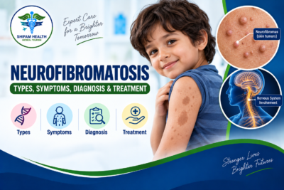
Neurofibromatosis (2026): Types, Symptoms, Diagnosis & Treatment
Filters & Insights
Learn about neurofibromatosis, including NF1 and NF2, symptoms, causes, diagnosis, treatment options, tumor risks, and management.

Hearing “neurofibromatosis” attached to your own or your child’s diagnosis often raises a specific kind of confusion, because the name itself doesn’t tell you much — and a lot of what’s easy to find online blurs together conditions that are, medically, genuinely different diseases with different genes, different tumor risks, and different treatment paths. This guide is built to untangle that from the start.
Table of Contents
ToggleWhat Is Neurofibromatosis?
Neurofibromatosis refers to a group of genetic conditions that primarily affect the nervous system, skin, and other tissues, and can increase the risk of certain tumors and other complications. It’s genetic — some cases are inherited from a parent, while many arise from a new genetic change with no family history at all. Symptoms and severity vary enormously between individuals, even within the same family.
Neurofibromatosis type 1 (NF1) and NF2-related schwannomatosis are different disorders, caused by different genes, with different typical features — despite the similar-sounding names, they are not simply two stages or severities of the same condition. Not everyone with neurofibromatosis develops tumors, and many people have relatively mild, manageable disease, while others face more significant neurological, visual, hearing, orthopedic, or tumor-related complications requiring lifelong specialist care.
Is Neurofibromatosis Genetic or Hereditary?
Yes, neurofibromatosis is genetic — but “genetic” and “inherited from a parent” aren’t the same thing, which is worth being precise about. NF-related conditions generally follow autosomal dominant inheritance, meaning a person carrying a pathogenic gene variant has roughly a 50% chance of passing it to each child. However, NF1 frequently arises from a de novo (new) genetic variant — meaning a significant proportion of people diagnosed with NF1 have no affected parent at all; the genetic change occurred spontaneously. NF2-related schwannomatosis can also be inherited or arise de novo.
Mosaic disease, where the genetic change occurs only after conception and affects some but not all of the body’s cells, adds further complexity to both inheritance patterns and genetic testing, discussed in more detail below. Genetic counseling is the right resource for understanding your specific situation and any implications for family planning — this article provides general education, not an individual inheritance risk assessment.
Is Neurofibromatosis Cancer?
Neurofibromatosis itself is not cancer. This is worth stating plainly and directly, because it’s a common source of unnecessary fear. What is true is that certain forms of neurofibromatosis increase the risk of specific tumors:
- NF1 is associated with increased risk of plexiform neurofibroma (generally benign), optic pathway glioma, malignant peripheral nerve sheath tumor (MPNST — the malignant tumor type genuinely worth knowing about), and, per current evidence, certain other malignancies at somewhat elevated rates.
- NF2-related schwannomatosis is associated with vestibular schwannoma, meningioma, ependymoma, and other schwannomas — the large majority of which are benign.
Having neurofibromatosis does not mean a person has cancer — it means ongoing surveillance is part of responsible long-term care, precisely so that if something concerning does develop, it’s caught and evaluated early rather than being a surprise.
The Genetic Basis: Three Distinct Conditions
Current international consensus recognizes neurofibromatosis as encompassing three genetically and clinically distinct conditions, not one disease with variants:
NF1 — caused by pathogenic variants in the NF1 gene on chromosome 17, which encodes neurofibromin, a protein that normally acts as a negative regulator of the Ras/MAPK cell-signaling pathway. When neurofibromin doesn’t function properly, this pathway becomes overactive, contributing to tumor development. NF1 accounts for roughly 96% of all neurofibromatosis cases and has an estimated birth incidence of approximately 1 in 2,600-3,000.
NF2-related schwannomatosis — the condition historically called “neurofibromatosis type 2” is now more precisely termed NF2-related schwannomatosis, reflecting updated international consensus terminology. It’s caused by pathogenic variants in the NF2 gene, which encodes merlin (schwannomin), a different tumor-suppressor protein. This accounts for roughly 3% of cases.
Other (non-NF2) schwannomatosis — a genetically distinct group, with some cases linked to SMARCB1 or LZTR1 gene variants, and others where the genetic cause remains unresolved by current testing. This accounts for less than 1% of cases. Genetic classification in this area continues to evolve as research advances.
These three conditions are not the same disease at different severities — they have different genetic causes, different characteristic tumor spectrums, and different management priorities, which is precisely why an accurate diagnosis of which condition you or your child has matters enormously for what comes next.
NF1 vs. NF2-Related Schwannomatosis
| Feature | NF1 | NF2-Related Schwannomatosis |
|---|---|---|
| Main gene | NF1 (chromosome 17) | NF2 (merlin/schwannomin) |
| Approximate share of NF cases | ~96% | ~3% |
| Hallmark feature | Café-au-lait macules, neurofibromas | Bilateral vestibular schwannomas |
| Plexiform neurofibroma | Characteristic | Not typical |
| Optic pathway glioma | Associated, particularly in childhood | Not typical |
| Hearing loss | Not a defining feature | Common, often bilateral |
| Meningioma | Possible but less characteristic | More characteristic |
| Bone abnormalities (scoliosis, tibial dysplasia) | More common | Less characteristic |
| Typical age of clinical recognition | Often early childhood (skin findings) | Often young adulthood (hearing symptoms) |
Neurofibromatosis Type 1 (NF1)
NF1 is the most common of the three conditions and typically becomes clinically apparent in early childhood through characteristic skin findings, well before any tumor-related symptoms appear.
Skin Signs
Café-au-lait macules are flat, light-brown skin patches that often appear during infancy or early childhood — multiple characteristic lesions (six or more, per diagnostic criteria) are an important clue, though café-au-lait spots alone do not diagnose NF1, since isolated spots are common in the general population too. Axillary and inguinal freckling — freckle-like spots in the armpit or groin folds — is a distinctive feature of NF1 that typically appears somewhat later than café-au-lait macules, often not present at birth.
Neurofibromas
Ordinary neurofibromas are generally benign tumors involving peripheral nerves, occurring as cutaneous (skin-surface), subcutaneous (beneath the skin), or deeper lesions. They can cause cosmetic concern, tenderness, or, depending on location, functional impact — but standard neurofibromas are not cancer and don’t require treatment simply because they exist.
Plexiform Neurofibromas
This deserves serious, dedicated attention. Plexiform neurofibromas are complex tumors involving multiple nerve branches and surrounding tissue, occurring in up to 30-50% of people with NF1, most often identified in childhood. Unlike ordinary neurofibromas, they tend to grow diffusely along nerve structures rather than as a discrete lump, which makes them genuinely difficult to remove completely — they can intertwine with nerves and blood vessels in ways that make clean surgical margins hard or impossible to achieve.
Plexiform neurofibromas can cause pain (reported in the majority of symptomatic pediatric cases in published survey data), visible disfigurement, motor dysfunction, and, in some locations, more serious functional problems like airway or bowel compression. They also carry a lifetime risk of malignant transformation into MPNST — not a common outcome for any individual tumor, but a real one, which is why ongoing monitoring matters even for plexiform neurofibromas that seem stable.
Malignant Peripheral Nerve Sheath Tumor (MPNST)
MPNST is an uncommon but genuinely serious malignant tumor that can arise in people with NF1, particularly from a pre-existing plexiform neurofibroma. Warning signs that warrant prompt specialist evaluation — though none of these prove malignancy on their own — include new persistent pain in a known neurofibroma, rapid growth, new weakness, new neurological symptoms, or a noticeable change in a tumor’s texture or firmness. When malignant transformation is suspected, FDG-PET or PET-MRI imaging can be useful, since MPNST tends to show higher metabolic activity than benign plexiform tissue — but elevated metabolic activity alone doesn’t confirm MPNST; biopsy and pathology remain the definitive diagnostic step.
Optic Pathway Glioma
Strongly associated with NF1 and usually identified during childhood, optic pathway gliomas are tumors along the visual pathway. Many remain stable and don’t require treatment — regular monitoring, particularly of vision, is the standard approach for asymptomatic or stable tumors. Treatment becomes relevant specifically when there’s progressive visual loss or clinically significant tumor growth, not simply because the tumor is present. Not every child with NF1 develops an optic pathway glioma.
Lisch Nodules
These are benign iris hamartomas — small, harmless growths on the colored part of the eye — associated with NF1. They don’t typically affect vision and are useful primarily as a diagnostic feature during eye examination, not as something requiring treatment in their own right.
Bone and Orthopedic Problems
NF1 can be associated with scoliosis, tibial dysplasia (abnormal shin-bone development), bowing, and, in some cases, difficulty with fracture healing (non-union). Severity varies enormously between individuals — some people with NF1 have no notable bone involvement, while others require ongoing orthopedic management.
Learning and Behavioral Features
Learning difficulties are genuinely common in NF1 — published data reports learning disabilities in over half of patients and at least one cognitive impairment in the large majority — alongside a higher rate of ADHD, executive-function challenges, and speech or language differences. This does not indicate low intelligence, and it’s worth stating clearly: these are recognized, manageable features of the condition, and appropriate educational and neuropsychological support can make a genuine, meaningful difference — this is a resource-and-support conversation, not a limitation to be quietly accepted.
Hypertension and Vascular Complications
People with NF1 have some increased risk of hypertension and, less commonly, vascular abnormalities including renal artery stenosis or, rarely, pheochromocytoma/paraganglioma (adrenal-related tumors that can affect blood pressure). This is part of why regular blood pressure monitoring is a standard part of NF1 follow-up, though it doesn’t mean every rare associated complication requires universal screening for every patient — your specialist team will guide what’s appropriate based on your specific presentation.
NF2-Related Schwannomatosis
This condition’s hallmark feature is fundamentally different from NF1: bilateral vestibular schwannomas — tumors on both sides affecting the nerve responsible for hearing and balance (the same tumor type discussed independently as “acoustic neuroma” for sporadic, unilateral cases). Beyond this hallmark feature, people with NF2-related schwannomatosis may develop other schwannomas (affecting peripheral, spinal, or other cranial nerves), meningiomas, and ependymomas.
Vestibular Schwannomas in NF2-Related Disease
Symptoms typically include hearing loss (often progressive and, notably, potentially affecting both ears given the bilateral nature of this disease), tinnitus, balance problems, and, with larger tumors, facial nerve effects. Because both hearing nerves may ultimately be involved, preserving hearing on at least one side becomes an especially high management priority — treatment decisions for each tumor (observation, microsurgery, stereotactic radiosurgery, or, in selected progressive cases, bevacizumab) are made individually per tumor rather than assuming both need identical, simultaneous treatment.
Meningiomas and Ependymomas
Meningiomas — tumors of the tissue surrounding the brain and spinal cord — are a recognized association with NF2-related schwannomatosis and are often slow-growing; management (observation, surgery, or radiation) depends on location, growth, and symptoms rather than automatic treatment simply because a meningioma is present. Spinal ependymomas and other central nervous system tumors can also occur, again managed based on individual growth and symptom pattern rather than a universal protocol.
Other (Non-NF2) Schwannomatosis
This genetically distinct, less common group is characterized primarily by multiple schwannomas, often causing significant pain, without the bilateral vestibular schwannomas that define NF2-related disease. Some cases are linked to SMARCB1 or LZTR1 gene variants; others remain genetically unresolved by current testing methods. Not every form of schwannomatosis causes vestibular schwannomas — this is a genuinely distinct clinical pattern worth not conflating with NF2-related disease.
Symptoms Summary by Condition
| Feature | More Strongly Associated With |
|---|---|
| Café-au-lait macules | NF1 |
| Axillary/inguinal freckling | NF1 |
| Neurofibromas / plexiform neurofibroma | NF1 |
| Optic pathway glioma | NF1 |
| Scoliosis / tibial dysplasia | NF1 |
| Bilateral vestibular schwannoma | NF2-related schwannomatosis |
| Progressive hearing loss | NF2-related schwannomatosis |
| Meningiomas | NF2-related schwannomatosis |
| Multiple, often painful schwannomas without vestibular involvement | Other schwannomatosis |
How Neurofibromatosis Is Diagnosed
The diagnostic pathway typically includes medical and family history, skin examination, neurological examination, blood pressure assessment, eye examination, hearing assessment, imaging where clinically indicated, genetic testing where appropriate, and specialist review.
NF1 Diagnostic Criteria (2021 Revision)
NF1 diagnostic criteria were substantially revised in 2021 through international consensus, updating criteria that had remained largely unchanged for roughly 25 years. Diagnosis considers a combination of features including café-au-lait macules, characteristic freckling, neurofibromas, plexiform neurofibroma, optic pathway glioma, specific bone findings, Lisch nodules or choroidal abnormalities on eye examination, identification of a pathogenic NF1 variant on genetic testing, and family history of an affected first-degree relative. The 2021 revision specifically incorporated genetic testing results alongside clinical features and added choroidal abnormalities as a recognized criterion, reflecting how much diagnostic precision has advanced. Diagnosis can be genuinely more difficult in very young children (since some features develop over time rather than being present from birth) and in mosaic disease.
Mosaic NF1
Mosaic NF1 occurs when the pathogenic NF1 variant arises after conception, meaning it’s present in some but not all of the body’s cells. This can result in findings limited to certain body regions rather than the more generalized pattern typical of classic NF1, and it can make both clinical diagnosis and genetic testing genuinely more challenging — a standard blood test may not detect the variant if it’s not well-represented in blood cells, sometimes requiring testing of affected tissue directly. Mosaic NF1 is not automatically mild — presentation varies depending on which tissues are affected and to what extent.
Genetic Testing
Testing may target the NF1 gene, the NF2 gene, or, for other schwannomatosis, SMARCB1, LZTR1, or broader multigene panels depending on clinical presentation. A negative genetic test does not always exclude a clinical diagnosis — mosaicism, technical limitations of current testing methods, and difficulty interpreting some genetic variants can all contribute to this. Genetic counseling helps interpret what a specific result — positive, negative, or a variant of uncertain significance — actually means for you and your family.
Imaging
Imaging needs depend heavily on the specific NF type and presenting symptoms — potentially including brain MRI, spinal MRI, MRI of a known plexiform neurofibroma, and, in selected patients, whole-body MRI to assess overall tumor burden. Not every person with neurofibromatosis needs routine whole-body MRI — this is used selectively, not as a universal screening tool for every diagnosed patient.
Treatment Overview
There is no single treatment for neurofibromatosis, because it is a group of genetic conditions, and treatment is directed mainly at managing symptoms, specific tumors, and complications as they arise — not at “curing” the underlying genetic change itself, which current medicine cannot do. Treatment may involve observation and structured surveillance, surgery, targeted drug therapy, radiation for selected tumors, pain management, hearing and vision support, orthopedic care, educational support, and genetic counseling — the specific combination depends entirely on which condition you have and which complications you’re managing.
NF1 Treatment by Complication
| Complication | Possible Management Approach |
|---|---|
| Ordinary neurofibroma | Observation, or surgical removal if symptomatic |
| Plexiform neurofibroma | Observation, surgery when feasible, or targeted MEK-inhibitor therapy |
| Optic pathway glioma | Observation for stable disease; systemic treatment if progressive or vision-threatening |
| Bone/orthopedic disease | Orthopedic management, tailored to severity |
| Hypertension | Standard medical evaluation and treatment |
| Learning difficulties | Educational and neuropsychological support |
| MPNST | Specialist sarcoma-team cancer treatment |
Plexiform Neurofibroma Treatment in Detail
Observation is appropriate for stable, minimally symptomatic tumors — most plexiform neurofibromas don’t require immediate intervention simply because they’re identified.
Surgery may be considered when the tumor is resectable, symptoms are significant, function is meaningfully impaired, cosmetic effects are severe, or malignancy is suspected. As noted above, complete removal is often genuinely difficult given how these tumors intertwine with nerve and vascular structures, and recurrence after partial removal is common.
Targeted (MEK-inhibitor) therapy represents a genuine, relatively recent treatment advance. Selumetinib was the first approved MEK inhibitor for symptomatic, inoperable NF1-associated plexiform neurofibromas, initially approved for pediatric patients and since studied and used in adults as well. More recently, mirdametinib has also received approval as an additional MEK-inhibitor option, per current guidance identifying MEK inhibitors as the current standard of care specifically for symptomatic, inoperable plexiform neurofibromas.
On effectiveness — an honest picture: published trial data (the international KOMET study in adults) reported an objective response rate of roughly 20% by cycle 16 with selumetinib versus 5% with placebo — a real, statistically significant benefit, but one that should be understood accurately rather than oversold: this is meaningful tumor shrinkage in a minority of treated patients, alongside broader reported benefits in pain and function for some patients, not a treatment that shrinks every tumor dramatically. It is not a cure, and it requires ongoing specialist monitoring given potential adverse effects, which can include rash, diarrhea, muscle-related symptoms, cardiac effects, eye-related toxicity, and liver-related changes — real considerations requiring regular follow-up testing during treatment, not decisions to make lightly.
Optic Pathway Glioma Treatment
Many NF1-associated optic pathway gliomas are simply observed with regular monitoring, particularly of visual function. Active treatment — generally following pediatric neuro-oncology protocols rather than adult glioma treatment approaches — is considered specifically for progressive visual loss or clinically significant tumor growth, not for every identified tumor.
NF2-Related Schwannomatosis Treatment
Management is highly individualized per tumor and per patient, potentially including active surveillance, surgery, stereotactic radiosurgery in selected situations, and, for selected progressive vestibular schwannomas where hearing preservation is a clinical priority, bevacizumab — a systemic (antibody-based) therapy that has shown tumor growth control and, in some patients, hearing stabilization or improvement in published evidence. This is not standard treatment for every NF2-related tumor; it applies to specific, progressive situations and requires specialist assessment given potential systemic side effects.
MPNST Treatment
MPNST management generally requires a specialist sarcoma team and typically centers on surgery, with radiation therapy and, for high-risk or advanced disease, chemotherapy considered as part of a comprehensive plan. Complete surgical resection when technically feasible is often central to the treatment approach, though this must be balanced against preserving function, similar to other complex nerve-sheath tumor surgery.
Radiation Therapy — A Word of Caution
Radiation can be appropriate for selected NF-associated tumors, but treatment decisions in this population require particular care given potential long-term toxicity, a recognized risk of secondary malignancy specifically relevant to NF1 biology, patient age, and available alternatives. It is not absolutely forbidden in NF1 — but it’s approached with more caution and individualized weighing of risks and benefits than it might be for an unrelated tumor in someone without NF1.
Pain Management
Pain in neurofibromatosis can arise from neurofibromas, plexiform neurofibromas, nerve compression, bone problems, or, when present, malignant transformation. A multidisciplinary approach — treating the underlying cause where possible, physical therapy, pain specialist involvement, psychological support, and appropriate medication under medical supervision — generally serves patients better than pain medication alone.
Surveillance and Long-Term Follow-Up
Ongoing monitoring is a core, lifelong part of managing neurofibromatosis, not a one-time diagnostic step.
For NF1, this typically includes blood pressure checks, skin examination for new or changing lesions, growth and developmental monitoring in children, learning assessment, vision screening (particularly in childhood, given optic pathway glioma risk), neurological symptom review, pain assessment, and bone health monitoring.
For NF2-related schwannomatosis, monitoring focuses on hearing, vestibular (balance) symptoms, MRI where indicated to track known tumors, and assessment for new spinal or other tumors.
There is no single universal MRI or monitoring schedule — your specific surveillance plan should be set by your specialist team based on your specific NF type, tumor history, and risk factors, generally reviewed at least annually with adjustments based on findings.
Children and Pregnancy
Children with neurofibromatosis benefit from coordinated, multidisciplinary care — pediatrician, geneticist, pediatric neurologist, ophthalmologist, orthopedist, and, when relevant, pediatric oncologist and developmental/educational specialists — since early identification of complications generally allows for better management than addressing problems only once they become significant.
Pregnancy in someone with NF1 requires attentive, specialist-coordinated care: blood pressure monitoring takes on added importance, existing neurofibromas may change during pregnancy, and certain complications may need specific attention. Genetic counseling before or during pregnancy can help clarify inheritance risk for the child. This is general information — individualized pregnancy management should be discussed directly with your obstetric and NF specialist team together.
Cancer Risk and Prognosis
NF1 is associated with increased risk of certain malignancies beyond MPNST, including, per current evidence, some hematologic malignancies and breast cancer at a younger-than-average age in women with NF1 — which is part of why some current guidance suggests earlier or additional breast screening consideration for women with NF1, though the specific screening approach should be confirmed with your own specialist team against current guidelines rather than assumed from general information here.
We will not give you one universal life-expectancy or prognosis number — and you should treat any source offering one (particularly any percentage framed as a “survival rate” for neurofibromatosis broadly, which conflates fundamentally different tumor risks across three different conditions) with real skepticism. Prognosis genuinely depends on which NF type you have, age, tumor burden, whether MPNST or other malignancy occurs, degree of neurological involvement, vision and hearing status, and vascular complications if present. What current evidence does support: many people with neurofibromatosis live long lives, particularly with NF1 specifically, provided lifelong monitoring identifies and addresses complications as they arise — this is a manageable, lifelong condition for the majority of patients, not a uniformly grim prognosis.
Neurofibromatosis Treatment Cost in India
There is no single, fixed treatment price for neurofibromatosis — and this is truer here than for almost any other condition in this content set, since cost depends entirely on which complications require treatment for your specific case over your lifetime, not a single procedure.
| Component | Typical Range (International Patient, Indicative) | Notes |
|---|---|---|
| Genetic consultation and testing | $300 – $1,200 | Depends on whether targeted single-gene or broader panel testing is needed |
| MRI/imaging (per study) | $300 – $900 | Whole-body MRI, where used, runs toward the higher end |
| Ophthalmology/audiology assessment | $100 – $400 | Routine surveillance component |
| Neurofibroma removal surgery | $1,500 – $4,000 | For symptomatic, straightforward lesions |
| Plexiform neurofibroma surgery | $4,000 – $10,000+ | Highly variable given surgical complexity and nerve involvement |
| MEK-inhibitor therapy (selumetinib/mirdametinib, per month) | Substantial — likely $3,000+ | Patent-protected, likely imported medication; significant ongoing cost requiring direct verification |
| MPNST treatment (surgery + oncology care) | Highly variable | Requires individualized specialist evaluation; not a fixed package |
Neurofibromatosis Treatment in India for International Patients
India offers relevant multidisciplinary infrastructure for neurofibromatosis care — medical genetics, neurology, neurosurgery, neuro-oncology, pediatric oncology, neurotology (for NF2-related disease), ophthalmology, orthopedics, and radiation oncology are available at major centers. We are not going to claim India is automatically “the best” country for neurofibromatosis treatment — what genuinely matters when comparing centers includes:
- Genuine multidisciplinary team coordination, not a single specialist working in isolation across a condition that touches so many organ systems
- Genetic testing capability and turnaround time
- Documented experience with plexiform neurofibroma surgery and, if relevant to your case, MEK-inhibitor treatment access
- MPNST management experience specifically, given how serious and uncommon this complication is
- Skull-base and neurotology expertise if NF2-related disease is your diagnosis
- Pediatric-specific services if the patient is a child
- Transparent, itemized cost estimates rather than a vague single figure
Choosing a Treatment Center
Rather than a superficial “top hospitals” list, look for a center offering: medical genetics, neurology, neurosurgery, neuro-oncology, pediatric oncology capability if relevant, ophthalmology, orthopedics, radiation oncology, genetic testing, MRI, molecular pathology, MEK-inhibitor access if plexiform neurofibroma treatment is needed, and rehabilitation services. If a hospital is named to you, verify these capabilities directly rather than relying on marketing claims — and be appropriately skeptical of any source citing a single “survival rate” for neurofibromatosis broadly, since this conflates genuinely different conditions and tumor risks.
International Patient Journey
- Share medical records, MRI/CT, and any pathology reports
- Identify suspected NF type and review any existing genetic testing
- Identify current symptoms and complications requiring attention
- Multidisciplinary specialist review
- Treatment recommendation and transparent, itemized cost estimate
- Medical visa assistance
- Travel and accommodation planning
- Treatment
- Rehabilitation where needed
- Long-term monitoring plan and coordination with your home-country follow-up care
We coordinate each of these steps but cannot guarantee cure, tumor removal, genetic correction, treatment success, exact treatment duration, exact cost, or visa approval before your case has been medically evaluated.
Frequently Asked Questions
1. What is neurofibromatosis?
Neurofibromatosis is a group of genetic conditions affecting the nervous system and skin, with an increased risk of certain tumors.
2. Is neurofibromatosis genetic?
Yes. It results from pathogenic gene variants that may be inherited or arise as new genetic changes.
3. What are the types of neurofibromatosis?
The main types include NF1, NF2-related schwannomatosis, and other schwannomatosis.
4. What is NF1?
NF1 is the most common type, often causing café-au-lait spots, neurofibromas, and an increased risk of certain tumors.
5. What is NF2-related schwannomatosis?
It is a distinct genetic condition commonly associated with bilateral vestibular schwannomas, which can affect hearing and balance.
6. Is neurofibromatosis cancer?
No. Neurofibromatosis itself is not cancer, although it can increase the risk of certain benign and malignant tumors.
7. What are café-au-lait spots?
These are flat, light-brown skin patches commonly associated with NF1. Multiple spots may support diagnosis but do not confirm it alone.
8. Can neurofibromatosis cause learning difficulties?
Yes. NF1 can be associated with learning difficulties, ADHD, and other cognitive or behavioral challenges.
9. Can neurofibromatosis cause tumors?
Yes. NF1 can cause neurofibromas and plexiform neurofibromas, while NF2-related disease commonly involves vestibular schwannomas.
10. Can neurofibromatosis be cured?
There is currently no cure for the underlying genetic condition. Treatment focuses on managing symptoms, tumors, and complications.
12. Can neurofibromatosis shorten life expectancy?
Life expectancy varies according to the NF type and complications. Regular lifelong monitoring can help identify and manage serious problems early.
How Shifam Health Can Help
A neurofibromatosis diagnosis touches multiple specialties over a lifetime — genetics, neurology, surgery, oncology, ophthalmology, audiology — which makes finding genuinely coordinated care more important than for a single-procedure condition. We help connect you with multidisciplinary teams experienced with your specific NF type, coordinate genetic testing and imaging review before you travel, and build a treatment estimate around your actual current complications rather than a vague, one-size-fits-all number.
Reach out on WhatsApp or submit a quick inquiry — our team responds within 24 hours, with no obligation to proceed.
This article is for general informational purposes and does not constitute medical advice or individualized genetic counseling. Neurofibromatosis diagnosis, genetic test interpretation, and treatment planning should be individualized by an experienced multidisciplinary specialist team, ideally including a medical geneticist. Cost figures are indicative component estimates and require case-specific verification, particularly for MEK-inhibitor therapy given its patent-protected pricing.


Popular Posts From Last Week
- September 21, 2026
- shifamhealth
Hearing “neurofibromatosis” attached to your own or your child’s diagnosis often raises a specific kind of confusion, because…
- September 18, 2026
- shifamhealth
Written by: Shifam Health Editorial Team — Orthopedic Content Division Medically reviewed by: Shifam Health Clinical Advisory Panel…
- September 16, 2026
- shifamhealth
Ankle valgus deformity is an abnormal alignment in which the ankle tilts toward the outside of the body,…
- September 20, 2026
- shifamhealth
Medically reviewed content · Published 2026 · Last reviewed September 2026 A limb deformity in a child is…
- September 15, 2026
- shifamhealth
Medically reviewed content · Published 2026 · Last reviewed September 2026 Bow legs medically called genu varum —…
- September 15, 2026
- shifamhealth
Medically reviewed content · Published 2026 · Last reviewed August 2026 A burst fracture is a spinal vertebra…
- September 16, 2026
- shifamhealth
Epithelial ovarian cancer is the most common broad category of ovarian cancer, arising from the cells covering the…
- September 17, 2026
- shifamhealth
Hepatoblastoma is a rare malignant liver tumor that develops primarily in young children, most commonly before age 5….
- September 19, 2026
- shifamhealth
Finding a purple or reddish patch on your skin that won’t fade, or hearing “Kaposi sarcoma” mentioned during…
- September 18, 2026
- shifamhealth
Kyphosis means forward curvature of the spine — and some kyphosis is completely normal anatomy in the mid-back…
- September 19, 2026
- shifamhealth
Lumbar radiculopathy is a neurological syndrome caused by irritation, inflammation, or compression of a nerve root in the…
- September 20, 2026
- shifamhealth
Still having pain after spine surgery — whether it never fully went away, or came back after a…