



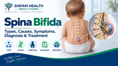
Spina Bifida: Types, Causes, Symptoms, Diagnosis & Treatment
Filters & Insights
Spina bifida ranges from mild, symptom-free forms to myelomeningocele with major complications. Learn about types, fetal surgery, and lifelong care.

Spina bifida is a group of neural tube defects caused by incomplete closure or development of the neural tube during early embryonic development. It varies enormously in severity from forms that cause no symptoms at all to open forms associated with significant neurological, urological, and orthopedic complications and no single description applies to everyone diagnosed with it.
A few essential distinctions worth understanding from the start:
- Spina bifida is the general term for this group of conditions
- Neural tube defects is the broader category that spina bifida belongs to, which also includes conditions affecting brain development
- Open spinal dysraphism refers to forms where neural tissue is exposed, not covered by skin (myelomeningocele is the classic example)
- Closed spinal dysraphism refers to forms where the abnormality is covered by skin, generally associated with a different, often milder, clinical picture
- Spina bifida occulta — often entirely asymptomatic — should not be confused with myelomeningocele, the most severe, open form
Not everyone with spina bifida has paralysis, and not everyone has intellectual disability — these are common misconceptions worth correcting immediately, since severity depends heavily on the specific type and level of the condition, discussed throughout this guide.
Table of Contents
ToggleTypes of Spina Bifida
The three main categories — spina bifida occulta, meningocele, and myelomeningocele — represent a spectrum from a frequently asymptomatic incidental finding to a serious open condition requiring urgent newborn surgery, and terminology can vary somewhat between classification systems and clinical settings.
Spina bifida occulta
- A defect in the vertebral arch (the bony ring surrounding the spinal cord) without protrusion of the spinal cord or its coverings
- Often entirely asymptomatic, and sometimes discovered incidentally on imaging done for an unrelated reason
- Possible cutaneous (skin) markers overlying the area such as a dimple, hair tuft, or birthmark can sometimes signal an underlying issue and may warrant further evaluation
- Not every incidental vertebral arch defect requires treatment — many are simply variants with no clinical significance, though findings that suggest an associated spinal cord abnormality warrant assessment
Meningocele
- The meninges (protective membranes surrounding the spinal cord) protrude through the vertebral defect, forming a sac
- Neural tissue itself is relatively preserved compared to myelomeningocele
- Neurological consequences can still vary between individuals, though generally less severe than myelomeningocele
Myelomeningocele
The most severe and clinically significant form, and the focus of much of this guide:
- Both the meninges and neural tissue protrude through the defect, exposed rather than covered by skin
- Associated with meaningful neurological impairment, including lower-extremity weakness or paralysis (extent depending heavily on the lesion level)
- Sensory abnormalities below the level of the lesion
- Bladder and bowel dysfunction (discussed in detail later)
- Strongly associated with hydrocephalus and Chiari II malformation (both discussed in dedicated sections below)
- Associated with various orthopedic abnormalities
Closed spinal dysraphism (broader category)
A broader category of spinal cord and vertebral abnormalities where the defect is covered by skin — this includes several distinct conditions with varying clinical significance, generally requiring individual evaluation rather than being treated as one uniform entity.
Causes and Embryology
Spina bifida results from failure of the neural tube to close properly during very early pregnancy, generally considered a multifactorial condition involving both genetic susceptibility and environmental/nutritional factors not purely genetic and not purely nutritional.
In simple terms: early in embryonic development, a structure called the neural tube forms and normally closes to become the brain and spinal cord. When this closure process fails at the spinal level, spina bifida results — the specific location and completeness of the closure failure determines the type and severity of the resulting condition.
This is not simply a genetic condition, nor simply a nutritional one — current understanding treats it as multifactorial, involving genetic susceptibility interacting with environmental and nutritional factors, most notably folate status (discussed next).
Folic Acid and Prevention
Adequate folate/folic acid intake before and during very early pregnancy meaningfully reduces the risk of neural tube defects including spina bifida — this is one of the most well-established findings in preventive medicine — but folic acid does not prevent every case, and it’s important to distinguish general prevention from recurrence prevention in higher-risk situations.
Key distinctions:
- Primary prevention — folic acid supplementation for the general population of women who could become pregnant, aimed at reducing first-occurrence risk
- Recurrence prevention — for women who’ve had a previous pregnancy affected by a neural tube defect, higher-dose supplementation is commonly recommended under specialist guidance, given their elevated recurrence risk
- Management after a fetal diagnosis — a separate matter altogether, discussed later in this guide; folic acid is not a treatment for an already-affected pregnancy
Why timing matters so much: neural tube closure occurs very early in pregnancy — often before many women even know they’re pregnant — which is exactly why standard guidance focuses on adequate folate status before conception, not simply once pregnancy is confirmed.
On specific dosing: standard recommendations for women of reproductive age generally involve daily folic acid supplementation as part of general preconception and early pregnancy care, with higher doses specifically recommended for women with a previous neural tube defect-affected pregnancy or certain other risk factors (such as being on certain antiseizure medications) — the specific dose appropriate for your situation should be confirmed with your own physician or a maternal-fetal medicine specialist, since recommendations are risk-stratified and this article isn’t a substitute for that individualized guidance.
Folic acid does not prevent every case of spina bifida — it meaningfully reduces population-level risk, but spina bifida can still occur despite adequate folate status, reflecting the condition’s multifactorial nature.
Genetics and Recurrence Risk
Spina bifida is generally not caused by a single gene — it reflects multifactorial inheritance, meaning genetic susceptibility interacts with environmental factors, and most cases occur without another affected family member.
- A family history of neural tube defects does increase recurrence risk for future pregnancies, which is exactly why higher-dose folic acid is specifically recommended for this group
- Genetic counseling and more detailed genetic evaluation may be particularly relevant when additional congenital anomalies are present alongside the spina bifida, when there’s a broader syndromic presentation, when family history is significant, or when another specific genetic condition is suspected
- Not every child with isolated spina bifida requires extensive genetic testing — this is generally reserved for situations with these additional concerning features, not applied universally
Symptoms and Clinical Features
Symptoms and findings vary enormously by type and, for myelomeningocele specifically, by the level and completeness of the spinal cord involvement:
- Spina bifida occulta: often no symptoms at all; occasionally a skin marking overlying the spine
- Meningocele: variable, generally less severe neurological findings than myelomeningocele
- Myelomeningocele: can include lower-extremity weakness or paralysis, sensory loss below the lesion level, bladder and bowel dysfunction, and, related to associated hydrocephalus and Chiari II malformation, additional neurological findings discussed in their own sections below
No single symptom list applies to every patient — this variability is central to understanding spina bifida, and it’s exactly why individualized assessment, not a generic checklist, determines a specific person’s actual clinical picture.
Prenatal Diagnosis
Spina bifida, particularly the open myelomeningocele form, can often be detected during pregnancy through maternal serum screening and, more definitively, prenatal ultrasound but prenatal imaging cannot perfectly predict exactly how a specific child will function after birth.
Maternal serum screening — including elevated alpha-fetoprotein — can raise suspicion and prompt further evaluation, though it’s a screening tool, not a diagnostic one on its own.
Prenatal ultrasound is central to diagnosis, assessing:
- Fetal spinal anatomy directly
- Indirect and direct signs suggestive of an open spinal defect
- The level of the lesion
- Ventriculomegaly (enlarged brain ventricles, relevant to hydrocephalus risk)
- Findings associated with Chiari II malformation
- Any additional associated anomalies
This evaluation is generally coordinated through maternal-fetal medicine specialists and often involves broader multidisciplinary counseling once a diagnosis is suspected or confirmed.
A genuinely important caveat: prenatal imaging cannot always precisely predict individual postnatal neurological function. Lesion level and imaging findings provide meaningful information and general expectations, but the same lesion level can produce somewhat different functional outcomes between individual children — this uncertainty is a real, honest part of prenatal counseling, not something to gloss over.
Fetal MRI
Fetal MRI may be considered to provide additional anatomical detail beyond what ultrasound alone shows, complementing rather than replacing ultrasound. It is not mandatory for every suspected case — its use depends on the specific clinical situation and what additional information is genuinely needed for counseling or surgical planning.
Prenatal Counseling
After a prenatal diagnosis, families generally need to understand and discuss:
- The specific lesion level and whether it’s an open or closed defect
- Associated brain findings on imaging
- Ventriculomegaly/hydrocephalus risk
- The genuine uncertainty regarding precise individual neurological function — this should be communicated honestly, not glossed over
- Whether fetal intervention is an option in this specific case (discussed next)
- What postnatal surgery would involve if fetal surgery isn’t pursued or isn’t an option
- The reality of lifelong bladder and bowel care needs
- Mobility implications, again with appropriate uncertainty
- Rehabilitation and long-term follow-up needs
Deterministic statements — such as telling a family “this baby will never walk” — should be avoided. Prenatal imaging provides meaningful information, but it cannot precisely predict individual functional outcomes with the certainty such statements imply, and responsible prenatal counseling reflects this genuine uncertainty rather than false precision in either direction.
Fetal Surgery
Fetal (prenatal) surgical repair is a specialized treatment option for selected pregnancies with open spina bifida (myelomeningocele) — not a universal treatment for every diagnosis. Based on the landmark MOMS trial, prenatal repair reduces the likelihood of needing a postnatal shunt and is associated with improved motor outcomes for some children compared to standard postnatal repair but it carries real maternal and fetal risks and does not reverse all neurological deficits already present.
What fetal surgery does and doesn’t do: prenatal repair aims to close the exposed spinal defect before birth, which can favorably alter the course of certain complications (notably reducing shunt-dependent hydrocephalus in many cases) — but it is not a cure for spina bifida, and it does not reverse neurological damage to the spinal cord that has already occurred by the time of surgery.
Long-term follow-up data from the MOMS trial’s childhood cohort (MOMS2) found meaningfully reduced — but not eliminated — rates of needing clean intermittent catheterization for bladder management after prenatal repair (roughly 62%) compared to standard postnatal repair (roughly 87%). This is genuinely encouraging, but it’s also honest evidence that fetal surgery reduces rather than eliminates the likelihood of needing ongoing bladder management — presenting it as eliminating this need would overstate what the evidence actually shows.
Two surgical approaches currently exist, with different risk-benefit profiles that remain an actively studied comparative question rather than a settled matter:
| Approach | Key Features |
|---|---|
| Open fetal surgery | Involves a maternal hysterotomy (uterine incision) to directly access and repair the fetal defect; this is the approach studied in the original MOMS trial, generally performed between roughly 19 and 26 weeks gestation |
| Fetoscopic (minimally invasive) repair | A newer, evolving approach using smaller access points rather than a full hysterotomy; some data suggest lower risk of uterine dehiscence and potential for future vaginal delivery, but current evidence also shows higher rates of certain complications (including preterm rupture of membranes) compared to open surgery in some published series |
Eligibility is genuinely selective, generally based on criteria similar to those used in the MOMS trial, including specific gestational age windows, lesion characteristics, absence of severe additional fetal anomalies, and maternal health factors — for example, maternal obesity (BMI ≥35) is a recognized exclusion criterion at most centers under MOMS-based eligibility frameworks, reflecting a genuine, current clinical consideration rather than a hypothetical one.
Maternal and fetal risks are real and should never be minimized:
- Preterm birth
- Premature rupture of membranes
- Uterine complications, including a risk of uterine dehiscence or rupture at delivery for the current pregnancy and, importantly, for future pregnancies (open surgery specifically mandates cesarean delivery for the current and all future pregnancies) — though recent analysis of the original MOMS trial participants found the risk of uterine rupture in a subsequent pregnancy comparable to the risk after a standard prior classical cesarean, which is a genuinely reassuring finding worth including, without overstating it as risk-free
- Placental complications
- Maternal surgical risk generally
- Fetal risks, including the possibility of fetal demise, though this is uncommon
- The clear need for delivery planning at a center equipped to manage a post-hysterotomy or post-fetoscopic pregnancy
Fetal surgery requires a center with genuine, specific multidisciplinary fetal-surgery expertise — not simply a general hospital with obstetric and neurosurgical departments separately. This is a highly specialized field requiring a coordinated team (maternal-fetal medicine, pediatric neurosurgery, and others) with specific experience in this exact procedure, and this distinction matters enormously when evaluating where such surgery might be performed.
Postnatal Diagnosis and Immediate Newborn Management
For a newborn with suspected or confirmed open myelomeningocele not previously treated with fetal surgery, this requires urgent specialist neonatal and neurosurgical care from birth — this should not be delayed for any reason, including travel planning.
Initial newborn evaluation includes a thorough neurological examination, assessment of the lesion itself, head circumference measurement, fontanelle assessment, evaluation of motor and sensory function in the lower limbs, assessment of anal and bladder function, and an orthopedic examination.
General principles of immediate management (this is not a procedural guide for caregivers — it describes what the treating medical team does):
- Protection of the exposed neural tissue from injury and infection
- Careful, specific positioning and handling to protect the defect until surgical closure
- Infection prevention measures
- Thorough neurological assessment
- Evaluation for hydrocephalus
- Early neurosurgical planning for closure, generally within the first day or so of life
Imaging in the newborn period may include spinal ultrasound in selected cases, MRI, and brain imaging to assess for hydrocephalus and Chiari II-related findings.
Neurosurgical Closure
Surgical closure of an open myelomeningocele, if not already performed prenatally, aims to cover the exposed neural tissue, reduce infection risk, and prevent further injury to already-vulnerable neural structures but it does not restore spinal cord function that has already been lost.
- The goal is protective and preventive: closing the defect, reducing infection risk, and preventing additional injury to exposed tissue — not restoring already-damaged spinal cord function
- Timing considerations depend on the specific clinical situation, generally addressed promptly after birth when fetal surgery hasn’t already been performed
- Potential complications include wound-related issues, infection, and cerebrospinal fluid-related complications (such as CSF leak)
It should never be implied that closure surgery guarantees normal subsequent neurological development — it addresses the anatomical defect and protects against further injury and infection, which is itself an important and necessary goal, but it doesn’t reverse neurological function already lost before surgery.
Hydrocephalus
Hydrocephalus — excess cerebrospinal fluid accumulation affecting the brain — occurs in a substantial proportion of children with myelomeningocele, closely related to the associated Chiari II malformation, and management options include observation in selected cases, or CSF diversion procedures like ventriculoperitoneal shunting or endoscopic third ventriculostomy.
Why it occurs: hydrocephalus in myelomeningocele is closely linked to the associated Chiari II malformation (discussed next), which affects normal CSF circulation patterns.
Management options — and importantly, no single method is universally superior; the appropriate choice depends on individual anatomy and clinical circumstances:
- Observation in selected, carefully monitored cases without significant or progressive ventriculomegaly
- Ventriculoperitoneal (VP) shunt — a device diverting excess CSF, historically the most common approach
- Endoscopic third ventriculostomy, with or without choroid plexus cauterization in selected patients and centers — an alternative approach in appropriately selected cases
Shunt-related realities worth understanding honestly: shunts can create ongoing shunt-dependence, and they carry real risks of obstruction, infection, and the eventual need for revision surgery over a child’s lifetime — this isn’t a “fix it once” device for most patients, but rather a long-term component of care requiring ongoing monitoring.
Shunt failure and emergency warning signs
This is one of the most important safety sections in this entire guide. Signs that may suggest shunt malfunction or increased intracranial pressure — which vary by age and individual, and should prompt urgent medical assessment, not a wait-and-see approach:
- Worsening or new headache
- Vomiting, particularly if persistent or unexplained
- Unusual sleepiness or lethargy
- Altered consciousness
- Rapid increase in head size in infants
- A bulging fontanelle in infants
- Visual changes
- Worsening balance or coordination
- Seizures
- Any notable behavioral or neurological change from the child’s or adult’s baseline
Parents and caregivers are not expected to definitively diagnose shunt failure themselves but recognizing these warning signs and seeking urgent medical assessment when they appear is genuinely important, and delay can have serious consequences.
Chiari II Malformation
Chiari II malformation — downward displacement of specific hindbrain structures — is strongly associated with myelomeningocele and related to the hydrocephalus discussed above. Not every imaging finding of Chiari II requires surgery, but certain symptoms warrant prompt neurosurgical assessment.
Possible symptoms associated with symptomatic Chiari II — these don’t occur in every patient, and their presence and severity vary substantially:
- Swallowing difficulty
- Breathing abnormalities, including stridor
- Vocal cord dysfunction
- Weakness, particularly in the upper limbs in some cases
- Abnormal eye movements
- Feeding difficulties, particularly relevant in infants
- Sleep-disordered breathing
Not every child with imaging evidence of Chiari II needs surgical intervention — many have the anatomical finding without significant symptoms requiring treatment. Neurosurgical assessment becomes appropriate specifically when symptoms suggestive of brainstem compression or dysfunction are present, evaluated by a specialist familiar with this specific condition.
Tethered Cord
Tethered spinal cord — abnormal fixation of the spinal cord, restricting its normal movement — can develop after spina bifida repair, sometimes years later, and typically presents with progressive (not sudden) neurological, orthopedic, or urological changes rather than a static picture.
Why it can occur: scar tissue from the original repair (prenatal or postnatal) can cause the spinal cord to become abnormally fixed in position, which becomes a functional problem as the child grows and the spine lengthens relative to the fixed cord.
Possible warning signs of a developing tethered cord, generally progressive rather than sudden:
- Worsening leg weakness
- A change in gait pattern
- New sensory changes
- Increasing scoliosis
- Worsening bladder function
- Worsening bowel function
- New or worsening back or leg pain
Evaluation typically involves neurological assessment, urodynamic testing (given how commonly bladder function changes with tethering), MRI, and multidisciplinary review integrating findings across specialties.
Detethering surgery may be considered when these findings are present and progressive, but this decision is genuinely individualized — not every finding suggestive of tethering on imaging alone, without clinical progression, automatically requires surgery.
Neurogenic Bladder and Kidney Protection
Spinal cord involvement in myelomeningocele commonly affects bladder control, which can lead to urinary retention, incontinence, abnormally high bladder pressure, and recurrent urinary tract infections — and if unmanaged, this can eventually cause kidney damage. Protecting kidney function is a central, lifelong goal of spina bifida care, not a secondary concern.
Why this matters so much: nerve involvement affecting bladder function can create a cycle of urinary retention, high-pressure bladder, and, in some cases, vesicoureteral reflux (urine flowing backward toward the kidneys) — over time, this can cause kidney scarring and impaired kidney function if not properly managed.
Assessment and monitoring:
- Urodynamic testing — assessing how the bladder fills and empties, and at what pressures
- Renal ultrasound
- Ongoing renal function monitoring
Management strategies, individualized to the specific pattern of bladder dysfunction:
- Clean intermittent catheterization — a core management strategy for many patients
- Anticholinergic or other medications where appropriate, to help manage bladder pressure and function
- Other bladder-management strategies as clinically indicated
- Surgery in selected, more complex cases
A genuinely important safety point: apparently stable urinary symptoms do not necessarily mean the kidneys are safe. High bladder pressure can cause silent, progressive kidney damage even without dramatic symptoms — this is exactly why structured, ongoing surveillance under specialist guidance matters throughout life, not just reactive treatment when symptoms become noticeable.
This article does not provide instructions for starting catheterization or bladder medication independently — this requires specific instruction and ongoing supervision from a urology/neuro-urology specialist familiar with the individual patient.
Neurogenic Bowel
Bowel-related issues in spina bifida commonly include constipation, fecal incontinence, and reduced bowel sensation or voluntary control, reflecting the same underlying nerve involvement affecting bladder function.
Management approaches, individualized to the specific pattern:
- Structured bowel programs (a consistent, planned routine for bowel management)
- Dietary and fluid considerations
- Medications where appropriate
- Rectal interventions where indicated
- Antegrade continence procedures (a surgical option allowing bowel irrigation through a created access point) in selected patients where other measures haven’t adequately achieved continence
The focus of bowel management genuinely extends beyond the purely medical — function, dignity, and quality of life are central goals, since bowel continence has real, significant implications for social participation, independence, and self-esteem, particularly as children grow into adolescence and adulthood.
Mobility, Rehabilitation, and Orthopedic Complications
Mobility in spina bifida depends on multiple factors together — lesion level, actual neurological function (which doesn’t always track perfectly with lesion level alone), muscle strength, orthopedic deformities, the extent of hydrocephalus/brain involvement, and rehabilitation — not lesion level in isolation, which is why specific promises about walking ability based on lesion level alone should be avoided.
Supporting mobility involves physiotherapy, strengthening, gait training, orthoses (braces), walkers, wheelchairs, standing programs (supporting bone health and other benefits even for children who primarily use a wheelchair), and other adaptive equipment — matched to the individual’s specific abilities and goals, using respectful, person-centered language rather than treating mobility level as a simple hierarchy of outcomes.
Orthopedic complications that can occur, related to the underlying neuromuscular imbalance:
- Clubfoot
- Hip dislocation
- Joint contractures
- Scoliosis and kyphosis (spinal deformity, discussed further below)
- Other lower-limb deformities
- Increased fracture risk, related in part to reduced weight-bearing and bone density
- Gait abnormalities
Management ranges from observation to bracing to physiotherapy to serial casting to surgery — not every orthopedic finding requires surgical correction, and the appropriate approach depends on the specific deformity, its functional impact, and individual goals.
Scoliosis and spinal deformity specifically deserve particular mention: both congenital and neuromuscular factors can contribute, progression requires monitoring, bracing may be appropriate in selected cases, and spinal surgery is considered in select cases. Importantly, spinal surgery decisions in this population must account for interactions with any previous spinal repair, tethered cord status, existing neurological function, and respiratory function — this is genuinely more complex than scoliosis surgery in a child without spina bifida, given these additional, interacting considerations.
Development, Learning, and Quality of Life
Spina bifida does not automatically cause intellectual disability, though some individuals, particularly related to hydrocephalus, may experience specific challenges with attention, executive function, learning, visual-spatial processing, or memory — these are specific, sometimes subtle patterns, not a uniform intellectual impairment.
Developmental and educational support that may be relevant includes formal developmental assessment, educational support and accommodations, and neuropsychological evaluation where indicated by specific concerns — matched to the individual child’s actual profile rather than assumed based on the spina bifida diagnosis alone.
On quality of life more broadly: independence, education, employment, mobility, relationships, accessibility, pain management, bladder/bowel management, mental wellbeing, and social participation all factor into overall quality of life for people with spina bifida — using respectful, disability-informed language throughout, and not framing disability itself as inherently a tragedy. Many people with spina bifida, across the full range of severity, live full, meaningful lives, pursue education and careers, form relationships, and participate fully in their communities — genuine challenges exist and deserve honest acknowledgment, but they don’t define the totality of what’s possible.
Adult Spina Bifida
Spina bifida is a lifelong condition, and adult care involves its own set of considerations — including the transition from pediatric to adult care systems, ongoing neurogenic bladder/bowel management, kidney health surveillance, tethered cord risk, spinal deformity, pain, skin integrity, sexuality, fertility, and pregnancy planning — that shouldn’t be treated as an afterthought to pediatric care.
Transition from pediatric to adult care is a genuinely important, sometimes under-supported process — many adults with spina bifida report gaps in care specifically around this transition, making proactive planning and identifying appropriate adult specialists genuinely important.
Ongoing adult considerations:
- Continued neurogenic bladder and bowel management
- Ongoing kidney health surveillance — this doesn’t stop being important in adulthood
- Continued risk of tethered cord, which can develop or recur at any age
- Spinal deformity monitoring
- Chronic pain, which affects a meaningful proportion of adults with spina bifida
- Mobility, which can change over time (sometimes improving with continued rehabilitation, sometimes requiring adaptation as an individual ages)
- Skin problems and pressure injury risk, discussed further below
- Sexuality and sexual function — genuinely individual, and not something to assume based on the diagnosis
- Fertility considerations
- Pregnancy, for those who become pregnant, which requires coordinated maternal-fetal medicine involvement given the relevant urinary, bowel, and mobility considerations, along with careful delivery and anesthetic planning
- Employment and accessibility considerations
- Independent living
- Mental wellbeing, which deserves genuine attention as part of comprehensive adult care, not an afterthought
Skin and pressure injury prevention: reduced sensation and mobility in some individuals can increase the risk of pressure sores, unnoticed injuries, and skin breakdown. Reasonable general practices include regular skin inspection, appropriate pressure relief, well-fitted seating and footwear, appropriate mobility equipment, and prompt medical assessment of any new wound or skin change — this is general awareness information, not individualized nursing instruction, which should come from your own care team familiar with your specific situation.
On anesthesia and surgery more broadly: because of Chiari II malformation, any hydrocephalus, cervical or spinal abnormalities, and sometimes restrictive respiratory issues, anesthesia for any procedure in a person with spina bifida may require specific planning by an anesthesiologist familiar with these considerations — this is worth proactively mentioning before any surgery or procedure, including ones unrelated to spina bifida itself.
Prognosis
There is no single prognosis for “spina bifida” — outcomes depend on the specific type, lesion level, actual neurological function, presence and management of hydrocephalus, Chiari II-related complications, bladder and kidney function, orthopedic complications, and access to coordinated multidisciplinary care. Modern management can substantially influence outcomes, but individual results genuinely vary.
Deterministic statements about walking ability, intelligence, independence, or life expectancy should be avoided — these outcomes depend on the specific combination of factors above, assessed individually, not predicted from the general “spina bifida” label alone.
What can be said honestly: modern, coordinated multidisciplinary management — spanning neurosurgery, urology, orthopedics, rehabilitation, and other specialties as needed — has meaningfully improved health and functional outcomes for people with spina bifida over recent decades.
This doesn’t mean every outcome is guaranteed to be favorable, but it does mean that access to appropriate, ongoing, coordinated care genuinely matters for long-term outcomes, which is exactly why the “lifelong multidisciplinary team” concept — spanning neurosurgery, urology, nephrology, orthopedics, rehabilitation, neurology, developmental medicine, gastroenterology/colorectal care, and psychological/social support, with the specific team composition depending on the individual’s age and complications — is so central to this condition’s management.
Spina Bifida Treatment in India and Cost
India offers relevant infrastructure for various aspects of spina bifida care, including maternal-fetal medicine, pediatric neurosurgery, pediatric urology, orthopedics, and rehabilitation services — though not every center offers every service, and highly specialized care like fetal surgery specifically requires a center with genuine, verified multidisciplinary fetal-surgery expertise, not simply a general hospital.
Potentially available services include prenatal diagnosis and maternal-fetal medicine consultation, pediatric neurosurgery for postnatal closure and hydrocephalus management, pediatric urology, nephrology, orthopedics, rehabilitation services, relevant imaging, and neonatal intensive care. We are not claiming that every hospital offers every one of these services, nor naming any specific hospital as “the best” — this requires direct, current verification with the specific center being considered, and for anything as specialized as fetal surgery, confirming genuine, demonstrated experience with this exact procedure is essential rather than assuming general pediatric surgical capability extends to it.
On cost: there is no single generic “spina bifida treatment price.” Potential cost components include prenatal consultation, fetal imaging, genetic evaluation where indicated, fetal surgery where appropriate and available, delivery, neonatal intensive care, neurosurgical closure, hydrocephalus treatment (including shunt surgery), urological evaluation and urodynamics, orthopedic surgery, rehabilitation, long-term medications, and ongoing follow-up.
We were unable to verify reliable, current, India-specific pricing across this full range of services with sufficient confidence to present specific figures here — costs vary enormously by which specific interventions are needed, hospital, city, and case complexity, and any figure presented without direct verification against actual current hospital quotes would risk being misleading. A personalized estimate, based on your specific clinical situation and the specific interventions needed, requires direct consultation with the treating hospital.
International Patient Journey and Travel Safety
Practical journey for international families:
- Medical record collection — prenatal ultrasound reports, fetal MRI where available, genetic reports, neonatal records, MRI/CT, operative reports, shunt history, urodynamic studies, renal imaging, current medications, and rehabilitation records
- Specialist review — matching the specific case to appropriate specialists based on the actual clinical picture
- Diagnostic clarification — determining whether existing imaging and records are sufficient, or whether additional evaluation is needed
- Treatment plan discussion — clarifying what surgery, if any, is indicated; bladder/bowel management approach; hydrocephalus management; rehabilitation plan; and follow-up needs
- Cost estimate — specific to the actual treatment plan, from the treating hospital directly
- Medical visa support, where applicable
- Travel planning — including an honest assessment of medical stability before travel is appropriate (discussed further below)
- Local logistics — airport transfer, accessible accommodation, caregiver requirements, hospital appointment coordination, and any needed mobility equipment
- Treatment and rehabilitation, coordinated across the relevant specialties
- Continuity after returning home — complete records and follow-up recommendations provided to the patient’s local care team, genuinely important given this is a lifelong condition requiring ongoing local follow-up between any specialist visits
Travel safety — this is a mandatory consideration, not an afterthought. International travel for elective evaluation or treatment should not be pursued when urgent local care is actually needed. Situations where urgent local evaluation should take priority over travel planning include:
- Suspected shunt failure or signs of increased intracranial pressure
- Acute neurological deterioration
- Severe respiratory problems, particularly relevant given Chiari II-related risks
- Serious infection
- Acute urinary obstruction
- Severe kidney complications
- Rapidly worsening neurological symptoms
- Neonatal instability
For newborns with open, untreated myelomeningocele specifically: urgent specialist neonatal and neurosurgical care should never be delayed for the sake of international travel planning. This is a situation requiring the nearest appropriate specialist care immediately, not a scenario to coordinate around travel logistics.
Shifam Health is an international healthcare coordination and medical travel support company — not a treating physician, fetal medicine specialist, neurosurgeon, hospital, or diagnostic provider. They may assist with medical record organization, specialist coordination, hospital coordination, appointment scheduling, treatment estimates, medical visa support and invitation documentation, travel logistics, accessible accommodation coordination, interpreter and local support, and follow-up communication.
Shifam Health does not diagnose spina bifida, independently determine neurological prognosis, prescribe treatment, or perform surgery, and does not replace fetal medicine or neurosurgical specialist involvement. They does not guarantee fetal-surgery eligibility, surgical outcome, walking ability, neurological recovery, cure, hospital acceptance, visa approval, or exact treatment cost.
Myths vs. Facts
| Myth | Fact |
|---|---|
| “Spina bifida always causes paralysis.” | Severity varies enormously by type and lesion level — many people with spina bifida have significant mobility and function. |
| “Everyone with spina bifida has intellectual disability.” | Intellectual disability is not universal; some individuals, particularly related to hydrocephalus, may have specific learning or attention-related challenges rather than global intellectual impairment. |
| “A prenatal diagnosis tells you exactly how a child will function.” | Prenatal imaging provides meaningful information but cannot precisely predict individual functional outcomes with certainty. |
| “Fetal surgery cures spina bifida.” | Fetal surgery can favorably alter the course of certain complications (like reducing shunt need) but does not reverse spinal cord damage already present, nor eliminate all lifelong care needs. |
| “A shunt permanently fixes hydrocephalus.” | Shunts require lifelong monitoring and carry ongoing risks of obstruction, infection, and the eventual need for revision. |
| “If bladder symptoms are mild, the kidneys are safe.” | High bladder pressure can silently damage kidneys even without dramatic symptoms — structured surveillance matters regardless of apparent symptom severity. |
| “People with spina bifida cannot walk.” | Many people with spina bifida do walk, with or without assistive devices, depending on lesion level and individual factors. |
| “Spina bifida only affects children.” | It’s a lifelong condition requiring ongoing adult care, including bladder/bowel, kidney, orthopedic, and other management. |
| “Surgery restores damaged spinal cord function.” | Both fetal and postnatal closure surgery protect against further damage and address the anatomical defect, but do not reverse neurological function already lost. |
| “People with spina bifida cannot have children.” | Fertility and pregnancy are individual considerations requiring specialist counseling, not something to assume is impossible. |
| “Spina bifida does not require lifelong follow-up.” | Ongoing, coordinated multidisciplinary follow-up throughout life is genuinely important for managing bladder, kidney, orthopedic, and neurological considerations. |
Frequently Asked Questions
What is spina bifida?
A born with a disability caused by incomplete closure of the spine during early pregnancy.
What are the main types of spina bifida?
Spina bifida occulta, meningocele, and myelomeningocele, the most severe open form.
What causes spina bifida?
Genetic and environmental factors, particularly inadequate folate during early pregnancy, contribute to its development.
Can spina bifida be detected before birth?
Yes. Prenatal ultrasound and maternal blood screening can identify many cases.
How is spina bifida treated?
Treatment may include prenatal or postnatal surgery, rehabilitation, and management of neurological, bladder, and orthopedic complications.
Can spina bifida be cured?
There is no complete cure. Surgery and ongoing care can manage complications and improve function.
Is fetal surgery available for spina bifida?
Yes, selected pregnancies with open myelomeningocele may qualify after specialist evaluation.
Can children with spina bifida walk?
Many can walk independently or with assistive devices, depending on neurological function and lesion level.
Does spina bifida affect intelligence?
Not always. Some children experience learning or attention difficulties, particularly when hydrocephalus is present.
Can spina bifida cause kidney problems?
Yes. Neurogenic bladder can damage the kidneys without appropriate monitoring and treatment.
Is spina bifida treatment available in India?
Yes. Specialist centers offer neurosurgery, urology, orthopedic treatment, and rehabilitation. Capabilities vary by hospital.
Conclusion
Spina bifida is genuinely not one condition with one outcome — it spans a spectrum from an incidental, symptom-free finding to a complex, lifelong condition requiring coordinated care across multiple medical specialties. Whether the conversation is about a prenatal diagnosis, a newborn’s immediate care, a growing child’s development, or an adult managing this condition throughout their life, the most valuable thing a family or patient can do is seek genuinely individualized, honest guidance from specialists familiar with the actual clinical details not a generic prognosis based on a diagnosis label alone.
If you’re evaluating care options, including care in India, sharing complete medical records and imaging with appropriate specialists is the necessary first step toward an accurate, individualized understanding of your specific situation.
This article is educational and does not replace clinical examination, diagnosis, prenatal counseling by qualified fetal-medicine specialists, neurosurgical or urological assessment, or individualized treatment planning. Spina bifida varies substantially between individuals, and treatment decisions require specialist evaluation of your specific case. If you suspect a medical emergency including shunt failure, acute neurological deterioration, or an untreated newborn with open spina bifida seek urgent local medical care immediately rather than relying on this article or delaying for travel planning.



Popular Posts From Last Week
- September 21, 2026
- shifamhealth
Hearing “neurofibromatosis” attached to your own or your child’s diagnosis often raises a specific kind of confusion, because…
- September 20, 2026
- shifamhealth
Medically reviewed content · Published 2026 · Last reviewed September 2026 A limb deformity in a child is…
- September 24, 2026
- shifamhealth
Osteopenia means bone mineral density lower than the young-adult reference range but not low enough to meet the…
- September 25, 2026
- shifamhealth
Spina bifida is a group of neural tube defects caused by incomplete closure or development of the neural…
- September 19, 2026
- shifamhealth
Finding a purple or reddish patch on your skin that won’t fade, or hearing “Kaposi sarcoma” mentioned during…
- September 18, 2026
- shifamhealth
Kyphosis means forward curvature of the spine — and some kyphosis is completely normal anatomy in the mid-back…
- September 21, 2026
- shifamhealth
Medically reviewed content · Published 2026 · Last reviewed September 2026 Hip dysplasia describes a hip joint where…
- September 19, 2026
- shifamhealth
Lumbar radiculopathy is a neurological syndrome caused by irritation, inflammation, or compression of a nerve root in the…
- September 20, 2026
- shifamhealth
Still having pain after spine surgery — whether it never fully went away, or came back after a…
- September 22, 2026
- shifamhealth
Medically reviewed content · Published 2026 · Last reviewed September 2026 The meniscus is a C-shaped piece of…
- September 24, 2026
- shifamhealth
Osteoarthritis is a whole-joint disorder not simply cartilage “wearing out” involving changes to cartilage, the underlying bone, the…