



Meniscus Tear: Causes, Symptoms, Diagnosis and Treatment (2026 Guide)
Filters & Insights
Learn about meniscus tears, including causes, symptoms, diagnosis, treatment options, and recovery for knee injuries.

Medically reviewed content · Published 2026 · Last reviewed September 2026
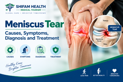
The meniscus is a C-shaped piece of fibrocartilage in the knee — each knee has two, a medial meniscus (inner) and a lateral meniscus (outer) — that helps distribute load, absorb shock, and stabilize the joint between the thigh bone and shin bone. A meniscus tear is a disruption in this tissue, and tears vary enormously by location, pattern, size, and cause: some result from a sudden twisting injury during sports (traumatic tears), while others develop gradually as the tissue weakens with age, often alongside osteoarthritis (degenerative tears).
A meniscus tear seen on MRI does not automatically mean that tear is the cause of a patient’s pain — degenerative meniscal changes are extremely common with aging and frequently show up as an incidental finding, unrelated to whatever symptom actually brought someone in for imaging. Not every tear needs surgery, and MRI alone should never be the sole basis for a treatment decision.
This guide explains the different types of meniscus tears and what they mean, how diagnosis actually works, when conservative treatment makes sense versus when surgery is genuinely indicated, the real difference between meniscus repair and partial meniscectomy, and what treatment and its cost look like for international patients considering care in India. This is general medical education, not a diagnosis, and does not replace individualized evaluation by a qualified specialist.
Table of Contents
ToggleMeniscus Anatomy
| Structure | Location | Clinical Relevance |
|---|---|---|
| Medial meniscus | Inner side of the knee | More commonly torn than the lateral meniscus; more firmly attached to surrounding structures, which may contribute to this |
| Lateral meniscus | Outer side of the knee | More mobile than the medial meniscus; the site of the discoid meniscus variant, discussed below |
| Anterior horn / body / posterior horn | Front, middle, and back regions of each meniscus | Tear location within these regions affects surgical accessibility and repair feasibility |
| Peripheral (outer) zone | Outer rim of the meniscus, closest to the capsule | Better blood supply — tears here generally have greater healing potential |
| Central (inner) zone | Inner portion of the meniscus | Little to no direct blood supply — tears here have limited intrinsic healing capacity |
| Meniscocapsular junction | Where the meniscus attaches to the joint capsule | A specific tear location relevant to certain repair techniques |
The meniscus’s blood supply pattern — richer at the periphery, essentially absent toward the center — is one of the most clinically important facts in this entire topic, since it directly determines which tears have a realistic chance of healing (on their own or after surgical repair) and which don’t. The meniscus also works closely with the ACL, PCL, collateral ligaments, and articular cartilage to keep the knee stable and properly loaded, which is why meniscus tears frequently occur alongside — not instead of — other knee injuries.
Traumatic vs. Degenerative Meniscus Tears
| Feature | Traumatic Tear | Degenerative Tear |
|---|---|---|
| Typical age | Younger, often active/athletic individuals | Older adults, though can occur earlier with existing joint wear |
| Onset | Sudden, tied to a specific incident | Gradual, sometimes without a clear inciting event |
| Typical mechanism | Twisting, pivoting, deep squatting, sudden direction change, contact injury | Age-related tissue weakening, repetitive loading, often alongside osteoarthritis |
| Common associated injuries | ACL tear, other ligament injuries | Osteoarthritis, cartilage wear |
| Initial treatment approach | Often conservative first, with repair considered if mechanical symptoms or instability are present | Often conservative first; surgery reserved for persistent, clearly correlated symptoms |
| Role of surgery | More often has repair potential, particularly in younger patients with peripheral tears | Often less amenable to repair; surgery, when needed, more often involves limited debridement |
Age alone doesn’t determine treatment — a younger patient can have a tear pattern poorly suited to repair, and an older patient with a degenerative tear can still be an appropriate repair or even surgical candidate depending on the specific tear and their activity goals. The traumatic/degenerative distinction is a starting framework, not a rigid rule.
Types of Meniscus Tears
| Tear Pattern | What It Looks Like | Healing / Repair Potential |
|---|---|---|
| Vertical longitudinal | A tear running parallel to the meniscus’s curved edge | Often good repair potential, especially if peripheral |
| Horizontal | Splits the meniscus into upper and lower layers | Often associated with degeneration; repair potential varies |
| Radial | Runs perpendicular to the meniscus’s curve, toward the center | Can disrupt the meniscus’s ability to distribute load (hoop stress) significantly, especially near the root |
| Oblique / flap | A diagonal tear creating a flap of loose tissue | The flap can catch in the joint, causing mechanical symptoms |
| Complex | A combination of multiple tear patterns together | Generally more difficult to repair given the mixed pattern |
| Bucket-handle | A displaced vertical longitudinal tear where the torn inner fragment flips like a bucket handle | Can cause mechanical locking and may require prompt assessment |
| Root tear | A tear at the very back attachment point of the meniscus | Functionally important because it can substantially impair meniscal load distribution |
| Ramp lesion | A tear at the back of the medial meniscus near its capsular attachment | Frequently occurs alongside ACL injuries and can be easy to miss on standard imaging |
| Parrot-beak | A specific oblique tear pattern resembling a bird’s beak shape | Can cause catching or locking depending on stability |
| Meniscocapsular | A tear specifically at the junction where the meniscus attaches to the joint capsule | Generally has good healing potential given proximity to blood supply |
Meniscus root tears
A meniscus root tear occurs at the point where the meniscus firmly attaches to the bone at its front or back end — because this attachment is what allows the meniscus to resist being pushed outward under load (a mechanism called hoop stress), a complete root tear functionally behaves similarly to having no meniscus at all in that compartment, allowing the meniscus to extrude (bulge out from its normal position) and substantially increasing the load reaching the underlying cartilage. Root tears can be acute/traumatic or degenerative, occur in either the medial or lateral meniscus, and are associated with accelerated cartilage wear if left unaddressed.
Not every root tear requires surgery — treatment depends on the patient’s age, activity level, current cartilage condition, leg alignment, whether osteoarthritis is already present, and the specific tear characteristics, but root tears deserve specific consideration precisely because of how much mechanical function is lost when this particular attachment fails.
Bucket-handle tears
A bucket-handle tear is a displaced vertical longitudinal tear where the inner torn fragment flips into the middle of the joint, like the handle of a bucket lifting up from its base. This specific pattern commonly causes mechanical locking — a genuine inability to fully straighten the knee because the displaced fragment is physically blocking normal joint movement — and is frequently associated with ACL injuries occurring at the same time.
A knee that’s genuinely locked and won’t fully extend, particularly after a twisting injury, warrants prompt orthopedic assessment; that said, not every knee that feels “stuck” or stiff represents a bucket-handle tear — true mechanical locking (discussed further below) is a specific finding distinguished from general stiffness or reluctance to move due to pain and swelling.
Causes and Risk Factors
Meniscus tears result from sports injuries involving twisting, pivoting, deep squatting, or sudden changes in direction; contact injuries; and, separately, gradual degenerative changes associated with aging, osteoarthritis, and repetitive loading over years. Some degenerative tears occur with relatively minor movements — a simple squat or twist that wouldn’t injure a healthy meniscus can tear one that’s already weakened by degeneration, which is part of why some patients are surprised their “minor” movement caused a genuine tear.
Risk factors include age, participation in pivoting sports, occupations involving frequent kneeling or squatting, obesity, existing osteoarthritis, previous knee injury (particularly ACL injury, which significantly increases meniscus tear risk), ligament instability, and previous meniscal surgery. These describe increased likelihood, not direct, guaranteed causation for any individual.
Symptoms
Common symptoms include knee pain (often localized to the joint line), swelling, stiffness, a sensation of catching or clicking, reduced tolerance for twisting, squatting, or stairs, and, in some cases, a feeling of instability. Clicking alone does not prove a meniscus tear — many knees click or pop without any tear present, and this sensation alone isn’t diagnostic.
Locking vs. catching: a genuinely important distinction
True mechanical locking means the knee becomes physically blocked and cannot move through its full normal range — it’s stuck, not just stiff or painful to move. Catching or clicking describes intermittent sensations that can have multiple causes and don’t represent the knee being physically blocked. This distinction matters clinically: persistent inability to fully extend the knee deserves prompt evaluation, since it can indicate a displaced tear (like a bucket-handle tear) mechanically obstructing the joint, while general clicking or occasional catching, especially without pain or swelling, is a much less urgent finding.
Swelling
Immediate swelling after an injury (within hours) more often suggests an associated injury like an ACL tear, since ACL injuries typically bleed into the joint quickly, while meniscal injury can cause more delayed swelling, sometimes appearing the day after activity. Recurrent effusion (fluid buildup) can occur with an ongoing meniscal problem. Swelling by itself is not specific to meniscal tears — many knee conditions cause swelling, and its presence or timing is one clue among several, not a stand-alone diagnostic sign.
Meniscus tear vs. ACL injury
| Feature | Meniscus Tear | ACL Injury |
|---|---|---|
| Typical Mechanism | Twisting, squatting, direct trauma | Sudden deceleration, pivoting, landing awkwardly |
| Swelling | Variable, sometimes delayed | Often rapid, significant swelling from bleeding into the joint |
| Instability | Less typical unless combined with ligament injury | A hallmark symptom — knee “giving way” |
| Locking | Can occur with displaced tears | Not typical unless a meniscus tear coexists |
| Examination | Joint-line tenderness, meniscal provocative tests | Positive Lachman, anterior drawer, and pivot-shift tests |
These two injuries frequently occur together — a significant proportion of ACL injuries involve a concurrent meniscus tear, which is exactly why a knee injury with any suggestion of instability deserves a full ligament and meniscus evaluation together, not an assumption that only one structure is involved.
Meniscus tear vs. osteoarthritis
Degenerative meniscal tears frequently coexist with knee osteoarthritis — they often share the same underlying cause (aging, cumulative joint wear) rather than one causing the other. Treating an MRI-visible meniscus tear surgically does not necessarily treat the underlying osteoarthritis, and in a knee with significant existing arthritis, removing torn meniscal tissue may provide limited or no additional benefit beyond what appropriate arthritis management would achieve on its own — this is a genuinely important distinction for older patients specifically, discussed further below.
When to Seek Urgent Care
Most meniscus tear symptoms warrant a specialist appointment, not an emergency room visit. Seek urgent evaluation for: a knee locked in a fixed position and unable to fully extend, inability to bear weight after major trauma, visible deformity, rapidly increasing swelling, suspected fracture, severe pain after significant injury, neurovascular symptoms (numbness, color change, or coldness in the lower leg or foot), or — importantly — a hot, swollen knee accompanied by fever, which can represent joint infection (septic arthritis), a genuine emergency requiring prompt medical evaluation, not international travel planning.
How a Meniscus Tear Is Diagnosed
Diagnosis combines history, physical examination, and imaging when appropriate — not any one of these alone. History covers the injury mechanism (or lack of one, for degenerative tears), timing of onset, pain location, swelling pattern, locking or catching, any sense of instability, previous knee injuries, sports or occupational activity, and existing osteoarthritis.
Physical examination assesses gait, alignment, range of motion, joint-line tenderness, effusion, ligament stability, and specific meniscal provocative tests — the McMurray test and Thessaly test are commonly used, though it’s worth knowing that individual physical exam tests have real limitations and shouldn’t be interpreted in isolation — a positive or negative single test doesn’t confirm or rule out a tear on its own; the overall clinical picture matters more than any single maneuver.
Imaging
X-rays don’t directly visualize the meniscus well, but remain useful for assessing osteoarthritis, alignment, joint-space narrowing, and ruling out fracture — they’re often obtained before or alongside MRI, particularly for degenerative knee symptoms in older patients. MRI is the preferred imaging modality for evaluating meniscal structure in most clinical scenarios where imaging is warranted, showing tear location and pattern, associated ligament injuries, cartilage damage, bone marrow abnormalities, and joint effusion. MRI is not required for every suspected tear — whether it’s obtained depends on age, mechanism, symptom severity, exam findings, suspected associated injury, and whether the result would actually change the treatment plan.
A crucial point: MRI findings must be interpreted together with symptoms and examination, not read in isolation. Degenerative meniscal signal changes and tears are extremely common incidental findings on knee MRI, especially with increasing age — a meaningful proportion of people with no knee pain at all show meniscal abnormalities on MRI if scanned. Report terminology worth understanding without over-alarm: horizontal, radial, complex, and flap tears describe pattern (as above); extrusion refers to the meniscus bulging outward beyond its normal position, associated with root tears and degeneration, though extrusion alone doesn’t automatically determine treatment; a parameniscal cyst is a fluid collection sometimes associated with a horizontal tear; and degenerative signal describes tissue changes that may or may not represent a clinically significant tear. None of this terminology should be read as automatically more severe than the patient’s actual symptoms suggest.
Differential Diagnosis
Knee pain resembling a meniscus tear can also arise from an ACL or collateral ligament injury, patellofemoral pain, chondral (cartilage) injury or an osteochondral lesion, a bone bruise, bursitis, tendinopathy, synovitis, inflammatory arthritis, septic arthritis (a genuine emergency, distinguished by fever and a hot, significantly swollen joint), or referred pain from the hip.
A thorough history and exam — not assuming every knee pain is a meniscus tear simply because that’s a common diagnosis — is what actually sorts through these possibilities.
Can a Meniscus Tear Heal on Its Own?
It depends heavily on where the tear is located. Tears in the peripheral (outer) zone, where blood supply is good, have genuine healing potential — some smaller peripheral tears can improve or stabilize without surgery. Tears in the central (inner) zone, where blood supply is minimal to absent, have limited intrinsic healing capacity and are unlikely to heal on their own regardless of treatment. Degenerative tears behave somewhat differently from acute traumatic tears, given the underlying tissue quality.
Importantly, symptoms can improve substantially even when the anatomical tear remains visible on repeat imaging — treatment decisions should be based on symptoms and function, not solely on whether an MRI still shows a tear. Not all tears heal, but many patients achieve meaningful, lasting symptom improvement regardless of whether the tear itself has anatomically resolved.
Non-Surgical Treatment
For many meniscus tears—especially degenerative tears without locking and stable traumatic tears—conservative treatment is often the first and sufficient approach. It may include activity modification, ice, compression, elevation, pain-relieving medication, and structured physiotherapy to improve knee movement, strength, balance, and function. Prolonged rest or immobilization is generally unnecessary unless specifically advised.
Corticosteroid injections may provide short-term pain relief but do not repair the torn meniscus. NSAIDs, paracetamol, or topical NSAIDs may also help manage symptoms when medically appropriate.
PRP and stem-cell treatments remain emerging options as of 2026. Evidence is still limited and inconsistent, and they should not be promoted as guaranteed methods to regrow or repair meniscal tissue.
Conservative treatment is particularly appropriate when symptoms are manageable, there is no true mechanical locking, and no associated injury requires surgery.
When Is Surgery Considered?
Surgery may be appropriate for persistent, disabling symptoms despite an adequate trial of conservative treatment; traumatic tears with genuine repair potential (particularly in younger, active patients with peripheral tears); displaced tears causing mechanical obstruction (like a bucket-handle tear preventing full extension); selected unstable tears; selected root tears where the clinical picture supports intervention; associated ligament injury requiring surgical treatment anyway; and situations where symptoms clearly correlate with the specific tear seen on imaging — not simply because a tear is present. There’s no single universal timeframe (like “six weeks of failed conservative treatment”) that applies as a strict cutoff for every patient; the decision is individualized based on tear type, symptoms, and how conservative treatment has actually gone.
Arthroscopy
Knee arthroscopy — a minimally invasive, camera-guided surgical technique — is how most meniscus surgery is performed, whether repair or partial meniscectomy. It allows direct visualization and treatment of the tear through small incisions. Arthroscopy is not automatically needed simply because MRI shows a tear — the decision to operate depends on the full clinical picture described throughout this guide, not the imaging finding alone.
Meniscus Repair vs. Partial Meniscectomy
Meniscus repair preserves the meniscus by stitching the torn tissue so it can heal. The technique depends on the tear’s location, pattern, tissue quality, age, and associated injuries. Not every tear is suitable for repair, particularly degenerative or poorly vascularized tears.
Partial meniscectomy removes only the damaged or unstable portion of the meniscus while preserving as much healthy tissue as possible. Total meniscectomy, which removes the entire meniscus, is rarely performed because of its importance in protecting the knee joint.
| Feature | Meniscus Repair | Partial Meniscectomy |
|---|---|---|
| Main Goal | Preserve meniscal tissue and function | Remove damaged or unstable tissue to relieve symptoms |
| Suitable Tear Types | Peripheral, vascularized, appropriate pattern and tissue quality | Central-zone, degenerative, or otherwise irreparable tears |
| Early Recovery | Generally slower — often includes weight-bearing and range-of-motion restrictions to protect healing | Generally faster — often quicker return to activity |
| Long-Term Joint Consideration | Preserving tissue is generally associated with better long-term joint mechanics | Removing tissue, even partially, reduces the meniscus’s shock-absorbing and load-distributing function over time |
A faster early recovery after meniscectomy does not automatically mean better long-term joint outcomes — this is a genuinely important point that’s easy to miss when recovery speed is the only thing being compared. The evidence generally supports that, when a tear is genuinely repairable, preserving meniscal tissue is preferable for long-term joint health compared to removing it, even though meniscectomy typically offers a quicker return to activity in the short term. Neither procedure is universally “better” — the right choice depends on whether the specific tear is actually repairable in the first place, which is a tissue and pattern question, not a preference question.
Root repair
Meniscus root repair — specifically reattaching a torn root to bone — aims to restore the meniscus’s ability to resist hoop stress and reduce the accelerated cartilage wear associated with untreated root tears. Patient selection depends on cartilage status, leg alignment, and overall joint condition; in some cases, an alignment-correcting procedure may be considered alongside root repair if malalignment is contributing to the problem. Root repair does not promise restoration to a completely normal meniscus — the goal is improved function and reduced progression of joint damage, not a guarantee of full anatomical or functional normalcy.
Combined ACL and meniscus injury
When ACL reconstruction and meniscus repair are needed together, repair is often performed at the same time as the ACL surgery, since the biological environment created during ACL reconstruction (including bleeding into the joint) may actually support meniscal healing. This combined approach affects overall rehabilitation timing and return-to-sport planning, which your surgical team will coordinate as one integrated plan rather than two separate recoveries.
Surgical Risks
Risks of meniscus surgery include infection, bleeding, blood clots, anesthesia-related complications, joint stiffness, persistent pain, recurrent tear, repair failure (the sutured tissue doesn’t heal), rare neurovascular injury, and — a genuine long-term consideration regardless of which procedure is performed — progression of cartilage degeneration over time, particularly after meniscectomy given the reduced tissue available to protect the joint surface. Risk level varies by the specific procedure, extent of tissue involved, and individual patient factors.
Recovery
Recovery differs meaningfully between repair and meniscectomy, and by individual factors including extent of tissue treated, associated injuries, and overall health.
| Feature | Meniscus Repair | Partial Meniscectomy |
|---|---|---|
| Weight-Bearing | Often restricted or protected for a period to allow healing | Often allowed sooner |
| Bracing | Sometimes used to protect the repair during early healing | Less commonly needed |
| General Pace | Slower — healing tissue needs time and protection | Generally faster early recovery |
| Rehabilitation Focus | Progressive, staged loading respecting the healing timeline | Earlier progression to strengthening and function |
Both pathways involve structured physiotherapy — restoring range of motion, rebuilding quadriceps, hamstring, and hip strength, and progressively returning to functional movement — with the specific pace and restrictions set by the treating surgeon based on what was actually done during surgery, not a fixed universal calendar. Return to sport should be based on pain resolution, swelling control, full or near-full range of motion, strength, neuromuscular control, and, where available, sport-specific functional testing — not simply counting weeks since surgery or waiting until pain disappears, since pain resolving doesn’t necessarily mean the knee has regained the strength and control needed to safely handle sport-specific demands.
Can you walk or exercise with a meniscus tear?
Many people can walk with a meniscus tear, sometimes with pain or swelling; a displaced tear causing locking can genuinely limit normal walking, and inability to bear weight after significant trauma warrants evaluation rather than being pushed through. For exercise, low-impact activities like cycling (as tolerated) and swimming, along with guided strengthening, are generally reasonable, while pivoting, twisting, deep loaded squats, and high-impact activity warrant more caution depending on the specific tear — exercise selection genuinely depends on tear characteristics and current symptoms, not a blanket rule for every meniscus tear.
Prevention
Reasonable, evidence-supported risk reduction includes strength and neuromuscular training (particularly for pivoting-sport athletes), attention to sports technique, gradual training progression rather than sudden increases in load, adequate recovery between intense activity, appropriate management of previous knee injuries, and maintaining a healthy body weight where relevant to individual circumstances. Meniscus tears cannot always be prevented — some result from injuries or degenerative processes that aren’t fully within an individual’s control.
Meniscus Tears in Older Adult
Degenerative meniscal changes are extremely common in older adults, frequently found incidentally on MRI performed for another reason, and frequently coexist with osteoarthritis. Surgery should not be chosen automatically based on imaging findings alone in this population — exercise-based treatment is often appropriate as a first approach, and in a knee with significant existing arthritis, addressing the meniscus tear surgically may provide limited additional benefit beyond appropriate arthritis management. This isn’t a statement that older patients shouldn’t have surgery when genuinely indicated — it’s a caution against treating an incidental MRI finding as an automatic surgical indication regardless of age.
Meniscus Tears in Children and Adolescents
Meniscus tears in younger patients are most often sports-related and traumatic, and meniscal preservation is generally prioritized even more strongly than in adults, given the greater cumulative joint stress a young person’s knee will experience over a longer remaining lifetime. Growth considerations and pediatric-specific evaluation matter for this age group, and specialist pediatric or adolescent sports medicine orthopedic input is valuable rather than applying adult treatment algorithms by default.
Discoid meniscus
A discoid meniscus is a congenital anatomical variant — usually affecting the lateral meniscus — where the meniscus is abnormally thick and disc-shaped rather than the normal C-shape. It may be entirely asymptomatic and found incidentally, or it can become symptomatic, typically causing snapping or clicking, and is more prone to tearing than a normally shaped meniscus given its unusual structure. A discoid meniscus by itself is not a disease requiring treatment — treatment is considered only when it becomes genuinely symptomatic or torn, not simply because the variant is present on imaging.
Long-Term Outlook
Long-term outlook depends on tear type, whether repair or meniscectomy was performed (or whether treatment stayed conservative), the extent of any tissue removed, associated ligament injury, rehabilitation, and ongoing activity modification. Meniscal preservation, when the specific tear allows it, is generally associated with better long-term joint mechanics than tissue removal.
This does not mean every meniscus tear inevitably leads to arthritis — many people, particularly those with smaller tears or successful repairs, have good long-term function without significant joint degeneration, while more extensive tissue loss, especially combined with existing risk factors, does carry a meaningfully higher long-term joint-wear risk.
When to Get a Second Opinion
A second opinion is particularly reasonable when surgery is being recommended, when meniscectomy is proposed but repair might be possible, when a root tear diagnosis is involved, when your MRI findings and actual symptoms don’t clearly match, when significant osteoarthritis is present alongside the tear (raising the “would surgery actually help” question discussed above), when multiple procedures are being proposed at once, or when symptoms persist after a prior surgery. Bring your MRI images (not just the report), X-rays, any prior operative reports, physiotherapy records, and your medication/injection history so a second specialist has the complete picture rather than starting from scratch.
Meniscus Tear Treatment Cost in India
Cost depends heavily on whether treatment stays conservative or progresses to surgery, and, if surgical, whether repair or meniscectomy is performed and how complex the case is.
| Component | Approximate Cost (USD) |
|---|---|
| Orthopedic/Sports Medicine Consultation | $30–$150 |
| X-ray | $20–$80 |
| MRI | $150–$500 |
| Physiotherapy Course | $150–$500 |
| Corticosteroid Injection | $50–$200 |
| Arthroscopic Partial Meniscectomy (Domestic Indian Pricing) | Roughly $650–$950 based on multiple published domestic sources |
| Arthroscopic Meniscus Repair (Domestic Indian Pricing) | Roughly $950–$4,200 based on published domestic sources, with figures varying considerably by complexity and hospital |
| Combined ACL Reconstruction + Meniscus Repair | Adds meaningfully to cost given the combined procedure and hardware |
Meniscus Tear Treatment in India for International Patients
India has substantial orthopedic and sports medicine capability, including arthroscopic meniscus repair, partial meniscectomy, and combined ACL/meniscus surgery — for international patients, the appeal is typically this capability combined with costs generally lower than the US, UK, or much of Europe. India isn’t automatically the cheapest or best destination for every patient, and this shouldn’t be assumed without direct comparison for your specific situation. For any acute emergency presentation — a genuinely locked knee, inability to bear weight after major trauma, or a hot, feverish, swollen knee — appropriate local emergency evaluation should always come first, before international travel planning.
The international patient journey
- Share medical records and existing imaging (MRI/X-ray)
- Specialist review, with treatment recommendation
- Receive a cost estimate
- Hospital and appointment coordination
- Travel planning and medical visa documentation, if travel is appropriate
- In-person consultation and any needed additional investigation
- Treatment
- Rehabilitation
- Follow-up
Treatment plans can change after an in-person examination or a specialist’s direct review of your actual imaging — a preliminary remote opinion is a reasonable starting point, not a final, fixed plan.
How Shifam Health Helps International Patients
Shifam Health is a medical tourism facilitator/coordinator, not a hospital, orthopedic surgeon, or diagnostic provider — we don’t diagnose meniscus tears or determine your treatment plan. What we do is help international patients navigate the practical side of accessing meniscus tear evaluation and treatment in India: coordinating the sharing of medical records and imaging with appropriate orthopedic and sports medicine specialists, helping identify hospitals with genuine relevant experience, obtaining written treatment and cost estimates, assisting with medical visa documentation, arranging airport pickup and accommodation, providing interpreter support where needed, and staying in touch for follow-up communication once you return home.
We don’t guarantee repair success, pain relief, return to sport, or exact cost — those depend entirely on your specific tear and treatment, determined by your treating medical team.
If you have a suspected or confirmed meniscus tear and you’re exploring evaluation or treatment options in India, share your available reports and imaging with our team on WhatsApp or through a quick inquiry form — there’s no obligation, and we typically respond within 24 hours.
Frequently Asked Questions
What is a meniscus tear? A meniscus tear is an injury to the C-shaped cartilage that cushions and stabilizes the knee.
Can a meniscus tear heal without surgery? Yes. Some small tears, especially in the well-blood-supplied outer zone, may heal or become less symptomatic with conservative treatment.
What are the symptoms of a meniscus tear? Common symptoms include knee pain, swelling, stiffness, clicking, and difficulty fully bending or straightening the knee.
Can you walk with a torn meniscus? Yes, many people can walk with a meniscus tear, although pain and swelling may occur. Inability to bear weight or true locking needs prompt evaluation.
Can a meniscus tear cause locking? Yes. A displaced tear, particularly a bucket-handle tear, can cause true mechanical locking of the knee.
How is a meniscus tear diagnosed? Diagnosis involves medical history, physical examination, and, when needed, an MRI to assess the meniscus and associated knee injuries.
Can a meniscus tear be treated without surgery? Yes. Physiotherapy, activity modification, pain management, and selected injections may help, particularly for degenerative tears without locking.
When is meniscus surgery needed? Surgery may be considered for persistent symptoms, displaced tears causing locking, or certain traumatic tears that are suitable for repair.
What is meniscus repair? Meniscus repair uses stitches or specialized techniques to preserve and heal the torn tissue when the tear pattern and blood supply are suitable.
What is partial meniscectomy? It involves removing only the unstable or irreparable portion of the torn meniscus while preserving as much healthy tissue as possible.
Which is better: meniscus repair or meniscectomy? When possible, repair is generally preferred because preserving meniscal tissue supports long-term knee health.
How much does meniscus tear treatment cost in India? Costs vary by procedure and hospital; published estimates range from about $650–$4,200. A personalized hospital quote is recommended.
Conclusion
A meniscus tear is not one uniform diagnosis — location, pattern, blood supply, and cause together determine whether a specific tear can heal on its own, is a good candidate for repair, or is better managed with limited tissue removal or conservative care altogether. The single most protective idea in this guide is that an MRI finding a tear doesn’t automatically mean that tear is causing your symptoms or that surgery is the right next step — getting an accurate, correlated diagnosis, and understanding whether meniscal preservation is realistically possible for your specific tear, is the most useful step before deciding on a treatment path, in India or anywhere else.
This article is for general medical education and does not replace individualized advice from a qualified orthopedic surgeon or sports medicine specialist. It is not a diagnosis, treatment recommendation, or guarantee of any outcome.



Popular Posts From Last Week
- September 21, 2026
- shifamhealth
Hearing “neurofibromatosis” attached to your own or your child’s diagnosis often raises a specific kind of confusion, because…
- September 18, 2026
- shifamhealth
Written by: Shifam Health Editorial Team — Orthopedic Content Division Medically reviewed by: Shifam Health Clinical Advisory Panel…
- September 16, 2026
- shifamhealth
Ankle valgus deformity is an abnormal alignment in which the ankle tilts toward the outside of the body,…
- September 20, 2026
- shifamhealth
Medically reviewed content · Published 2026 · Last reviewed September 2026 A limb deformity in a child is…
- September 16, 2026
- shifamhealth
Epithelial ovarian cancer is the most common broad category of ovarian cancer, arising from the cells covering the…
- September 17, 2026
- shifamhealth
Hepatoblastoma is a rare malignant liver tumor that develops primarily in young children, most commonly before age 5….
- September 19, 2026
- shifamhealth
Finding a purple or reddish patch on your skin that won’t fade, or hearing “Kaposi sarcoma” mentioned during…
- September 18, 2026
- shifamhealth
Kyphosis means forward curvature of the spine — and some kyphosis is completely normal anatomy in the mid-back…
- September 21, 2026
- shifamhealth
Medically reviewed content · Published 2026 · Last reviewed September 2026 Hip dysplasia describes a hip joint where…
- September 19, 2026
- shifamhealth
Lumbar radiculopathy is a neurological syndrome caused by irritation, inflammation, or compression of a nerve root in the…
- September 20, 2026
- shifamhealth
Still having pain after spine surgery — whether it never fully went away, or came back after a…
- September 22, 2026
- shifamhealth
Medically reviewed content · Published 2026 · Last reviewed September 2026 The meniscus is a C-shaped piece of…