



Home > Blog > Rheumatoid Arthritis of the Spine: Symptoms, Causes, Diagnosis and Treatment (2026 Guide)
Rheumatoid Arthritis of the Spine: Symptoms, Causes, Diagnosis and Treatment (2026 Guide)
Filters & Insights
Learn about rheumatoid arthritis of the spine including symptoms, diagnosis, treatment options, complications & ways to manage spinal inflammation.

Medically reviewed content · Published 2026 · Last reviewed September 2026
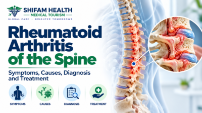
Rheumatoid arthritis (RA) primarily affects small joints, but in some patients, the chronic inflammation of RA also involves the spine, most importantly the upper cervical spine (the neck, particularly the junction between the skull and the top two vertebrae). There, inflammatory tissue (pannus) and erosive damage can weaken the ligaments and bone that normally keep this region stable, potentially leading to instability, and in more advanced cases, compression of the spinal cord or, rarely, the brainstem.
This is genuinely different from ordinary neck pain or common age-related cervical spondylosis, and it deserves to be taken seriously but it’s equally important not to panic: not every RA patient with neck pain has cervical instability, and modern RA treatment (particularly DMARDs and biologic therapy that control inflammation early and effectively) has meaningfully changed how often severe cervical disease develops compared to decades past, when RA was often treated less aggressively.
This guide explains how RA can affect the spine, which symptoms deserve urgent versus routine attention, how instability and cord involvement are actually diagnosed, current treatment approaches — including the genuinely important anesthesia and perioperative considerations unique to this condition and what treatment and its cost look like for international patients considering care in India. This is general medical education, not a diagnosis, and does not replace individualized evaluation by a qualified rheumatologist and spine specialist.
Table of Contents
ToggleWhy the Upper Cervical Spine Matters So Much
The atlas (C1) and axis (C2) — the top two vertebrae — have a unique job: they support the skull and allow the head to rotate, held together and to the skull by a specific set of ligaments rather than relying primarily on disc and bone structure the way the rest of the spine does. The transverse ligament in particular holds the odontoid process (a bony peg projecting up from C2, also called the dens) snugly against the back of C1’s front arch, preventing the dens from shifting backward toward the spinal cord and, just above it, the brainstem.
RA’s chronic synovial inflammation can form pannus (abnormal, destructive inflammatory tissue) around this joint, eroding bone and weakening or destroying the transverse ligament — because this specific region sits so close to the spinal cord, brainstem, and vertebral arteries, even relatively modest instability here carries more potential consequence than similar-looking instability lower in the spine.
The subaxial cervical spine (C3–C7) can also be affected by RA, through instability, deformity, and canal narrowing at these lower levels, though the upper cervical region is generally where the most clinically significant rheumatoid spinal disease occurs.
Patterns of Rheumatoid Spinal Involvement
Atlantoaxial instability
The most common pattern: erosion and inflammation damage the transverse ligament and surrounding bone at the C1–C2 joint, allowing the dens to shift further backward than normal relative to C1 (typically during neck flexion), narrowing the space available for the spinal cord.
This can range from mild, asymptomatic laxity found incidentally on imaging to significant instability causing neurological symptoms.
Cranial settling (vertical subluxation / basilar invagination)
A more advanced pattern where erosive destruction allows the skull to actually settle downward onto the upper cervical spine, causing the odontoid process to migrate upward — potentially toward or into the space where the brainstem sits.
This is generally considered a more concerning pattern than atlantoaxial instability alone, given its closer relationship to brainstem structures, and is assessed using specific radiographic measurements discussed below.
Subaxial cervical instability
Involvement of the lower cervical spine (C3–C7), causing vertebral instability, subluxation (vertebrae shifting out of normal alignment relative to each other), deformity, and, in some cases, narrowing of the spinal canal at these levels — mechanically different from the upper cervical patterns above but capable of producing similar neurological consequences if the spinal cord is compressed.
Other spinal involvement
RA can, less commonly, be associated with changes elsewhere in the spine, but the thoracic and lumbar spine are not the major clinical concern in rheumatoid spinal disease the way the cervical spine is — this guide focuses on cervical disease because that’s where the evidence and clinical significance concentrate.
Causes and Risk Factors
Not every person with RA develops clinically significant spinal disease. Factors associated with a higher likelihood include longstanding RA, persistent or poorly controlled inflammatory disease activity over time, erosive disease (visible joint damage on imaging elsewhere in the body), seropositive disease (positive rheumatoid factor or anti-CCP antibodies) in some studies, and more severe systemic disease overall.
No single factor guarantees that a given patient will develop cervical instability — these are population-level associations, not individual predictions. Importantly, modern treat-to-target RA management — using DMARDs and, where needed, biologic or targeted synthetic therapies to achieve early, sustained control of inflammation — appears to have reduced the frequency and severity of significant cervical spine involvement compared to the treatment era before these therapies were widely available or used as aggressively; older studies describing cervical disease frequency largely reflect a different treatment landscape than current practice.
Who Should Be Assessed, and When?
| Patient Characteristic | Why It Matters | What Should Prompt Assessment |
|---|---|---|
| Longstanding, especially poorly controlled, RA | Greater cumulative risk of erosive cervical involvement | New or worsening neck symptoms |
| Known existing cervical instability | Baseline risk for progression or acute decompensation | Any new neurological symptom |
| Planned surgery requiring general anesthesia | Airway management risk if cervical instability is present | Pre-anesthesia evaluation should specifically consider cervical spine status |
| Previous cervical spine surgery | Altered anatomy, potential adjacent-level issues | New symptoms at or near the prior surgical level |
| Severe, longstanding neck pain with occipital headache | Possible upper cervical involvement | Specialist evaluation |
| Hand clumsiness, dropping objects, gait changes | Possible myelopathy (spinal cord dysfunction) | Prompt neurological assessment |
Routine screening imaging is not automatically required for every RA patient — assessment is generally triggered by specific symptoms, known risk factors, or the specific context of planned surgery/anesthesia, not applied as a blanket screening protocol to everyone with an RA diagnosis.
Symptoms
Common symptoms include neck pain, neck stiffness, reduced range of neck motion, occipital headaches (at the base of the skull), and pain radiating into the shoulders or arms, sometimes with numbness or tingling.
Neurological symptoms — which change the clinical significance and urgency considerably — include hand clumsiness, dropping objects, general weakness, difficulty walking, balance problems, gait disturbance, and changes in fine motor control or sensation. In more advanced disease, myelopathy (spinal cord dysfunction from compression) can develop, and, in severe cranial settling specifically, brainstem involvement is a recognized, serious possibility.
Not every headache, dizziness, or vague neurological complaint in someone with RA is caused by cervical instability — these symptoms have many possible causes, and appropriate evaluation, not assumption in either direction, is what actually determines the cause.
Emergency Red Flags: When Is This a Medical Emergency?
Urgent medical assessment is needed for: rapidly worsening weakness, new difficulty walking, repeated falls, new loss of hand function, significant new numbness, progressive neurological symptoms of any kind, new bowel or bladder dysfunction, severe neurological deterioration, or severe symptoms following trauma in a patient with known or suspected cervical instability.
These findings can indicate spinal cord or, in severe cases, brainstem compromise — situations where the timing of evaluation and treatment genuinely matters and delay can allow permanent neurological injury to occur or worsen.
Suspected spinal cord or brainstem compromise should never be managed by waiting for an elective overseas consultation or planning international medical travel — appropriate urgent local medical care comes first, always, regardless of any treatment plans being considered elsewhere.
RA Cervical Disease vs. Other Causes of Neck Pain
| Condition | Key Distinguishing Features |
|---|---|
| RA cervical spine disease | Inflammatory erosion and instability, most prominent at the upper cervical spine, in a patient with known RA |
| Cervical spondylosis (age-related wear) | Gradual degenerative change, not inflammatory/erosive in the RA sense, can occur in anyone regardless of RA status |
| Cervical disc herniation | Focal disc material compressing a nerve root, typically causing more localized radicular symptoms |
| Ankylosing spondylitis | A different inflammatory arthritis, generally causing progressive stiffening/fusion rather than the instability pattern typical of RA |
| Osteoporotic cervical fracture | Related to bone fragility rather than joint/ligament erosion, often with a clear traumatic or minimal-trauma event |
| Infection (discitis/osteomyelitis) | Fever, systemic illness, elevated inflammatory markers with a different pattern; discussed in more detail in our companion discitis guide |
| Malignancy | Progressive symptoms, sometimes with weight loss or other systemic features, requiring its own specific workup |
Clinical evaluation combined with imaging is what actually differentiates these possibilities — RA cervical disease and ordinary age-related cervical spondylosis, for instance, can genuinely coexist in the same patient, and distinguishing which is driving a particular symptom requires the full diagnostic picture, not assumption based on the RA diagnosis alone.
How Diagnosis Works
Diagnosis combines history (RA duration and disease activity, medication history, previous cervical imaging or surgery, trauma, neurological symptoms, systemic symptoms) with physical and neurological examination — cervical range of motion, strength, sensation, reflexes, gait, coordination, and specific signs suggesting spinal cord involvement (upper motor neuron signs) — and imaging.
This examination should be performed by a trained clinician; patients should not attempt to test their own neck stability through forceful movement.
Imaging
Plain X-rays, including flexion-extension views when clinically appropriate and properly supervised, can show instability and specific measurements discussed below — but dynamic flexion-extension imaging must be ordered and supervised by the treating team, never attempted independently by a patient forcefully flexing or extending their own neck to “test” stability.
CT provides detailed bony anatomy, useful for assessing erosions and for surgical planning when needed.
MRI is central for evaluating the spinal cord itself, the degree of neural compression, soft tissue and pannus, and — most importantly — for identifying or excluding myelopathy and brainstem involvement; whether contrast is useful depends on the specific clinical question being asked.
Key radiographic measurements
The atlanto-dental interval (ADI) measures the space between the front arch of C1 and the odontoid process — increased ADI suggests atlantoaxial instability. The posterior atlanto-dental interval (PADI), also called the space available for the cord, measures the space behind the odontoid — this is often considered more directly relevant to actual cord safety than ADI alone, since it more directly reflects how much room the spinal cord has.
Specific measurements are also used to assess cranial settling. No single measurement threshold should be treated as an absolute, stand-alone guarantee of safety or danger — these values are interpreted alongside the patient’s actual neurological status and the full imaging picture, not as an isolated pass/fail number.
Laboratory tests
ESR, CRP, complete blood count, rheumatoid factor, and anti-CCP antibodies help assess overall RA disease activity and support diagnosis and differential diagnosis generally, but blood tests alone do not diagnose cervical instability — a patient can have well-controlled systemic disease on blood work and still have structural cervical instability, or vice versa; imaging and clinical/neurological assessment specifically address the spine question.
Differential Diagnosis
Neck pain in someone with RA can have many causes beyond RA cervical disease itself — cervical osteoarthritis, degenerative disc disease, disc herniation, spinal stenosis unrelated to RA, muscular pain, shoulder pathology referring pain to the neck, peripheral neuropathy, fracture, infection, malignancy, and, in some cases, medication-related issues.
Not every symptom in an RA patient should be assumed to be RA-related — this differential consideration is exactly why a proper clinical workup matters rather than attributing every complaint to the underlying disease.
Treatment Overview
Treatment is organized around several interacting factors: current systemic inflammatory disease activity, whether structural instability is present and how significant it is, whether there’s neurological involvement (and how severe), deformity, pain severity and functional impact, and overall surgical risk if intervention is being considered.
There is no single treatment pathway — management genuinely differs for a patient with RA and no spinal symptoms, versus one with mild instability and no neurological findings, versus one with myelopathy.
Medical management of RA
Controlling the underlying systemic disease is foundational, using conventional synthetic DMARDs (such as methotrexate, hydroxychloroquine, sulfasalazine, or leflunomide), biologic DMARDs, and targeted synthetic DMARDs as appropriate to the individual’s disease, guided by a treating rheumatologist rather than a fixed universal regimen. Corticosteroids may have a role in appropriate circumstances, with the usual considerations about long-term use.
It’s essential to understand a genuine limitation here: medication that controls systemic inflammation does not reliably reverse structural instability or spinal cord compression that has already developed — good disease control going forward can help prevent further progression, but it doesn’t undo mechanical damage already present.
Non-surgical spinal management
Supervised physiotherapy, attention to posture, general conditioning, and safe, appropriately guided strengthening can support function and comfort. Neck bracing may be considered in selected situations, though it has real limitations and isn’t a universal or long-term solution, and prolonged use carries its own considerations requiring specialist guidance.
A specific, important caution: patients with known or suspected cervical instability should not undergo aggressive, high-velocity cervical manipulation (as used in some chiropractic techniques) unless specifically assessed and cleared by an appropriate specialist first — this isn’t a blanket statement that all physiotherapy is dangerous, but high-velocity manipulation specifically carries theoretical risk in a spine that may already have compromised stability.
When Is Surgery Needed?
Surgery is not automatically required simply because an X-ray shows an abnormality — including a measurable degree of instability. Surgery may be considered for symptomatic instability, progressive neurological deficit, confirmed spinal cord compression or myelopathy, significant deformity, cranial settling with genuine risk to neural structures, or structural instability that, taken together with clinical findings, creates meaningful neurological risk. The decision integrates clinical symptoms, neurological examination findings, the specific structural measurements discussed above, MRI/CT findings, and overall surgical and anesthesia risk — not any single finding in isolation.
Types of surgery
C1–C2 fusion stabilizes the atlantoaxial joint specifically, appropriate when instability is confined to this level without significant cranial settling. Occipitocervical fusion extends stabilization from the skull (occiput) down into the cervical spine, used when instability involves cranial settling or when C1–C2 alone doesn’t adequately address the problem.
Subaxial cervical surgery decompression, stabilization, or both addresses lower cervical involvement when present. An important technical point: in an unstable rheumatoid cervical spine, decompression alone (simply removing pressure on the spinal cord) may not be sufficient without also addressing the underlying instability — surgical planning has to account for the spine’s biomechanics as a whole, not just the area of direct neural compression.
Surgical risks
| Risk | Consideration |
|---|---|
| Neurological/spinal cord injury | A serious, though not routine, risk given proximity to critical structures |
| Vertebral artery injury | Relevant specifically to upper cervical surgery given the vessels’ course through this region |
| Infection, bleeding | General surgical risks, with RA-related immunosuppression as an additional consideration |
| Nonunion/pseudarthrosis | The fusion fails to solidly heal |
| Hardware failure, adjacent-segment problems | Long-term mechanical considerations |
| Swallowing difficulties | Can occur, particularly with certain surgical approaches |
| Reduced neck motion | An expected consequence of fusion, proportional to the levels involved |
| Anesthesia complications | Discussed in detail below, given specific RA-related airway and systemic considerations |
No surgery is “100% safe,” and this procedure category carries genuinely serious potential risks alongside its potential benefits — a realistic, specific risk discussion with your surgical and anesthesia team is essential before proceeding.
Anesthesia and Perioperative Considerations: A Genuinely Distinctive Concern
This deserves particular emphasis because it’s an area general spine-surgery content often overlooks, and it’s specifically relevant to RA. RA can affect far more than the joints being operated on. Relevant considerations include: cervical instability itself creating airway management risk during intubation (since standard head positioning for airway access could theoretically stress an unstable neck); limited neck movement from disease itself, independent of instability; temporomandibular joint (jaw) involvement, which can limit mouth opening and complicate airway access; cricoarytenoid joint involvement (a less commonly known RA manifestation affecting small joints in the voice box, which can narrow the airway); associated pulmonary disease (RA can affect the lungs in some patients); cardiovascular disease; anemia (common in chronic inflammatory disease); and immunosuppression from RA medications, affecting infection risk around the time of surgery.
Because of these combined factors, airway management for a patient with known or suspected cervical instability often requires specific specialized planning — this is exactly why anesthesia teams experienced with RA and cervical spine surgery specifically matter for this population, not simply general anesthesia experience.
Perioperative medication management — whether and when to pause DMARDs, biologics, targeted therapies, or adjust corticosteroids around the time of surgery — must be individualized by the treating rheumatologist, surgeon, and anesthesia team together, weighing infection risk against disease flare risk.
There is no universal stop/start schedule that applies to every patient and every medication — this decision requires genuine coordination between these specialists specific to your case, not a generic rule found online.
Recovery
Recovery depends on neurological status before surgery, the specific procedure performed, disease severity, age, other health conditions, bone quality, how fusion healing progresses, and rehabilitation. General elements include a hospital stay (typically several days, though this varies), early guided mobility once medically appropriate, wound care, structured rehabilitation, and gradual return to normal activities including driving and work, generally following the treating team’s specific guidance rather than a universal timeline this guide could specify. Follow-up imaging monitors fusion healing over time. This guide will not promise a fixed recovery timeline, given how much these individual factors genuinely vary the course.
Bone health and osteoporosis
Chronic RA inflammation, long-term corticosteroid use, and reduced mobility from disease can all contribute to reduced bone density, which is relevant both to overall fracture risk and specifically to how well surgical fixation (screws, hardware) will hold in the bone.
Bone health assessment is a reasonable and often important part of preparation before major cervical fusion surgery, particularly occipitocervical fusion, given how much successful fixation depends on adequate bone quality.
Long-Term Outlook
Outlook depends on the severity of structural and neurological involvement, how promptly it was identified and treated, ongoing control of systemic RA, age, and other health factors.
It’s honest to note that neurological damage present before surgery — particularly significant, longstanding myelopathy — may not always fully reverse even with appropriate, successful surgical stabilization; surgery aims primarily to stop further progression and address existing compression, and some patients do experience meaningful improvement, but this varies and can’t be promised for every case.
Ongoing rheumatology follow-up and continued disease control remain important long-term, both to protect the surgically treated segments from further RA-related change and to manage the systemic disease overall.
Special Situations
Older adults
Frailty, osteoporosis, cardiovascular and pulmonary disease, polypharmacy, and higher anesthesia risk are all more common considerations in older RA patients, alongside genuine rehabilitation and fall-risk factors after surgery — none of this means older age automatically rules out appropriate treatment, but it does mean risk assessment and planning are more individualized and layered in this population.
Pregnancy
Pregnancy planning for a patient with RA and known or suspected cervical spine involvement should involve both the treating rheumatologist and an obstetric team, given medication considerations (some RA medications aren’t appropriate during pregnancy), imaging considerations (some imaging is avoided or modified during pregnancy), and, where relevant, anesthesia planning if cervical instability is a factor — this is a genuinely individualized conversation, not something with a single universal answer.
Travel and flying
Whether travel, including international medical travel, is appropriate depends on whether disease is stable, any recent surgery, presence of neurological symptoms, and general medical clearance for travel — most people with stable, well-managed RA and no acute spinal concerns can travel normally.
The one point that bears repeating plainly: a patient with suspected spinal cord compression or an acute neurological emergency should not delay urgent local care in order to travel internationally for elective treatment, regardless of how appealing a specific treatment option elsewhere might be.
When to Seek a Second Opinion
A second opinion is particularly reasonable when surgery has been recommended, when cervical instability is suspected but the picture feels unclear, when imaging findings and reported symptoms don’t seem to match, when major fusion surgery (particularly occipitocervical fusion) is being proposed, when the diagnosis itself remains uncertain, or when neurological symptoms are progressing and you want broader input.
Gather your RA diagnosis and treatment history, rheumatology notes, all X-rays, CT, and MRI images (not just reports), blood test results, and any prior operative notes and implant details before seeking a second opinion, so a new specialist has the complete picture.
Rheumatoid Arthritis of the Spine Treatment Cost in India
Appropriate care for this condition benefits from a genuine multidisciplinary team — rheumatologist, spine surgeon or neurosurgeon, neurologist, radiologist, anesthesiologist with relevant experience, and rehabilitation specialists — and cost depends heavily on whether management stays medical/conservative or progresses to surgery, and, if surgical, which specific procedure is needed.
| Component | Approximate cost (USD) |
|---|---|
| Rheumatology + spine specialist consultation | $50–$200 |
| X-rays (including flexion-extension views) | $30–$100 |
| CT | $100–$300 |
| MRI | $150–$500 |
| Laboratory workup (inflammatory markers, RF, anti-CCP) | $50–$200 |
| Ongoing DMARD/biologic therapy (monthly) | Highly variable by specific medication — biologics in particular can be a significant, ongoing cost |
| Cervical spine fusion surgery (general/domestic Indian pricing, comparable procedure category) | Roughly $2,750–$6,000 based on published domestic sources for cervical fusion generally |
| Occipitocervical fusion specifically | More specialized and complex than routine cervical fusion; reliable India-specific pricing for this exact procedure wasn’t found in currently available published sources — expect cost at or above the higher end of general cervical fusion pricing, confirmed directly |
As with the domestic-vs-international pricing pattern seen throughout this content series, published figures above are generally domestic-market-facing prices — international-patient quotes, once diagnostics, longer hospital stay for a more complex procedure, ICU considerations if needed, and coordination are factored in, typically run higher.
Given how much this specific procedure category varies by complexity (C1-C2 fusion alone versus occipitocervical fusion, presence or absence of neurological compromise, revision versus primary surgery), a written, itemized quote based on your specific diagnosis, imaging, and proposed procedure is essential — more so than for routine degenerative spine surgery, given the added complexity RA-specific factors (bone quality, airway considerations, medication management) bring to both the surgery and its cost.
Rheumatoid Arthritis of the Spine Treatment in India for International Patients
India has meaningful multidisciplinary capability for this condition, including rheumatology, spine surgery/neurosurgery, and the imaging and anesthesia infrastructure needed for complex cervical spine surgery — for international patients, the appeal is typically this capability combined with costs generally lower than the US, UK, or much of Europe.
India isn’t automatically the best or cheapest option for every patient, and comparisons with other countries should be made directly for your specific situation rather than assumed.
For any acute neurological emergency — rapidly progressive weakness, new bowel/bladder dysfunction, or suspected spinal cord compromise — appropriate local emergency care should always come first, before any international travel planning.
The international patient journey
- Share medical records, RA history, medication history, and existing imaging
- Initial specialist review (rheumatology and spine specialist together)
- Confirm diagnosis and severity
- Determine whether management can remain medical/conservative or requires surgical evaluation
- Receive a preliminary treatment plan and cost estimate
- Arrange medical visa documentation, where applicable
- Travel and accommodation planning
- In-person consultation, completing any needed additional investigation
- Treatment (medical management continuation, or surgery where indicated)
- Rehabilitation and immediate follow-up
- Long-term follow-up coordination with your home-country rheumatologist and specialist team
Shifam Health does not diagnose, operate, prescribe, or replace your treating medical team at any point in this process.
Questions to Ask Your Specialist
- What exactly is unstable, and how significant is it based on my imaging?
- Is my spinal cord compressed? Is there evidence of myelopathy?
- Is surgery necessary now, or can this be monitored — and what happens if I delay?
- If surgery is recommended, what is its specific goal — decompression, stabilization, or both, and why this particular approach and fusion level?
- What are the major neurological and anesthesia-related risks specific to my situation?
- How will my RA medications be managed around the time of surgery?
- How will my airway be managed given my cervical spine status?
- What happens if the fusion doesn’t heal properly?
- How much neck movement might I realistically lose?
- What rehabilitation will I need, and what’s a realistic recovery expectation?
- What symptoms after treatment would require emergency attention?
How Shifam Health Helps International Patients
Shifam Health is an international healthcare coordination and medical travel support company, not a hospital, rheumatologist, spine surgeon, or treating medical provider — we don’t diagnose rheumatoid arthritis of the spine, determine treatment, or prescribe medication.
What we do is help international patients navigate the practical side of accessing multidisciplinary care in India: collecting and sharing medical records and imaging with appropriate specialists, coordinating specialist opinions and hospital logistics, helping obtain a written treatment and cost estimate, assisting with medical visa documentation, arranging airport pickup and accommodation, providing interpreter support where needed, and coordinating follow-up communication once you return home. We don’t guarantee treatment outcomes, surgical success, visa approval, or exact cost — those determinations and outcomes belong to your treating medical team.
If you or someone you’re caring for has RA and concerning neck symptoms, or a specific treatment plan you’d like reviewed, share your available reports and imaging with our team on WhatsApp or through a quick inquiry form — there’s no obligation, and we typically respond within 24 hours. For any acute neurological emergency, please seek urgent local medical care first.
Frequently Asked Questions
Can rheumatoid arthritis affect the spine?
Yes. RA can affect the upper cervical spine, causing inflammation, ligament damage, and instability.
What is atlantoaxial instability?
Abnormal movement between C1 and C2 caused by RA-related damage, potentially narrowing the space around the spinal cord.
What is cranial settling?
A more advanced condition where the skull settles downward onto the upper cervical spine, potentially affecting the brainstem or spinal cord.
What are the symptoms of cervical spine involvement?
Neck pain, stiffness, headaches, hand clumsiness, weakness, balance problems, or gait changes may occur.
How is RA cervical instability diagnosed?
Assessment may include neurological examination, X-rays with appropriate views, CT for bone detail, and MRI to evaluate the spinal cord and soft tissues.
Can RA medication reverse cervical instability?
No. RA treatment can control inflammation and help prevent progression but cannot reverse established structural instability.
Is physiotherapy safe?
Carefully supervised physiotherapy may be appropriate, but high-velocity neck manipulation should be avoided until cervical stability has been assessed.
When is surgery needed?
Surgery may be considered for symptomatic instability, spinal cord compression, myelopathy, significant deformity, or substantial neurological risk.
What is C1-C2 fusion?
A procedure that stabilizes the joint between the first two cervical vertebrae when instability is primarily at this level.
What is occipitocervical fusion?
A more extensive procedure connecting the skull base to the cervical spine when instability extends beyond C1-C2 or involves cranial settling.
Can neurological symptoms improve after surgery?
Many patients may improve, but recovery depends on the severity and duration of neurological damage. Longstanding damage may not completely reverse.
Is RA cervical spine disease an emergency?
Rapidly worsening weakness, new bowel or bladder dysfunction, or other signs of spinal cord or brainstem compromise require immediate medical assessment.
Conclusion
Rheumatoid arthritis of the spine is a genuinely distinct concern from ordinary neck pain — the upper cervical spine’s unique anatomy and RA’s specific inflammatory and erosive process combine to create real, if uncommon, risk to the spinal cord and, in advanced cases, the brainstem.
The most protective ideas in this guide are that neurological symptoms, not imaging findings alone, are what should drive urgency; that modern RA treatment has genuinely changed how often severe disease develops; and that both medical and surgical treatment decisions, including the genuinely important anesthesia planning this condition requires, need real coordination between rheumatology and spine specialists working together. If you or someone you’re caring for has RA and concerning spinal symptoms, connecting with that kind of coordinated, experienced team — not simply a general orthopedic or spine practice — is the most valuable step, in India or anywhere else.
This article is for general medical education and does not replace individualized assessment by a qualified rheumatologist, spine surgeon, neurosurgeon, or neurologist. It is not a diagnosis, treatment recommendation, or guarantee of any outcome. For neurological emergencies, seek urgent local medical care immediately.


Popular Posts From Last Week
- September 24, 2026
- shifamhealth
Osteopenia means bone mineral density lower than the young-adult reference range but not low enough to meet the…
- September 25, 2026
- shifamhealth
Spina bifida is a group of neural tube defects caused by incomplete closure or development of the neural…
- September 26, 2026
- shifamhealth
Skeletal dysplasia is not one disease — it’s an umbrella term for a large, genetically diverse group of…
- September 30, 2026
- shifamhealth
Medically reviewed content · Published 2026 · Last reviewed September 2026 Rheumatoid arthritis (RA) primarily affects small joints,…
- September 24, 2026
- shifamhealth
Osteoarthritis is a whole-joint disorder not simply cartilage “wearing out” involving changes to cartilage, the underlying bone, the…
- September 28, 2026
- shifamhealth
“Slipped disc,” “herniated disc,” “prolapsed disc” — these terms get used almost interchangeably by patients, and the imagery…
- September 29, 2026
- shifamhealth
A spinal fracture is a break in one or more of the bones (vertebrae) that make up the…
- September 27, 2026
- shifamhealth
Last reviewed: 27-Sept-26 · General information for patients from Eritrea planning treatment in India. Not legal or immigration…