



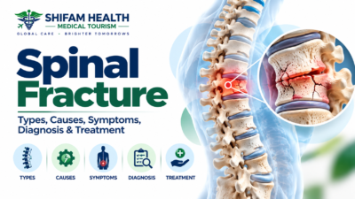
Spinal Fracture (2026): Types, Causes, Symptoms, Diagnosis & Treatment
Filters & Insights
Understand spinal fracture types, causes, emergency warning signs, diagnosis, and treatment from bracing to surgery including costs in India.

A spinal fracture is a break in one or more of the bones (vertebrae) that make up the spine, which can result from significant trauma, weakened bone from osteoporosis, or disease processes like cancer that damage bone from within.
Some spinal fractures are emergencies — particularly those involving spinal cord or nerve injury, or significant instability — while others, like many osteoporotic compression fractures, can often be managed without surgery.
A spinal fracture does not automatically mean the spinal cord has been damaged, and treatment depends on the fracture pattern, whether the spine is stable, neurological status, and the underlying cause — not on one imaging finding taken in isolation.
Table of Contents
ToggleWhat Is a Spinal Fracture?
A spinal fracture is a break in one or more vertebrae — the bones that stack to form the spine. Fractures can involve the main weight-bearing vertebral body at the front, the posterior elements (the bony arch, spinous process, and transverse processes at the back), or both, depending on the mechanism of injury. Common causes range from significant trauma (falls, motor vehicle collisions, sports injuries) to weakened bone from osteoporosis, which can fracture after relatively minor trauma, to pathological weakening of bone from cancer, infection, or other disease.
Some spinal fractures are genuine emergencies — particularly those involving spinal cord or nerve compromise, or significant mechanical instability — while others can often be managed without surgery.
Treatment depends on the specific fracture pattern, whether the spine remains mechanically stable, the patient’s neurological status, and the underlying cause of the fracture, evaluated together rather than any single factor in isolation.
A critical distinction to establish immediately: a bone fracture, spinal instability, spinal cord injury, and nerve-root injury are four different things, even though they’re related. A spinal fracture does not automatically mean the spinal cord has been damaged — many fractures occur with the neurological system completely intact.
Conversely, spinal cord injury can occasionally occur without an obvious fracture on imaging (particularly in certain injury patterns). Understanding which of these is actually present in a given case is central to appropriate treatment.
Spinal Anatomy in Plain Language
The spine is divided into the cervical spine (neck, 7 vertebrae), thoracic spine (mid-back, 12 vertebrae, supported by the rib cage), lumbar spine (lower back, 5 vertebrae), and sacrum (the fused bones at the base of the spine). Each vertebra has a vertebral body (the main weight-bearing block at the front) connected via pedicles to a bony arch formed by the laminae, with a spinous process projecting backward and transverse processes projecting to each side — together these posterior elements protect the spinal canal, which houses the spinal cord and the nerve roots that branch off at each level. Facet joints connect adjacent vertebrae and guide motion, intervertebral discs cushion the vertebral bodies, and ligaments provide overall structural stability.
The specific location and which anatomical structures are involved in a fracture directly affects stability, neurological risk, pain pattern, treatment approach, surgical planning (if needed), and expected recovery — this is why the same word, “fracture,” can describe injuries with vastly different implications depending on exactly what’s broken and where.
Types of Spinal Fractures
| Fracture Type | What Happens | Typical Clinical Significance |
|---|---|---|
| Compression/wedge fracture | Part of the vertebral body collapses, often at the front (anteriorly) | Common in osteoporosis and some trauma; often, though not always, stable |
| Burst fracture | The vertebral body fails in multiple directions, potentially involving the back wall and spinal canal | May threaten spinal stability or neural structures, depending on severity |
| Chance fracture | A horizontal splitting pattern through vertebral structures, classically from a flexion-distraction mechanism (e.g., a lap-belt injury) | Often associated with significant trauma and can accompany abdominal organ injury |
| Fracture-dislocation | Fracture combined with abnormal displacement/dislocation of vertebrae relative to each other | Often highly unstable and frequently neurologically significant |
| Transverse-process fracture | Fracture of a transverse process | Can indicate significant trauma but is often mechanically distinct from more unstable injury patterns |
| Spinous-process fracture | Fracture involving the spinous process | Generally assessed in the context of the overall injury pattern rather than in isolation |
Actual fracture classification requires imaging and specialist assessment — this table provides a general orientation, not a self-diagnosis tool.
Why Specialists Classify Fractures the Way They Do
Beyond basic fracture type, specialists assess additional factors including compression versus burst morphology, flexion-distraction mechanisms, translation (displacement) injuries, overall alignment, and — critically — whether the posterior ligamentous complex (the ligaments at the back of the spine that contribute significantly to stability) is intact or disrupted. Where clinically relevant, formal classification frameworks such as the AO Spine classification systems, the Thoracolumbar Injury Classification and Severity Score (TLICS), and subaxial cervical spine classification approaches are used by specialists to help standardize communication, estimate stability, and understand neurological risk. This article does not attempt to teach these scoring systems in detail, since they’re specialist tools intended for use alongside full clinical and imaging context — and importantly, no single classification score independently determines treatment on its own; it informs, rather than replaces, overall clinical judgment.
Traumatic vs. Osteoporotic vs. Pathological Fractures
This distinction is essential, since it fundamentally changes both the diagnostic workup and treatment approach.
Traumatic spinal fractures result from significant force — road traffic collisions, falls (particularly from height), sports injuries, workplace accidents, or other direct or high-energy impact — generally occurring in bone of normal strength.
Osteoporotic (fragility) fractures occur in bone weakened by osteoporosis, often after relatively minor trauma or, in some cases, no clearly identifiable trigger at all. Contributing factors include age-related bone loss, postmenopausal changes, and long-term corticosteroid exposure, among others.
Pathological fractures occur in bone weakened by an underlying disease process — most commonly metastatic cancer or multiple myeloma, but also certain benign bone lesions or infection. Pathological fractures often require a fundamentally different diagnostic and treatment pathway than either traumatic or straightforward osteoporotic fractures, since the underlying disease itself needs to be identified and addressed, not just the fracture.
Not every compression fracture in an older adult is automatically osteoporotic — this is a genuinely important point. Clinical features that should prompt consideration of a pathological cause include a known cancer history, unexplained weight loss, severe or worsening night pain, fracture at an unusual level, or fracture without clear osteoporosis risk factors.
Causes and Risk Factors
Trauma-related causes include high-speed collisions, falls from height, sports trauma, heavy direct impact, and other significant injury mechanisms. Bone-weakness-related causes include osteoporosis, osteopenia, long-term corticosteroid use, older age, postmenopausal bone loss, and other metabolic bone disorders.
Disease-related weakening includes cancer (primary bone tumors or metastatic disease), multiple myeloma, infection, and other structural bone disease. Additional relevant considerations include a previous fracture (which increases future fracture risk), reduced bone mineral density generally, frailty, certain medications, and conditions affecting balance or fall risk. Age alone does not cause spinal fracture — it’s a risk factor that interacts with bone quality, activity level, and fall risk, not a direct cause by itself.
How Spinal Fractures Cause Symptoms
Symptoms arise from several possible mechanisms: direct bone pain from the fracture itself, mechanical instability causing pain with movement, associated soft-tissue or ligament injury, nerve-root compression from bone fragments or resulting deformity, spinal canal narrowing, and, in more severe cases, spinal cord injury.
Symptom severity does not always indicate the full extent of structural injury — this is a genuinely important and sometimes counterintuitive point. A patient with a serious, potentially unstable fracture may initially have relatively limited symptoms, while another patient with a less structurally severe fracture may experience significant pain without any major neurological injury. This mismatch is exactly why proper clinical and imaging evaluation matters rather than judging severity from symptoms alone.
Symptoms
Common symptoms include sudden back or neck pain, localized spinal tenderness, pain that worsens with movement, muscle spasm, reduced mobility, difficulty standing or walking, and, when nerve structures are involved, radiating pain, numbness, tingling, or weakness.
For osteoporotic vertebral compression fractures specifically, presentation can differ somewhat: pain onset may be sudden or more gradual, and over time (particularly with multiple fractures), patients may notice height loss, increasing spinal curvature (kyphosis), reduced mobility, and, in some cases, chronic ongoing pain. Not every vertebral compression deformity seen on imaging represents an acute, new fracture — old, fully healed deformities can persist indefinitely on imaging, which is why symptom timing and, when needed, MRI (which can show marrow edema in genuinely acute fractures) matter for distinguishing new from old findings.
Neurological Symptoms
Neurological symptoms — weakness, numbness, tingling, loss of sensation, abnormal reflexes, difficulty walking, loss of coordination, or bowel/bladder dysfunction — can indicate involvement of the spinal cord, a specific nerve root, or, at the lower end of the spine, the cauda equina (a bundle of nerve roots). These represent distinct clinical situations: spinal cord injury affects the cord itself and can produce widespread effects below the level of injury; nerve-root injury typically produces symptoms in a specific, localized distribution; and cauda equina dysfunction classically involves bilateral leg symptoms, saddle numbness, and bladder/bowel dysfunction. Neurological symptoms of any kind may indicate a more urgent injury and should prompt specialist evaluation rather than being managed as routine back pain.
Emergency Red Flags
A suspected spinal fracture following significant trauma should always be medically assessed rather than managed at home. The following represent urgent or emergency warning signs:
- New or progressive weakness
- Loss of sensation, particularly involving the trunk or multiple limbs
- Difficulty walking
- Loss of bladder or bowel control
- Urinary retention
- Saddle/perineal numbness
- Severe neck or back pain following significant trauma
- Obvious spinal deformity
- Loss of consciousness or other major associated trauma
- Any other sign suggesting spinal cord compromise
A suspected spinal fracture, particularly after significant trauma, should generally be treated as a potentially serious injury until properly evaluated — this caution is appropriate even when initial symptoms seem manageable, given how symptom severity can understate actual structural injury, as discussed above.
Initial Response After Suspected Spinal Trauma
Safe, practical guidance following suspected spinal trauma: avoid unnecessary movement of the injured person, seek emergency medical assessment promptly, follow the instructions of emergency medical services once involved, and do not attempt forceful spinal manipulation or attempt to “put the spine back into place.” Avoid transporting a seriously injured person in an unsafe or improvised manner when professional emergency transport is available.
Emergency immobilization and handling should follow trained clinical/emergency protocols — this article does not provide detailed self-management or first-aid technique instructions beyond this general guidance, since improper handling by untrained bystanders can potentially worsen an unstable spinal injury.
How Is a Spinal Fracture Diagnosed?
Diagnosis integrates the mechanism of injury, clinical examination, neurological examination, imaging, bone health assessment where appropriate, and evaluation for underlying disease when clinically indicated. Clinicians assess pain, tenderness, spinal alignment, neurological status (strength, sensation, reflexes, gait), bowel/bladder function, and any associated injuries — trauma severe enough to fracture the spine often causes injuries elsewhere in the body as well, which need to be assessed concurrently.
X-Ray, CT, and MRI
| Imaging | Common Uses | Limitations |
|---|---|---|
| X-ray | Initial assessment of vertebral alignment, gross compression deformity | Limited soft-tissue detail; can’t reliably assess fracture age or spinal cord status |
| CT | Detailed bone morphology, spinal canal involvement, complex traumatic injury patterns, surgical planning | Limited direct visualization of the spinal cord and soft tissues; involves radiation |
| MRI | Spinal cord, nerve roots, ligaments, discs, other soft tissue, bone marrow (helpful for fracture acuity), occult fracture detection, neurological injury assessment | More limited availability/cost in some settings; not always necessary for straightforward cases |
MRI is particularly useful when neurological symptoms are present or when soft-tissue or spinal cord involvement needs direct evaluation — it provides information that X-ray and CT genuinely cannot. Bone density testing (DEXA/DXA) is relevant for evaluating and confirming osteoporosis after a suspected fragility fracture, supporting both diagnosis and subsequent treatment planning. Not every patient with a spinal fracture requires every imaging test — the specific combination needed depends on the clinical situation, injury mechanism, and initial findings.
Differential Diagnosis
Back or neck pain following trauma is not automatically a spinal fracture. Relevant alternative or coexisting diagnoses include muscle or ligament injury, disc injury, spinal cord injury without an obvious fracture on imaging (a recognized but less common pattern, particularly in certain populations), facet joint injury, rib fracture, hip or pelvic injury, kidney (renal) injury, vascular injury, spinal infection, and malignancy.
For an unexplained vertebral fracture — particularly one occurring without clear trauma or in an unexpected clinical context — clinicians may specifically investigate for osteoporosis, cancer, multiple myeloma, infection, or other metabolic bone conditions, rather than assuming a straightforward mechanical cause.
What Makes a Spinal Fracture Stable or Unstable?
This is one of the most important concepts in this entire article, because stability — more than fracture “type” alone — drives much of the treatment decision. Stability assessment considers fracture morphology, the number of structural spinal elements involved, the integrity of the posterior ligamentous complex, overall alignment, any translation (displacement) present, degree of deformity, neurological status, spinal canal compromise, and the patient’s overall condition.
This article deliberately does not present older, simplified rules (such as basic three-column models) as the singular modern standard — current specialist assessment considers multiple factors together rather than relying on one oversimplified framework, and this determination should be made by a spine specialist reviewing the actual imaging and clinical picture.
Stability directly influences whether treatment leans toward observation/conservative care, bracing, closed reduction, surgical stabilization, decompression, or some combination of these — which is why “is my fracture stable?” is often a more clinically useful question to ask your specialist than “what type of fracture do I have?” in isolation.
Non-Surgical Treatment
Non-operative treatment may be appropriate for many stable fractures without significant neurological deficit, and typically includes pain management, activity modification, appropriately guided mobilization, physical therapy, bracing in selected patients, ongoing monitoring, follow-up imaging where clinically indicated, and, for fragility fractures, osteoporosis treatment addressing the underlying bone weakness.
Prolonged bed rest can cause its own complications (deconditioning, blood clots, pressure injuries, further bone loss) and should not automatically be considered the default treatment — early, appropriately guided mobilization is generally favored once the clinical situation allows it. This article does not recommend a universal brace type or treatment duration, since these should be individualized to the specific fracture and patient.
Bracing
Spinal braces may be used to provide support and potentially limit painful movement during healing, though evidence and appropriate use differ between traumatic fractures and osteoporotic compression fractures — bracing decisions should be individualized rather than applied as a blanket rule. Limitations include skin irritation, muscle deconditioning with prolonged use, and variable patient compliance. Braces do not always accelerate bone healing — their primary role is generally symptomatic support and, in some traumatic fracture contexts, limiting harmful movement during the healing period.
Pain Management
Relevant medication categories include acetaminophen/paracetamol, NSAIDs where appropriate, short-term stronger analgesia in selected cases, muscle-relaxing medication in selected situations, and, for osteoporotic fractures, medications addressing the underlying bone disease. Relevant safety considerations include kidney disease, gastrointestinal disease, cardiovascular disease, sedation risk (particularly relevant to fall risk in older adults), and drug interactions. This article does not provide individualized dosing — medication selection should come from your treating physician based on your specific health profile.
Osteoporotic Vertebral Compression Fractures
Osteoporosis reduces bone strength throughout the skeleton, including the vertebrae, making them prone to compression under normal loads that healthy bone would withstand without injury. The typical fracture pattern involves anterior (front) height loss, producing a wedge-shaped deformity. Distinguishing acute from chronic fractures matters for treatment decisions and relies on symptom timing and, when needed, MRI evidence of bone marrow edema (present in genuinely recent fractures, generally absent in old, healed deformities).
Management centers on pain control, appropriately guided mobilization (rather than prolonged rest), and — critically — identifying and treating the underlying osteoporosis: bone density evaluation, bone-strengthening (osteoporosis) therapy selected based on individual fracture risk, fall-prevention strategies, and attention to preventing future fractures. Treating the fracture without addressing the underlying osteoporosis leaves the real problem — ongoing fracture risk — unresolved.
Vertebroplasty and Kyphoplasty
Vertebroplasty involves injecting bone cement directly into a fractured vertebral body to stabilize it and potentially relieve pain. Balloon kyphoplasty first creates a cavity using balloon inflation before cement injection, potentially allowing some height restoration. Both may be considered for selected painful vertebral compression fractures, most commonly osteoporotic.
This evidence deserves honest treatment rather than a confident sales pitch. Randomized trials comparing these procedures to sham (placebo) interventions have produced genuinely mixed results regarding pain benefit for vertebral compression fractures generally — this remains a debated area in the spine literature. Potential complications include cement leakage, infection, and, rarely, more serious complications from cement migration; there’s also ongoing research interest in whether these procedures affect adjacent-vertebra fracture risk, though this isn’t conclusively settled. Neither procedure should be presented as routine, default treatment for every compression fracture, and they are clearly distinct from surgical stabilization used for unstable traumatic fractures — these serve different purposes and are used in different clinical contexts.
When Is Surgery Considered?
Surgery may be considered depending on instability, significant or progressive deformity, neurological deficit, spinal cord or nerve compression, failure of appropriate conservative treatment, fracture-dislocation, severe spinal canal compromise in the appropriate clinical context, or a pathological fracture requiring stabilization alongside treatment of the underlying disease. Imaging findings alone do not automatically determine the need for surgery — treatment depends on the entire clinical picture, integrating symptoms, neurological status, stability, and imaging together.
Spinal Decompression
Decompression refers to relieving pressure on the spinal cord or nerve roots, typically by removing bone fragments, disc material, or other tissue compressing these structures. It is performed when neural compression is clinically significant — canal narrowing visible on imaging does not automatically mean decompression is required, since some degree of canal narrowing can be present without causing clinically significant neurological compromise. Decompression and stabilization address different problems (relieving pressure vs. providing mechanical stability) and are sometimes, though not always, performed together.
Spinal Fusion and Fixation
Fixation techniques — pedicle screws, rods, plates, and, where relevant, interbody techniques — aim to restore or maintain alignment, stabilize unstable spinal segments, protect neurological structures, and facilitate healing, generally combined with fusion to achieve long-term stability at the treated level. Implants and fusion are not universally necessary for every spinal fracture — many stable fractures heal without any surgical hardware at all.
Surgical Approaches
Depending on the fracture pattern, location, and other factors, surgical approaches can include anterior (front), posterior (back), or combined approaches, along with minimally invasive fixation techniques or, for more extensive injury, open reconstruction. Approach selection depends on fracture pattern, location, degree of stability, neurological status, bone quality, degree of deformity, associated injuries, and surgeon/institutional expertise and experience with a given technique. No single approach is universally superior — this decision is individualized to the specific injury and patient.
Cervical Spine Fractures
The cervical spine deserves particular attention because injuries here can be especially high-risk, given the close proximity to the brainstem and upper spinal cord and the region’s relatively greater mobility compared with other spinal segments.
This category includes upper cervical injuries (around the atlas/C1 and axis/C2, including specific patterns like odontoid fractures) and subaxial cervical fractures (C3 and below), each with distinct injury patterns, stability considerations, and treatment approaches.
Cervical fracture management should not be generalized from lumbar fracture principles — the anatomy, biomechanics, and neurological stakes differ meaningfully. For older adults specifically, odontoid fractures deserve particular attention, since they can occur after relatively minor trauma in the context of age-related bone and ligamentous changes, and management decisions in this population require careful consideration of both the fracture pattern and overall bone health and frailty. Treatment ranges from bracing/immobilization for appropriately selected stable injuries to surgical stabilization for unstable patterns or those with neurological compromise.
Thoracic and Thoracolumbar Fractures
Common mechanisms include falls and high-energy trauma, producing compression fractures, burst fractures, flexion-distraction injuries, and fracture-dislocations depending on the specific forces involved. The thoracolumbar junction (roughly T11–L2) is clinically important because it represents a transition zone between the more rigid, rib-supported thoracic spine and the more mobile lumbar spine, making it a common site for these injury patterns. Neurological risk and stability assessment follow the same general principles discussed above, and treatment ranges from conservative management for stable, neurologically intact injuries to surgical stabilization for unstable or neurologically significant patterns.
Lumbar Fractures
Lumbar spine fractures can involve the vertebral body, burst-type patterns, or posterior elements, with neurological complications depending on the degree of canal involvement and, at lower lumbar levels, potential cauda equina involvement. Stability assessment and treatment principles follow the same general framework outlined above. Not all lumbar fractures require surgery — many stable, neurologically intact lumbar fractures are managed successfully without operative treatment.
Pathological Spinal Fractures
Metastatic cancer, multiple myeloma, infection, and other bone-weakening conditions can cause pathological vertebral fractures. Because the underlying disease process, not just the mechanical fracture, needs to be addressed, evaluation typically requires oncology involvement, MRI/CT for detailed characterization, biopsy in selected cases to confirm the diagnosis, and treatment that may include radiation therapy in appropriate patients, surgical stabilization, systemic disease-specific treatment, and pain management coordinated across specialties.
Not every compression fracture in an older person should automatically be labeled osteoporotic without appropriate consideration of these alternative causes, particularly when clinical features raise concern (cancer history, unexplained weight loss, atypical presentation).
Spinal Fracture With Spinal Cord Injury
Spinal cord injury refers to damage to the spinal cord itself, which can be broadly categorized (at a general level) as complete (no function preserved below the level of injury) or incomplete (some function preserved), a distinction with significant implications for prognosis, though the full clinical picture is considerably more nuanced than this basic categorization suggests.
Neurological status is critical to management decisions, and this represents a genuine emergency requiring prompt specialist assessment, with decompression and/or stabilization considered based on the specific injury pattern, followed by intensive rehabilitation.
Fracture Healing: Not the Same as Pain Relief or Neurological Recovery
Bone healing depends on fracture stability, local blood supply, overall bone quality, and factors including smoking (which impairs healing), nutrition, diabetes and other relevant comorbidities, and certain medications. It’s important to distinguish three genuinely different things that are often conflated: fracture healing (the bone itself mending), pain improvement (which can occur even before complete healing, and can also persist after healing is technically complete), and neurological recovery (a separate process entirely, relevant only when nerve or spinal cord injury occurred, and following its own timeline independent of bone healing).
Recovery and Rehabilitation
It generally involves pain control, safe and appropriately guided mobilization, physiotherapy, strengthening, balance training, gait rehabilitation, and, where spinal cord or nerve injury occurred, dedicated neurological rehabilitation. Recovery varies substantially according to fracture type, patient age, bone quality, whether neurological injury occurred, whether surgery was performed, any associated injuries, and general health — there is no single universal recovery timeline that can be honestly applied across this range of situations.
Complications
Potential complications include neurological deterioration, spinal cord or nerve-root injury (if not already present, or worsening if present), spinal deformity, delayed healing or nonunion in relevant cases, chronic pain, height loss, kyphosis, reduced mobility, pressure injuries from prolonged immobility, blood clots, infection, hardware-related complications and failure (in surgically treated cases), adjacent-segment problems, and refracture. Complication risk depends on the specific fracture type, individual patient factors, the treatment approach chosen, and any associated injuries — this should be discussed specifically with your treating team rather than treated as a generic, uniform risk profile.
Special Populations
| Population | Key Considerations |
|---|---|
| Older adults | Higher osteoporosis prevalence, frailty, elevated fall risk, coexisting medical conditions, delirium risk during hospitalization, and rehabilitation needs that account for these factors |
| Patients with osteoporosis | Bone-health evaluation, anti-osteoporosis treatment, fall prevention, and secondary fracture prevention as core components of care |
| Children and adolescents | Different spinal anatomy and biomechanics, growth-related considerations, distinct fracture patterns from adults, and the need for pediatric spine specialist assessment rather than adult-based management |
| Athletes | Mechanism-specific evaluation, individualized rehabilitation, and appropriately cautious return-to-sport timing to avoid premature return before adequate healing and functional recovery |
| Cancer patients | Pathological fracture considerations, careful stability assessment, neurological risk evaluation, and close oncology coordination |
| Patients with spinal cord injury | Dedicated neurological rehabilitation, bladder/bowel management, mobility support, and long-term multidisciplinary follow-up |
Prevention
Preventing traumatic fractures involves measures like seat-belt use, general fall prevention, workplace safety practices, and appropriate safety measures in sports. Preventing fragility fractures involves osteoporosis screening and assessment where appropriate, bone-strengthening therapy when indicated, adequate nutrition, regular physical activity, fall-prevention strategies, and medication review where relevant (some medications affect bone density or fall risk). These measures reduce risk; they cannot eliminate fracture risk entirely — this should be communicated honestly rather than implying guaranteed prevention.
Travel After a Spinal Fracture
For patients — particularly international patients — considering travel after a spinal fracture or spinal surgery, relevant factors include whether the patient is medically stable for travel, current pain control, neurological status, mobility level, how recent any surgery was, blood clot risk (particularly relevant for long flights), the need for travel assistance, appropriate medical documentation, travel insurance considerations, specific airline requirements or restrictions, wheelchair or other transport arrangements, and follow-up care planning.
Spinal Fracture Treatment in India (2026)
Appropriately equipped centers in India provide orthopedic spine surgeons, neurosurgeons, radiologists, and rehabilitation specialists capable of evaluating and treating the range of spinal fracture types discussed in this article. The general pathway involves medical records submission, imaging review, specialist assessment, diagnosis and fracture classification, discussion of treatment options, hospital and specialist coordination, cost estimation, medical visa documentation where applicable, travel planning, in-person hospital evaluation, treatment, rehabilitation, follow-up, and return-home planning.
Not every spinal fracture should be treated in India — this is a decision that depends on the specific injury, urgency, and individual circumstances, and this article does not claim India is universally appropriate or superior for every case. This article also does not fabricate hospital rankings, surgeon rankings, success rates, specific technology claims, accreditation details, or treatment volumes for any specific center.
Spinal Fracture Treatment Cost in India
Reusing verified cost data relevant to spinal fracture treatment components researched elsewhere in this content area: vertebroplasty and kyphoplasty for compression fractures show published international-patient estimates in the range of roughly $1,300–$10,500 USD, with substantial variance across sources reflecting hospital tier and what’s included in a given quote. Spinal fusion/fixation surgery for more complex or unstable fractures shows published estimates in the range of roughly $4,500–$10,500 USD, though this can rise significantly for multilevel injuries, complex reconstruction, or cases requiring ICU care.
Complex trauma cases — particularly fracture-dislocation, multilevel injury, or fractures with spinal cord injury requiring intensive care — sit above these general ranges and cannot be responsibly estimated with a fixed published number, since cost in these situations depends heavily on injury severity, length of ICU stay, complications, and the specific reconstruction required. Relevant cost categories to expect as separately itemized include consultation, X-ray/CT/MRI, hospital admission, the specific treatment approach (conservative care, bracing, vertebral augmentation, fixation, decompression, or fusion), rehabilitation, and follow-up.
These figures should be treated as reference ranges, not firm quotes — an itemized, case-specific estimate from the treating hospital, based on actual imaging and diagnosis, is necessary for an accurate figure, and costs are never fabricated in this article.
International Patient Journey
The practical journey includes initial inquiry, medical record collection, imaging transfer, specialist opinion, treatment planning, cost estimation, hospital coordination, medical visa arrangements, travel, arrival, in-person consultation, treatment, hospital recovery, rehabilitation, follow-up, and return-home planning.
Urgent spinal trauma should receive immediate local emergency care rather than waiting for international travel arrangements to be completed — this is not a situation where medical tourism planning should take priority over local emergency evaluation and stabilization.
How Shifam Health Can Help
Shifam Health is a healthcare coordination and international patient support service — not the treating physician, spine surgeon, hospital, diagnostic center, or emergency service, and does not diagnose spinal fractures, independently interpret imaging as a treating clinician, perform surgery, or replace emergency services.
Shifam Health may assist with organizing medical records, coordinating specialist review, communicating with hospitals, obtaining treatment estimates, medical visa documentation support, travel and accommodation coordination, local patient support, and follow-up coordination. We cannot guarantee treatment outcomes, exact costs, exact recovery timelines, hospital acceptance, or visa approval. For acute spinal trauma, emergency medical care always takes priority over international treatment coordination.
International patients can share their medical records and imaging with Shifam Health to help coordinate an appropriate specialist review and understand potential treatment options in India.
When Should You Seek a Second Opinion for a Spinal Fracture?
A second opinion can be particularly valuable when surgery has been recommended, when fracture stability is genuinely unclear, when multiple different treatment options have been proposed, when neurological symptoms are present, when complex spinal reconstruction is being recommended, when a pathological fracture is suspected, when vertebral augmentation is being considered, or when you’re weighing treatment abroad against local options.
Useful records to bring include CT images, MRI images, X-rays, radiology reports, neurological examination findings, a description of the injury mechanism, any previous treatment received, bone-density results if available, cancer history where relevant, and a current medication list.
Frequently Asked Questions
What is a spinal fracture?
A break in one or more vertebrae caused by trauma, osteoporosis, or disease.
What are the symptoms?
Common symptoms include sudden back or neck pain, tenderness, muscle spasm, reduced mobility, and sometimes numbness or weakness.
Can a spinal fracture cause paralysis?
Severe fractures can damage the spinal cord and cause paralysis, although most fractures do not.
How is a spinal fracture diagnosed?
Diagnosis involves clinical and neurological examination with X-rays, CT, and/or MRI depending on the injury.
Can a spinal fracture heal without surgery?
Yes. Many stable fractures without significant neurological problems can be treated conservatively.
What is a compression fracture?
A fracture involving collapse of part of a vertebral body, commonly associated with osteoporosis.
What is a burst fracture?
A more severe vertebral fracture in which the vertebral body breaks in multiple directions and may affect the spinal canal.
When is spinal surgery necessary?
Surgery may be needed for unstable fractures, significant deformity, neurological compression, or failure of appropriate conservative treatment.
What are vertebroplasty and kyphoplasty?
Both are minimally invasive procedures that inject bone cement into a fractured vertebra. Kyphoplasty uses a balloon before cement injection.
Can cancer cause spinal fractures?
Yes. Cancer affecting the spine can weaken vertebrae and cause pathological fractures.
How long does a spinal fracture take to heal?
Healing varies significantly depending on the fracture type, treatment, bone health, and individual recovery.
Can spinal fractures happen again?
Yes, particularly when osteoporosis or other underlying risk factors remain untreated.
Final Takeaway
A spinal fracture is not one uniform injury — it ranges from a stable, minor compression fracture that heals with conservative care, to an unstable fracture-dislocation with spinal cord injury requiring emergency surgery, to a pathological fracture signaling an underlying disease that needs its own dedicated treatment.
A fracture does not automatically mean the spinal cord has been damaged, and symptom severity doesn’t always reflect the true extent of structural injury — which is why proper clinical and imaging evaluation matters more than assumptions in either direction. Stability, not fracture type alone, is often the central factor determining whether conservative management, bracing, or surgery is appropriate.
New weakness, numbness, bladder or bowel dysfunction, or other neurological symptoms after a spinal injury deserve urgent medical assessment, not a wait-and-see approach.
International patients can share their medical records and imaging with Shifam Health to help coordinate an appropriate specialist review and understand potential treatment options in India.
This article is educational and does not replace professional medical assessment. Suspected spinal fracture after significant trauma requires appropriate medical evaluation. Neurological symptoms may require emergency care. Treatment depends on the individual fracture and patient. International patients should not delay emergency treatment while arranging travel.


Popular Posts From Last Week
- September 24, 2026
- shifamhealth
Osteopenia means bone mineral density lower than the young-adult reference range but not low enough to meet the…
- September 25, 2026
- shifamhealth
Spina bifida is a group of neural tube defects caused by incomplete closure or development of the neural…
- September 26, 2026
- shifamhealth
Skeletal dysplasia is not one disease — it’s an umbrella term for a large, genetically diverse group of…
- September 24, 2026
- shifamhealth
Osteoarthritis is a whole-joint disorder not simply cartilage “wearing out” involving changes to cartilage, the underlying bone, the…
- September 28, 2026
- shifamhealth
“Slipped disc,” “herniated disc,” “prolapsed disc” — these terms get used almost interchangeably by patients, and the imagery…
- September 29, 2026
- shifamhealth
A spinal fracture is a break in one or more of the bones (vertebrae) that make up the…
- September 27, 2026
- shifamhealth
Last reviewed: 27-Sept-26 · General information for patients from Eritrea planning treatment in India. Not legal or immigration…