




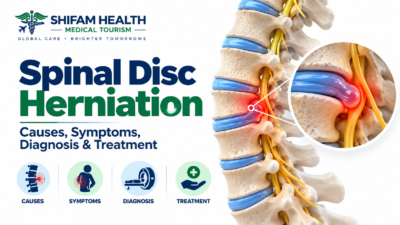
Spinal Disc Herniation (2026): Causes, Symptoms, Diagnosis & Treatment
Filters & Insights
Written By :
Medically Reviewed By :

Shifam Health Medical Team
Shifam Health - Medical Reviews Department
10+ Years Of Experience
A herniated disc can compress nearby nerves, causing pain, numbness, or weakness. Learn real causes, when surgery is needed & what usually helps most.
📅
Last updated: October 1, 2026

Spinal disc herniation occurs when the soft, gel-like center of an intervertebral disc pushes through a weakened or torn area of the disc’s tougher outer layer. This displaced material can sometimes press on or irritate a nearby spinal nerve, potentially causing pain, numbness, tingling, or weakness that may radiate along the nerve’s path — though many disc herniations cause no symptoms at all, and many cases that do cause symptoms improve without surgery over weeks to months.
The everyday term “slipped disc” is a bit misleading — the disc doesn’t actually slip out of place like a puck sliding across ice. What happens is more specific: the disc’s outer ring develops a tear or weak point, and inner disc material pushes outward through it, sometimes reaching far enough to contact a nearby nerve structure.
A principle worth holding onto throughout this entire article: an abnormal-looking disc on an MRI is not automatically the cause of a person’s pain. Studies of people without any back or leg symptoms have found disc bulges, protrusions, and even some herniations on imaging at meaningful rates, increasing with age. This is why diagnosis (covered in detail later) requires connecting imaging findings to your actual symptoms and examination — not simply reading an MRI report in isolation.
Table of Contents
ToggleAnatomy of the Spinal Disc
Understanding disc herniation requires understanding a few key structures:
- Vertebral bodies — the stacked bones forming the spinal column
- Intervertebral discs — cushioning structures between each pair of vertebrae, acting as shock absorbers and allowing spinal movement
- Nucleus pulposus — the soft, gel-like center of the disc
- Annulus fibrosus — the tougher, fibrous outer ring surrounding the nucleus pulposus, which can develop tears or weak points over time
- Vertebral endplates — the cartilage layers connecting the disc to the vertebral bone above and below
- Spinal canal — the central channel running through the vertebrae, housing the spinal cord (in the neck and upper back) and the cauda equina (the bundle of nerve roots continuing downward in the lower back)
- Neural foramina — the openings on each side of the spine where individual nerve roots exit toward the arms or legs
- Nerve roots — the individual nerves branching off the spinal cord/cauda equina, each supplying sensation and motor function to a specific, generally predictable area
Why herniation at different spinal levels causes symptoms in different body parts: each nerve root exiting the spine supplies a specific, largely predictable pattern of sensation and muscle function. A lumbar disc herniation can therefore cause symptoms in the lower back, buttock, leg, or foot, depending on which nerve root is affected; a cervical disc herniation can cause symptoms in the neck, shoulder, arm, or hand, depending on which cervical nerve root is involved. This is genuinely why “where it hurts” gives a clinician real diagnostic information about which spinal level and nerve root may be involved — though, as discussed later, real patients don’t always follow textbook patterns exactly.
Types and Terminology
Disc problems exist on a spectrum of severity and morphology — degeneration, bulge, protrusion, extrusion, and sequestration describe genuinely different structural findings, and the terminology matters for understanding your specific imaging report, though it should not be treated as automatically dictating treatment.
| Term | Meaning | Clinical Relevance |
|---|---|---|
| Disc degeneration | Structural changes within the disc (dehydration, height loss, wear) | May or may not cause symptoms; extremely common with age |
| Disc bulge | Broad, generalized extension of disc tissue beyond its normal margin | A common imaging finding, frequently seen even in people without symptoms |
| Disc protrusion | Focal (localized) displacement of disc material with a specific, contained morphology | May or may not compress a nerve, depending on location and size |
| Disc extrusion | Disc material extends beyond the disc space, with the displaced material having a narrower base/neck than the material itself | More likely to be associated with nerve compression than a bulge, though this still depends on location |
| Sequestered disc (disc sequestration) | A fragment of disc material has completely separated from the parent disc | Can migrate within the spinal canal, sometimes affecting a nerve root at a slightly different level than the disc space itself |
| Herniated disc | The general clinical term for displacement of disc material beyond its normal boundary (encompassing protrusion, extrusion, and sequestration) | May produce radiculopathy or other symptoms, depending on the specific case |
Avoid the trap of assuming that a more “severe-sounding” morphological term automatically determines treatment. An extrusion isn’t automatically treated more aggressively than a protrusion simply because of the terminology — actual nerve involvement, symptom severity, and clinical correlation matter more than the morphological label alone.
Cervical, Thoracic, and Lumbar Herniation
Disc herniation can occur at any spinal level, but lumbar herniation is the most common, cervical herniation is also frequent, and thoracic herniation is comparatively rare — each location has a genuinely different typical symptom pattern and, for the neck specifically, a distinct additional risk related to spinal cord involvement.
Lumbar disc herniation
The most common location, typically producing:
- Low back pain
- Buttock pain
- Leg pain, often described as sciatica when it follows the sciatic nerve distribution
- Numbness
- Tingling
- Weakness in specific muscle groups, depending on the nerve root involved
Cervical disc herniation
Typically produces:
- Neck pain
- Arm pain (cervical radiculopathy)
- Hand or finger numbness/tingling
- Weakness in specific arm or hand muscle groups
A distinction that genuinely matters and shouldn’t be glossed over: cervical radiculopathy vs. cervical myelopathy.
| Feature | Cervical Radiculopathy | Cervical Myelopathy |
|---|---|---|
| What’s affected | A specific nerve root | The spinal cord itself |
| Typical symptoms | Arm pain, numbness, weakness following a specific nerve root pattern | Broader symptoms — hand clumsiness, gait/balance difficulty, weakness in both legs, sometimes bladder/bowel changes |
| Urgency | Generally evaluated on a routine-to-prompt basis depending on severity | Can represent a more significant, sometimes progressive condition warranting more urgent specialist evaluation |
This distinction matters because myelopathy, when present, is generally treated with more urgency than radiculopathy alone — it reflects potential spinal cord involvement rather than an isolated nerve root, and progressive myelopathy symptoms deserve prompt specialist assessment.
Thoracic disc herniation
Genuinely less common than cervical or lumbar herniation, related to the thoracic spine’s greater structural stability from the rib cage limiting motion and stress at these levels. Symptoms can differ from lumbar or cervical disease and may include chest wall or band-like pain, and, in more significant cases, findings suggestive of spinal cord involvement given the thoracic spinal canal’s specific anatomy.
Not every episode of thoracic or chest-area pain is caused by a disc problem — this symptom location has a particularly broad differential diagnosis (including cardiac, pulmonary, and gastrointestinal causes) that deserves appropriate consideration, not an assumption of thoracic disc disease.
Causes and Risk Factors
Disc herniation is multifactorial — age-related disc degeneration is the underlying substrate in most cases, with mechanical stress, genetic susceptibility, and lifestyle factors contributing to when and whether symptomatic herniation occurs. It’s important to distinguish a risk factor, a direct cause, and a triggering event, since these are genuinely different concepts often conflated in popular health content.
| Factor Category | Examples |
|---|---|
| Age-related change | Progressive disc dehydration and structural weakening over years — the underlying substrate for most degenerative disc herniation |
| Mechanical/biomechanical | Repetitive loading, heavy lifting, twisting motions, sudden mechanical stress |
| Occupational | Jobs involving repetitive heavy lifting or awkward postures show some association with increased risk |
| Lifestyle | Smoking (associated with accelerated disc degeneration), and, where evidence supports it, obesity |
| Genetic | Family history and genetic susceptibility play a meaningful role in disc degeneration risk |
| Prior disc problems | A history of previous disc herniation is associated with increased risk of future herniation, including at adjacent levels |
| Trauma | Significant trauma can precipitate herniation, particularly relevant when acute onset follows a specific injury |
A risk factor is not the same as a direct cause, and a direct cause is not the same as a triggering event — this distinction matters for how patients understand their own situation. Someone who develops symptoms immediately after lifting a heavy object often already had underlying disc degeneration; the lifting incident may have been the trigger that caused a previously asymptomatic or minimally symptomatic disc to become acutely symptomatic, not necessarily the sole “cause” of the underlying disc changes themselves.
What this article will not claim: that poor posture, sitting for long periods, a single lifting incident, or any one isolated movement universally causes disc herniation. These are commonly repeated claims in popular health content that oversimplify a genuinely multifactorial process.
Why a Herniated Disc Causes Pain
Pain from a herniated disc reflects a combination of mechanical nerve-root compression and chemical/inflammatory irritation from the displaced disc material itself — and, importantly, the size of a herniation on MRI does not reliably predict symptom severity, which is one of the more counterintuitive but genuinely important facts about this condition.
Two overlapping mechanisms contribute:
- Mechanical compression — physical pressure on a nerve root or, in the cervical/thoracic spine, potentially the spinal cord
- Chemical/inflammatory irritation — displaced disc material can trigger a local inflammatory response, releasing chemical mediators that sensitize and irritate the adjacent nerve tissue, independent of purely mechanical pressure
Why herniation size doesn’t reliably predict symptom severity: a relatively small herniation positioned in a tight anatomical space, or affecting a nerve root that’s particularly sensitive to inflammation, can cause significant symptoms — while a larger herniation in a roomier area of the spinal canal, or one that’s been present long enough for some adaptation to occur, may produce comparatively few symptoms. This is genuinely one of the reasons imaging alone cannot be the sole basis for treatment decisions — the clinical picture, not the MRI measurement, is what actually matters most.
Symptoms
Common symptoms include localized back or neck pain, radiating limb pain (sciatica in the lumbar spine), numbness, tingling, burning or electric-type sensations, and weakness — but symptom patterns vary between individuals, and these descriptions represent typical patterns, not diagnostic certainty.
Common symptoms:
- Back or neck pain, localized to the affected spinal area
- Radiating limb pain — sciatica (lumbar) or radiating arm pain (cervical)
- Numbness
- Tingling
- Burning or electric shock-type pain
- Weakness in specific muscle groups
- Symptoms aggravated by certain movements or positions (commonly sitting, bending, coughing, or sneezing for lumbar disease)
- Reduced activity tolerance
Radiculopathy, explained
Radiculopathy refers to dysfunction of a specific nerve root, which can result from disc herniation among other causes. It explains why pain can travel a considerable distance from the actual spinal level involved — the nerve root’s territory extends along a specific, largely predictable path into the limb.
- Sensory symptoms: numbness, tingling, or altered sensation in the specific area supplied by the affected nerve root
- Motor symptoms: weakness in the specific muscles supplied by that nerve root
- Reflex changes: certain reflexes tested during a physical exam can be reduced or absent when a specific nerve root is affected
Symptoms by spinal level
| Level | Representative Pattern (General, Not Universal) |
|---|---|
| L4 nerve root | Pain/numbness toward the inner shin; weakness affecting the muscle used to lift the foot upward at the ankle; reduced knee reflex |
| L5 nerve root | Pain/numbness toward the top of the foot and big toe; weakness affecting big toe/foot extension |
| S1 nerve root | Pain/numbness toward the outer foot and sole; weakness affecting the calf/push-off strength; reduced ankle reflex |
| C6 nerve root | Pain/numbness toward the thumb side of the hand; weakness affecting the biceps/wrist extension |
| C7 nerve root | Pain/numbness toward the middle finger; weakness affecting the triceps |
Real patients frequently do not follow these textbook patterns exactly — overlap between nerve root territories, individual anatomical variation, and multi-level disease can all blur these classic patterns. This table is a general reference, not a self-diagnosis tool.
Red Flags and Medical Emergencies
This section is deliberately placed prominently and should not be skimmed.
Cauda equina syndrome
Cauda equina syndrome is a rare but genuine surgical emergency involving compression of the bundle of nerve roots in the lower spinal canal, most classically presenting with new bladder or bowel dysfunction and saddle (groin/perineal) numbness. It requires urgent evaluation because delayed treatment can result in permanent neurological damage — but ordinary back pain, even with sciatica, does not mean a person has cauda equina syndrome.
Symptoms requiring urgent, same-day medical assessment:
- New urinary retention or new difficulty urinating
- New loss of bladder control
- New bowel incontinence or loss of bowel control
- Saddle anesthesia — numbness in the groin, inner thighs, or perineal area
- Severe or rapidly progressive bilateral (both-sided) leg weakness
- Significant bilateral neurological symptoms
- Rapidly worsening neurological deficits generally
This is genuinely one of the few true spine emergencies — cauda equina syndrome is uncommon, but when these specific symptoms are present, urgent evaluation matters because the window for effective decompression before permanent damage can occur is time-sensitive.
Other important red flags for serious alternative causes
These don’t necessarily indicate an emergency on the same level as cauda equina syndrome, but warrant prompt (not necessarily same-day) evaluation and shouldn’t be assumed to represent routine disc herniation without appropriate assessment:
- Significant trauma preceding new spinal symptoms
- Fever combined with significant spinal pain (raising concern for infection)
- Known cancer history with new spinal symptoms
- Immunosuppression combined with new severe spinal symptoms
- Severe, unexplained constitutional symptoms (unexplained weight loss, night sweats) alongside spinal pain
No individual red flag has perfect predictive value on its own — these are reasons to seek prompt medical evaluation, not certainties of a serious underlying condition. The large majority of people with back or neck pain, including those with sciatica or arm radiculopathy, do not have any of these serious underlying causes.
When to See a Doctor
Medical evaluation is generally appropriate for:
- Persistent symptoms not improving with reasonable self-management
- Significant radiating pain
- Numbness
- Weakness
- Recurrent episodes
- Symptoms interfering with sleep, walking, work, or daily activities
- Progressive (worsening, not just persistent) neurological symptoms
- Any of the emergency red flags described above, which warrant urgent rather than routine care
This guidance is intended to help you decide whether and how urgently to seek care — it is not a substitute for that evaluation itself.
Diagnosis
Diagnosis combines medical history, symptom pattern, a focused neurological examination, and functional assessment — imaging is used selectively to confirm or clarify the clinical picture, not as the automatic first step for every episode of back or neck pain.
What a thorough evaluation typically includes:
- Medical history — symptom onset, duration, pattern, aggravating/relieving factors, prior episodes, red-flag screening
- Neurological examination — assessing strength, sensation, and reflexes in a pattern-specific way
- Functional assessment — gait, range of motion, and functional limitations
- Provocative testing — such as the straight-leg raise test (for lumbar disease) or other relevant maneuvers, which can support but not definitively confirm a diagnosis on their own
- Imaging when clinically indicated — discussed in detail below
MRI is not automatically required for every episode of back or neck pain. For many patients with a typical presentation and no red flags, a reasonable trial of conservative management is appropriate before imaging is pursued — imaging is generally reserved for persistent symptoms, red flags, planned intervention, or genuine diagnostic uncertainty.
MRI, CT, X-Ray, and EMG/NCS
| Test | What It Shows | When It’s Useful | Key Limitation |
|---|---|---|---|
| MRI | Disc morphology, nerve-root compression, spinal canal and neural foramina, soft tissue and neural structures in detail | Persistent or significant symptoms, red flags, or when intervention is being planned | Findings must correlate with symptoms and exam — imaging abnormalities are common even in people without pain |
| CT | Detailed bony anatomy | Particularly useful when detailed bone detail is needed, or when MRI is unsuitable (e.g., certain implanted devices) | Less detailed soft-tissue/neural visualization than MRI |
| X-ray | Bone alignment, vertebral height, degenerative changes, gross instability | Can still be useful for assessing alignment or ruling out certain bony pathology | Cannot directly visualize discs or nerve compression |
| EMG/NCS (electromyography/nerve conduction studies) | Electrical nerve and muscle function | Selected situations — clarifying nerve dysfunction, distinguishing radiculopathy from a separate peripheral neuropathy, or when the clinical picture and imaging don’t clearly correspond | Not needed for most straightforward cases; doesn’t replace clinical correlation |
No single test is automatically required for every patient — the appropriate combination of testing depends on the individual clinical picture, not a fixed protocol applied uniformly.
Differential Diagnosis
| Condition | Why It Can Resemble Disc Herniation | Key Distinguishing Context |
|---|---|---|
| Spinal stenosis | Can cause similar leg or arm symptoms | Often more posture/walking-dependent (neurogenic claudication pattern); frequently a different, often older, patient population |
| Facet joint disorders | Can cause localized and sometimes referred pain | Typically lacks the specific dermatomal (nerve-territory) pattern of true radiculopathy |
| Muscle-related pain | Common cause of back/neck pain with possible referred discomfort | Generally lacks true neurological findings (weakness, reflex changes, dermatomal numbness) |
| Sacroiliac joint disorders | Can cause buttock and leg pain resembling sciatica | Pain pattern and specific provocative tests differ from a nerve-root pattern |
| Peripheral nerve entrapment (e.g., piriformis-related, carpal tunnel for arm symptoms) | Symptoms can overlap with radiculopathy in the affected limb | Different anatomic location of compression, confirmed through targeted examination |
| Hip pathology | Can cause buttock, groin, or thigh pain sometimes confused with lumbar radiculopathy | Hip-specific examination findings, different pain triggers (hip rotation/loading) |
| Peripheral neuropathy | Can cause numbness/tingling in the limbs | Often bilateral, symmetric, and not following a single specific nerve root pattern |
| Vascular conditions | Can cause leg symptoms with exertion | Pulse findings and different relief pattern (not specifically postural, unlike neurogenic patterns) |
| Spinal infection | Can cause back pain with neurological symptoms | Fever, systemic illness, or specific risk factors are important context |
| Malignancy | Can cause spinal pain, sometimes with neurological findings | Cancer history, unexplained weight loss, or pain not following a typical mechanical pattern raise this concern |
| Vertebral fracture | Can cause acute back pain with possible neurological involvement | Trauma history, osteoporosis risk factors, or specific pain characteristics |
| Inflammatory disorders (e.g., inflammatory spondyloarthritis) | Can cause back pain, sometimes with morning stiffness | A different overall pattern — typically prolonged morning stiffness improving with activity, rather than the mechanical, position-dependent pattern more typical of disc-related pain |
Natural History and Prognosis
Many symptomatic disc herniations genuinely improve over time without surgery — this is well-supported by current evidence, including documented spontaneous regression of herniated disc material on repeat imaging in a meaningful proportion of patients. That said, recovery is variable between individuals, and it’s important to distinguish pain improvement, radiological improvement, and neurological recovery as three genuinely different things that don’t always track together.
What’s well-established:
- A substantial proportion of patients with symptomatic lumbar disc herniation experience meaningful improvement in pain and function over weeks to months with conservative management
- Repeat imaging studies have documented spontaneous resorption (natural shrinkage) of herniated disc material in a meaningful proportion of patients over time — this is a genuinely real, well-documented phenomenon, though the exact proportion and timeline vary across studies and populations, and larger extrusions/sequestrations have, in some research, shown a greater tendency toward resorption than smaller protrusions, somewhat counterintuitively
- Recurrence is possible, whether managed surgically or non-surgically
- Some patients have persistent symptoms despite reasonable conservative treatment
- Neurological deficits, when present, don’t always fully resolve, particularly if compression was significant or prolonged before treatment
Three genuinely different things worth distinguishing:
- Pain improvement — reduction in the patient’s actual pain experience
- Radiological improvement — visible shrinkage or resolution of the herniation on repeat imaging
- Neurological recovery — actual return of strength, sensation, or reflex function that was affected
These don’t always move together — a patient’s pain can improve substantially even while some residual disc abnormality remains visible on imaging, and conversely, radiological resorption doesn’t guarantee complete resolution of any neurological deficit that had already developed.
We are not providing a single universal recovery percentage or fixed timeline here — published rates and timelines vary by study population, herniation type, symptom severity, and how “improvement” or “resorption” is defined across different research, and presenting one number as if it applies to every patient would overstate the precision of what’s actually known. Your treating clinician can discuss what’s realistic for your specific presentation.
Non-Surgical Treatment
For most patients without significant neurological deficit or emergency red flags, non-surgical management is the appropriate starting point — combining reasonable activity, physical therapy, and individualized pain management, rather than prolonged bed rest or a one-size-fits-all exercise program.
Core principles:
- Maintaining appropriate activity — staying reasonably active is generally more helpful than complete inactivity for most patients
- Avoiding prolonged, unnecessary bed rest — extended bed rest tends to worsen overall conditioning and function without providing additional benefit for most patients
- Physical therapy — individualized assessment and exercise, discussed further below
- Activity modification — adjusting specific aggravating activities rather than avoiding activity altogether
- Pain-management strategies — individualized based on symptom severity and pattern
- Patient education — understanding the condition and realistic expectations genuinely supports engagement with treatment and reduces unnecessary anxiety
Treatment should be individualized — there is no single exercise program, activity plan, or conservative treatment protocol universally appropriate for every patient, given how much symptom pattern, severity, occupation, and overall fitness vary between individuals.
Medications
This section discusses medication categories for educational purposes only and does not provide individualized dosing recommendations. Any medication decision requires assessment by your own treating clinician.
| Category | General Role | Key Considerations |
|---|---|---|
| NSAIDs | Commonly used for pain and inflammation in appropriate candidates | Gastrointestinal risk, cardiovascular risk considerations, kidney disease considerations, and contraindications requiring clinician assessment |
| Acetaminophen/paracetamol | May be used for pain in appropriate patients, where clinically suitable | Dose limits related to liver safety; clinician guidance needed |
| Muscle relaxants | May be used short-term in selected situations | Sedation and interaction potential; not appropriate for every patient or for prolonged use |
| Neuropathic pain medications | May be considered where clinically appropriate, particularly for nerve-related (radicular) pain | Specific side-effect and interaction profiles requiring individualized assessment |
| Short-term medications generally | Selected patients only, based on clinical judgment | Should be reassessed regularly rather than continued indefinitely by default |
Particular caution is warranted with: opioids (given dependence and other risks, generally reserved for specific, limited circumstances rather than routine use), sedating medications generally, drug interactions, use in kidney disease, gastrointestinal risk with certain medication classes, cardiovascular risk considerations with certain medication classes, and any medication use during pregnancy, which requires specific, individualized guidance.
This article does not provide individualized dosing information — appropriate medication choice and dosing depend on your specific health profile and must be determined by your treating clinician.
Physical Therapy and Rehabilitation
Structured, individualized physical therapy plays a genuine, evidence-supported role in disc herniation recovery — supporting progressive activity, mobility, strength, and functional restoration — but it should be adapted to the individual, not delivered as a generic program, and it should not be oversold as something that reliably “cures” a herniated disc on its own.
What rehabilitation typically involves:
- Progressive, graded activity
- Core and trunk conditioning, where appropriate to the individual case
- Mobility work
- Strength training, adapted to the specific presentation
- Functional restoration — supporting return to meaningful daily activities
- Ergonomic guidance
- Return-to-work planning
- Return-to-sport planning, for appropriate patients
Avoid generic claims like “strengthening the core cures herniated discs” — this overstates what’s actually supported by evidence. Rehabilitation genuinely helps with function, symptom management, and supporting recovery, but it should be adapted to the individual’s specific symptoms, neurological findings, age, occupation, fitness level, and diagnosis — not applied as a one-size-fits-all program regardless of these factors.
Epidural Steroid Injections and Other Procedures
Epidural steroid injections and related procedures may provide meaningful short-term symptom relief for appropriately selected patients, particularly for radicular (nerve-related) pain, but they are a symptom-management tool, not a treatment that removes or structurally repairs the underlying herniation.
- Epidural steroid injections — delivering anti-inflammatory medication into the epidural space near the affected nerve structures
- Selective nerve-root injections — a more targeted approach in appropriate cases, sometimes also used diagnostically to help confirm which specific nerve root is symptomatic
- Potential benefit: meaningful, though often temporary, symptom relief for appropriately selected patients, which can support progress with rehabilitation during a particularly painful period
- Patient selection matters — not every patient with a herniated disc is an appropriate candidate, and benefit isn’t universal or guaranteed
- Limitations and risks — procedural risks exist and should be discussed directly with the performing physician; benefit, when present, is often temporary rather than permanent
This is a genuinely important distinction: injections manage symptoms — they do not remove the underlying herniated disc material or structurally repair the disc. They should not be presented as universally necessary, nor as a curative or structural treatment.
When Is Surgery Considered?
Surgery may be considered when there’s a significant neurological deficit, symptoms remain severe and functionally disabling despite an appropriate trial of conservative care, there’s clinically significant nerve compression correlating with the symptoms, or an emergency condition like cauda equina syndrome requires urgent intervention — the decision depends on the full clinical picture, not MRI severity alone.
Factors that genuinely inform this decision:
- Symptom severity and duration
- Neurological examination findings
- How well imaging findings correlate with actual symptoms and exam findings
- Duration of appropriate conservative treatment already tried
- Degree of functional impairment
- Patient preferences and goals
- Overall health and individual surgical risk
MRI severity alone does not determine whether surgery is needed. A large or severe-looking herniation on imaging, without significant correlating symptoms or neurological findings, is not automatically a surgical indication and conversely, a smaller herniation causing significant, persistent neurological compromise may reasonably prompt surgical consideration sooner. The clinical picture, not the imaging report in isolation, drives this decision.
Surgical Options
| Procedure | Basic Principle | Typical Indication | Key Considerations |
|---|---|---|---|
| Microdiscectomy/discectomy | Surgical removal of the herniated portion of disc material pressing on a nerve, using an operating microscope or loupe magnification through a small incision | Persistent radiculopathy with correlating nerve compression despite appropriate conservative treatment, or significant/progressive neurological deficit | Well-established, extensively studied approach with a long track record |
| Endoscopic spine surgery (transforaminal, interlaminar, or biportal techniques) | Uses a small camera and specialized instruments through very small access points | Similar general indications to microdiscectomy, in appropriately selected cases and anatomy | Discussed in detail in the dedicated comparison section below — genuinely evolving evidence, not simply “better” |
| Anterior cervical discectomy and fusion (ACDF) | Removal of the herniated cervical disc through an anterior (front) neck approach, followed by fusion of that spinal segment | Cervical radiculopathy or myelopathy not responding to appropriate conservative care, in appropriately selected patients | Long track record; fusion eliminates motion at the treated segment |
| Cervical disc arthroplasty (disc replacement) | Removal of the herniated cervical disc, replaced with an artificial disc device preserving motion at that segment, rather than fusing it | An alternative to ACDF in appropriately selected patients | Aims to preserve segmental motion; appropriate patient selection matters, and it isn’t suitable for every cervical case (for example, significant pre-existing facet arthritis or certain deformities may favor fusion instead) |
| Other decompression procedures | Various techniques addressing nerve compression depending on the specific anatomical situation | Selected based on the individual case | Chosen based on the specific pathology present, not a one-size-fits-all approach |
Endoscopic Discectomy vs. Microdiscectomy
Current, high-quality comparative evidence shows a genuinely mixed picture rather than one technique being clearly superior — endoscopic discectomy shows some favorable short-term perioperative outcomes, but recent large-scale data also show a signal toward higher reoperation rates within two years compared to microdiscectomy. “Less invasive” should not be equated with “automatically better.”
| Factor | Endoscopic Discectomy | Microdiscectomy |
|---|---|---|
| Incision | Very small access point(s) | Small incision, slightly larger than endoscopic |
| Tissue disruption | Generally less | Somewhat more, though still minimally invasive by broader surgical standards |
| Anesthesia considerations | Can sometimes be performed under local/regional anesthesia in select cases | Generally performed under general anesthesia |
| Hospital stay | Often shorter in comparative studies | Somewhat longer in some comparative studies |
| Early recovery/perioperative safety | Some current data show favorable early perioperative complication rates | Also has an established, extensively studied safety profile |
| Pain relief (short to mid-term) | Comparable to microdiscectomy in most current studies; some studies show earlier improvement | Comparable to endoscopic in most current studies |
| Recurrence/reoperation (longer-term) | A large, current (2025-2026) propensity-matched analysis found a higher 2-year rate of repeat lumbar decompression with endoscopic discectomy compared to microdiscectomy | Same analysis found a lower 2-year reoperation rate compared to endoscopic |
| Complications | Some current data show lower 90-day composite medical/surgical complication rates | Also has a well-established complication profile; direct comparisons vary across studies |
| Patient selection | Appropriate for specific anatomy and pathology; not every case is technically suitable | Broader applicability given the longer track record and technique flexibility |
| Surgeon expertise | Requires specific endoscopic training and experience — genuinely important given the technique’s learning curve | Widely practiced, though skill and experience still matter significantly for outcomes |
Cervical Disc Herniation Surgery
Cervical disc herniation surgery decisions are genuinely distinct from lumbar surgical decision-making, given the different anatomy, the possibility of spinal cord involvement (myelopathy), and the choice between fusion (ACDF) and motion-preserving disc replacement — this should not be combined into one generic “spine surgery” framework with lumbar decisions.
When cervical decompression is considered: persistent, significant radiculopathy or myelopathy not responding to appropriate conservative care, or a significant/progressive neurological deficit.
Key surgical considerations specific to the cervical spine:
- ACDF (anterior cervical discectomy and fusion) — a well-established, extensively studied approach; fusion eliminates motion at the treated level
- Cervical disc replacement (arthroplasty) — aims to preserve motion at the treated segment; appropriate patient selection genuinely matters, and it’s not suitable for every cervical case
- Posterior approaches — appropriate in selected cases depending on the specific pathology and anatomy
- Neurological risks — cervical spine surgery carries its own specific risk profile given the proximity to the spinal cord and critical neurovascular structures in the neck
- Adjacent-segment considerations — where evidence supports discussion, particularly relevant to the fusion-vs-motion-preservation conversation, since fusion can alter mechanical stress on neighboring spinal segments over time
Why lumbar and cervical decision-making shouldn’t be combined into one generic framework: the anatomy, the specific risks (including the unique consideration of spinal cord proximity in the neck), the surgical options (fusion vs. motion preservation is a distinctly cervical-relevant question), and the recovery considerations are genuinely different between these two regions, and treating them as interchangeable oversimplifies real clinical differences.
Surgery Risks and Complications
Being honest rather than minimizing: potential risks, which vary by the specific procedure performed, include:
- Infection
- Bleeding
- Dural tear/cerebrospinal fluid (CSF) leak
- Nerve injury
- Persistent pain despite technically successful surgery
- Recurrent disc herniation
- Need for reoperation
- Anesthesia-related complications
- Spinal instability, where relevant to the specific procedure
- Adjacent segment disease, particularly relevant to fusion procedures
- For appropriate cervical procedures specifically: vascular, airway, or swallowing-related complications, given the neck’s specific anatomy
Complication rates genuinely vary by the specific procedure, patient population studied, individual anatomy, surgeon experience, and study design — this is precisely why we’ve avoided quoting a single universal complication percentage in this article; specific rates should be discussed with your treating surgeon based on current evidence relevant to your specific procedure and situation, and avoided sensationalism in either direction (neither overstating risk nor minimizing it).
Recurrent Disc Herniation
Recurrent disc herniation — a new herniation at the same disc level after previous surgery — is a recognized possibility with any discectomy technique, distinct from simple residual disc material that may remain after initial surgery, and surgery does not eliminate future recurrence risk entirely.
- Recurrent vs. residual disc material: recurrence generally refers to a new herniation event after a period of clinical improvement following surgery, while residual material refers to disc tissue that may remain after the initial procedure without necessarily representing a new clinical event
- Risk factors, where evidence supports them, can include larger annular defects at the time of initial surgery, disc degeneration extent, and other individual factors
- Non-surgical management may be appropriate for some recurrences, following similar principles to initial conservative treatment
- Revision surgery may be considered when recurrence causes significant, persistent symptoms not responding to appropriate conservative measures
Surgery does not guarantee that recurrence will never happen — this is worth stating plainly rather than implying that surgical treatment eliminates future risk entirely.
Special Populations
| Population | Key Considerations |
|---|---|
| Older adults | Degenerative changes are often more extensive; multiple potential pain generators may coexist, complicating diagnosis; frailty, bone health, and comorbidities can affect treatment and surgical risk assessment |
| Younger adults | Activity and sport participation, occupational factors, and longer-term rehabilitation planning are often more central considerations |
| Athletes | Return-to-sport decisions require individualized load management and sport-specific rehabilitation, not a generic timeline |
| Pregnant patients | Evaluation and treatment require pregnancy-specific clinical considerations (imaging modality choice, medication safety, positioning) — this should not be generalized from routine adult care, and requires coordination with obstetric providers |
| Patients with osteoporosis or major comorbidities | Surgical planning and medication selection may differ meaningfully given bone quality and overall health considerations |
Prevention and Risk Reduction
Certain measures may reasonably support spinal health and potentially reduce recurrence risk, though no measure can guarantee avoidance of disc herniation given its genuinely multifactorial nature.
Reasonable, evidence-informed measures:
- Regular physical activity
- General strength and conditioning
- Maintaining a healthy body weight, where relevant to individual health
- Smoking cessation
- Using safe lifting techniques where practical
- Ergonomic adjustments to work and daily activities
- Avoiding prolonged inactivity
Prevention cannot guarantee avoidance of disc herniation — given the genuinely multifactorial nature of this condition (including genetic and age-related factors outside anyone’s control), these measures support general spinal health and may reduce risk, but they don’t provide certainty.
Work, Travel, and Daily Activities
Practical, general guidance — individualized advice should come from your treating clinician based on your specific situation:
- Sitting: reasonable breaks and position changes are generally more helpful than prolonged static sitting
- Standing: alternating positions is generally reasonable
- Lifting: using safe technique and avoiding activities that clearly and consistently aggravate symptoms
- Sleeping: finding a comfortable position; there’s no single universally “correct” sleep position for everyone
- Driving: generally reasonable once pain and any neurological symptoms allow safe control of the vehicle
- Air travel: generally reasonable for most patients with mild-to-moderate, stable symptoms, though prolonged sitting may warrant movement breaks where possible
- Work: return timing depends on job demands and symptom severity, individualized to your situation
- Exercise and return to normal activities: generally a gradual, progressive process rather than an abrupt return to full activity
For international patients specifically: long-distance travel with severe neurological symptoms, significant weakness, or uncontrolled pain should be discussed with a treating clinician before travel is undertaken. This article does not provide universal travel clearance — that assessment depends on your specific clinical situation at the time.
Spinal Disc Herniation Treatment in India
India offers relevant infrastructure for disc herniation evaluation and treatment, including spine specialist consultation, imaging, conservative management, injections, and a range of surgical options for both lumbar and cervical disease — though not every hospital offers every technique, and India should not be presented as universally superior to care available elsewhere.
A neutral overview of what the process generally involves:
- Initial medical-record review
- Spine specialist assessment
- Imaging review (existing imaging, or new imaging if needed)
- Diagnosis confirmation
- Treatment planning, weighing conservative versus surgical options based on the individual case
- Hospital selection based on genuine clinical need and the specific procedure required
- Cost estimation
- Pre-travel preparation
- Medical visa documentation
- Travel coordination
- Hospital admission, if treatment proceeds
- Rehabilitation
- Follow-up after returning home
We are not claiming India is universally superior to care available elsewhere, and we are not fabricating hospital rankings, surgeon rankings, success rates, treatment volumes, specific technology availability, accreditation status, or outcomes for any specific center. Evaluating a specific hospital’s actual capabilities for your specific procedure requires direct verification with that hospital, not general claims made in an educational article.
Cost of Treatment in India
There is no single universal price for “disc herniation treatment” — cost depends entirely on diagnosis, whether treatment is conservative or surgical, which specific surgical technique is used, hospital, surgeon, implants where relevant, investigations needed, room category, and length of stay. Based on internationally-quoted pricing patterns for comparable lumbar decompression/discectomy procedures researched elsewhere in our spine content, international-patient-facing costs for lumbar discectomy commonly fall in a roughly $3,200–$8,600 range, though this figure requires explicit case-specific confirmation and should not be treated as a quote.
| Cost Category | What’s Typically Involved |
|---|---|
| Consultation | Specialist evaluation, separate from any procedure cost |
| Diagnostic imaging | MRI, X-ray, CT, or EMG/NCS as clinically indicated |
| Conservative treatment | Physical therapy course, medication, structured pain management |
| Injections | Epidural steroid or selective nerve-root injections, where appropriate |
| Microdiscectomy/discectomy | Surgical procedure, anesthesia, hospitalization |
| Endoscopic surgery | Surgical procedure, anesthesia, hospitalization — cost can vary from microdiscectomy given specialized equipment involved |
| Cervical surgery (ACDF or disc arthroplasty) | Surgical procedure, implant (fusion hardware or artificial disc), anesthesia, hospitalization |
| Hospitalization | Varies by procedure complexity and length of stay |
| Rehabilitation | Postoperative physical therapy course |
| Follow-up | Postoperative visits and any follow-up imaging |
We have not invented a single specific number simply to satisfy a search query. Exact pricing requires case-specific assessment and a direct quotation from the treating hospital based on your actual diagnosis and recommended treatment plan — the range above is offered as a general planning reference drawn from comparable procedure pricing, not a specific quote for your situation.
International Patient Journey
- Share medical reports — imaging, prior treatment records, medication history
- Preliminary clinical review of the information provided
- Specialist opinion, based on the reviewed information
- Imaging review, determining whether existing imaging is sufficient or additional imaging is needed
- Treatment options discussion — conservative and, where relevant, surgical pathways
- Estimated cost, specific to the recommended plan
- Hospital/doctor selection, based on the specific clinical need
- Medical visa preparation, where applicable
- Travel planning
- Arrival and local coordination
- Consultation with the treating specialist in person
- Treatment, if recommended and agreed upon after in-person evaluation
- Recovery and rehabilitation
- Follow-up
- Return-home planning, including documentation for continued local follow-up
Medical decisions remain with qualified treating clinicians throughout this process — this journey describes coordination and logistics, not clinical decision-making itself.
Shifam Health’s Role
Shifam Health is a healthcare coordination partner and international patient support service — not a hospital, treating doctor, diagnostic authority, or surgeon, and not a substitute for medical assessment.
They can help international patients:
- Organize medical records
- Coordinate specialist opinions
- Help compare suitable treatment pathways based on information provided by treating specialists
- Facilitate hospital communication
- Obtain treatment estimates
- Support medical-visa documentation
- Coordinate travel and local logistics
- Assist during the treatment journey
Shifam Health does not guarantee treatment outcomes, surgical success, symptom relief, specific recovery timelines, visa approval, or hospital acceptance. All clinical decisions, diagnoses, and treatment recommendations come from qualified treating physicians directly.
Second Opinion
When should you seek a second opinion for a herniated disc?
Reasonable situations to consider one:
- Surgery has been recommended
- Your symptoms and MRI findings don’t seem to clearly match
- Symptoms persist despite appropriate treatment already tried
- Neurological deficits are present
- Multiple treatment options are being presented, and you want clarity on the reasoning behind the recommendation
- A major cervical procedure, or another complex procedure, has been proposed
- You want clarification before traveling internationally for treatment
Records worth providing for a second opinion:
- MRI/CT images, not only the written report
- The radiology report itself
- Neurological examination findings
- Medication history
- Previous treatment received
- Injection history, if applicable
- Any specific surgical recommendation already given, including the reasoning behind it
Patient Decision Framework
This table is educational, not individualized medical advice — it illustrates general categories of clinical situations, not a diagnostic tool for self-assessment.
| Clinical Situation | What It May Indicate | Typical Next Consideration |
|---|---|---|
| Mild symptoms without neurological deficit | Often suitable for conservative management | Appropriate activity, symptom control, individualized rehabilitation |
| Persistent, disabling radicular pain | Ongoing nerve irritation or compression | Specialist review and possible treatment escalation |
| Progressive weakness | Potential neurological compromise | Prompt specialist assessment |
| Cauda equina symptoms | Possible emergency | Urgent medical evaluation — do not delay |
| Imaging abnormality without matching symptoms | May be an incidental finding | Clinical correlation with your treating clinician, rather than treatment based on imaging alone |
Conclusion
Spinal disc herniation exists on a genuine spectrum — from an incidental finding on someone’s MRI that causes no symptoms at all, to a condition genuinely requiring urgent surgical evaluation. The single most important principle throughout this guide is that imaging findings must be interpreted alongside your actual symptoms and examination, not treated as a diagnosis in isolation.
Most symptomatic disc herniations improve with appropriate conservative care; when surgery is genuinely indicated, the right technique depends on your specific anatomy, symptoms, and clinical findings — not on which approach sounds most modern or minimally invasive.
If you’re evaluating your own situation, including treatment options in India, sharing your complete imaging and clinical history with a qualified spine specialist is the necessary first step toward an accurate, individualized plan.
Frequently Asked Questions
What is spinal disc herniation?
It occurs when the inner material of a spinal disc pushes through a weakened outer layer, sometimes compressing a nerve.
What causes a herniated disc?
Common factors include age-related disc degeneration, mechanical stress, genetic susceptibility, and sometimes trauma.
What are the symptoms?
Symptoms may include back or neck pain, radiating pain, numbness, tingling, or weakness when a nerve is affected.
Can a herniated disc heal on its own?
Yes. Many improve with conservative treatment, and some herniated material can naturally shrink over time.
Is MRI always necessary?
No. MRI is generally considered when symptoms persist, neurological problems or red flags are present, or surgery is being considered.
Can a herniated disc cause sciatica?
Yes. A lumbar herniated disc is a common cause of sciatica, although other conditions can also cause similar symptoms.
Can physiotherapy help?
Yes. Individualized physiotherapy can improve pain, strength, movement, and function.
Do epidural injections cure a herniated disc?
No. They may provide temporary symptom relief but do not repair or remove the herniated disc.
When is surgery needed?
Surgery may be considered for significant neurological deficits, persistent disabling symptoms despite appropriate treatment, or emergencies such as cauda equina syndrome.
What is a microdiscectomy?
A procedure that removes the portion of herniated disc compressing a spinal nerve.
Can a herniated disc recur after surgery?
Yes. Recurrence is possible even after successful surgery.
How much does herniated disc treatment cost in India?
Costs vary according to treatment type, surgical technique, hospital, and case complexity. A personalized estimate requires specialist assessment.

Popular Posts From Last Week
- September 25, 2026
- shifamhealth
Spina bifida is a group of neural tube defects caused by incomplete closure or development of the neural…
- September 26, 2026
- shifamhealth
Skeletal dysplasia is not one disease — it’s an umbrella term for a large, genetically diverse group of…
- September 30, 2026
- shifamhealth
Medically reviewed content · Published 2026 · Last reviewed September 2026 Rheumatoid arthritis (RA) primarily affects small joints,…
- September 28, 2026
- shifamhealth
“Slipped disc,” “herniated disc,” “prolapsed disc” — these terms get used almost interchangeably by patients, and the imagery…
- October 1, 2026
- shifamhealth
Spinal disc herniation occurs when the soft, gel-like center of an intervertebral disc pushes through a weakened or…
- September 29, 2026
- shifamhealth
A spinal fracture is a break in one or more of the bones (vertebrae) that make up the…
- September 27, 2026
- shifamhealth
Last reviewed: 27-Sept-26 · General information for patients from Eritrea planning treatment in India. Not legal or immigration…


