




Chiari Malformation (2026): Types, Symptoms, Diagnosis & Treatment
Filters & Insights
Written By :
Medically Reviewed By :

Shifam Health Medical Team
Shifam Health - Medical Reviews Department
10+ Years Of Experience
Learn about Chiari malformation, including its types, symptoms, diagnosis, and treatment options, from monitoring to surgical intervention.
📅
Last updated: October 12, 2026

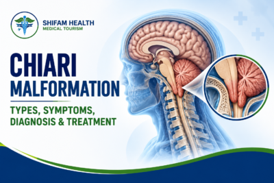
Chiari malformation is a structural condition in which part of the cerebellum (the brain’s balance and coordination center) sits low, pushing into the opening at the base of the skull. This can crowd the brainstem and upper spinal canal and may disturb the flow of cerebrospinal fluid. Some people have no symptoms; others have headaches, neck pain or neurological signs.
Does Chiari malformation always require surgery? No. Many people, particularly those with type I who have no symptoms, are only monitored. Surgery is considered when symptoms are significant or progressive, neurological deficits develop, or a syrinx (a fluid-filled cavity in the spinal cord) is present, and the decision rests on a neurosurgeon’s assessment, not an MRI measurement alone.
- Chiari malformation is about the relationship between the brain, the skull base and the upper spinal canal. It has several types, and type I is the most common.
- It may cause no symptoms and is sometimes found by chance on an MRI.
- Typical symptoms include headaches at the back of the head (often worse with coughing or straining), neck pain, dizziness and balance or sensory problems.
- Diagnosis uses a neurological examination and MRI. The measurement alone does not decide treatment.
- Treatment ranges from observation to decompression surgery, chosen individually.
- New or rapidly worsening weakness, trouble swallowing or breathing, or a sudden severe headache with neurological symptoms need urgent care.
Hearing the words “Chiari malformation” on an MRI report, or searching for them after months of headaches or dizziness, can be frightening. The reality is more nuanced than many online pages suggest: many people with the finding never need treatment, some have symptoms that respond to surgery, and some have symptoms that have a different cause entirely. This guide explains the types, symptoms, tests and treatment options in plain language. It is general education and cannot diagnose you or your child.
Table of Contents
ToggleWhat Is Chiari Malformation?
Chiari malformation describes a mismatch between the brain and the space available at the back of the skull. The cerebellum sits in the back part of the skull, called the posterior fossa. The brainstem passes down through a large opening at the skull’s base, the foramen magnum, into the spinal canal. In Chiari malformation, the lower parts of the cerebellum (the tonsils) extend downward through this opening.
That can crowd the area and, in some people, interfere with the flow of cerebrospinal fluid (CSF), the clear fluid that cushions the brain and spinal cord, and with the movement of that fluid with each heartbeat and with coughing or straining.
It is not always a problem. Many Chiari malformations are discovered by chance, when an MRI is done for another reason. An MRI shows an anatomical finding; whether it causes symptoms depends on the person. A low-lying cerebellar tonsil does not automatically mean a clinically significant Chiari malformation, and the diagnosis must be read alongside symptoms, the neurological examination and the imaging.
What Causes Chiari Malformation?
In most people, Chiari malformation is congenital, meaning it develops before birth. The leading explanation for type I is that the back of the skull (posterior fossa) is smaller or shaped differently than usual, leaving too little room for the cerebellum, so the tonsils are pushed down. The exact cause is not known in every case.
Other things that can be involved:
- Developmental differences in skull-base anatomy and the space available.
- Associated structural conditions in some people, for example some skull-base or connective tissue conditions.
- Less common acquired (secondary) forms, where the tonsils are pulled or pushed down by conditions that change CSF pressure, such as long-standing low pressure from a CSF leak or a space-occupying lesion.
Congenital Chiari malformation is not caused by ordinary activity, posture, a fall or any single event, and parents did not cause it.
Types of Chiari Malformation
The types are different conditions, not stages of one disease. Terminology varies between references.
Types of Chiari malformation
| Type | Key Features | Typical Presentation | Important Associations |
|---|---|---|---|
| Type I | Cerebellar tonsils descend below the foramen magnum | Often found in older children or adults; may be silent | Syringomyelia, sometimes hydrocephalus or scoliosis |
| Type II | Tonsils and the lower cerebellum and brainstem extend down; the brain structures are malformed | Found at or before birth | Myelomeningocele (spina bifida) and hydrocephalus |
| Type III | A rare, severe form with a defect in the skull or upper neck and herniation of brain tissue | Found at birth | Very serious neurological problems |
| Type IV | A historical label for severe cerebellar underdevelopment | Described in the early literature | Not used consistently today |
Chiari malformation type I
In type I, one or both cerebellar tonsils extend below the foramen magnum. Symptoms vary widely and often begin in adolescence or adulthood, and many people have none. The Congress of Neurological Surgeons (CNS) guideline defines it as tonsillar descent of 3 to 5 mm or more below the foramen magnum, and notes that imaging studies suggest it is present in roughly 0.24% to 2.6% of people when the 5 mm definition is used. Clinicians also describe Chiari 1.5, where the brainstem has descended as well, and Chiari 0, a syrinx with little or no tonsil descent.
Chiari malformation type II
Type II is almost always linked with myelomeningocele, a form of spina bifida in which the spinal cord and its coverings are exposed at birth, and often with hydrocephalus (fluid build-up in the brain). It needs specialist neurosurgical and multidisciplinary care from infancy. Children may have feeding or swallowing problems, noisy breathing and other neurological signs.
Chiari malformation type III
Type III is rare and severe. Part of the brain pushes through a defect at the back of the skull or upper neck (an encephalocele). It is found at birth and needs urgent specialist care.
Chiari malformation type IV
Type IV is a historical label from the original descriptions and refers to severe underdevelopment of the cerebellum. It is not used consistently in current practice and is not equivalent to types I to III, so the term should be treated with caution.
Type 1 vs type 2 at a glance
| Feature | Type 1 | Type 2 |
|---|---|---|
| Typical age at diagnosis | Often later childhood or adulthood, sometimes by chance | At or before birth |
| Associated conditions | Syringomyelia, sometimes hydrocephalus or scoliosis | Myelomeningocele and hydrocephalus |
| Typical symptoms | Back-of-head headaches, neck pain, balance or sensory problems; may be silent | Feeding and swallowing problems, breathing noise, limb weakness, depending on the spinal defect |
| General approach | Observation or decompression based on symptoms and syrinx | Specialist team managing the spinal defect, hydrocephalus and brainstem effects |
This is a general comparison and does not replace clinical evaluation.
Symptoms of Chiari Malformation
Symptoms depend on the type, how much CSF flow or the brainstem is affected, and any associated syrinx. None of these symptoms is specific to Chiari malformation; each has many other causes.
Possible symptoms and why they need clinical assessment
| Symptom | Why It May Occur | Why Assessment Matters |
|---|---|---|
| Headache at the back of the head, often worse with coughing, sneezing, straining or exertion | Pressure changes at the skull base | Many other conditions cause headache; strain-related headache is the symptom that most often improves with surgery |
| Neck pain | Crowding at the craniocervical junction | Common from many causes |
| Dizziness, balance or coordination problems | Cerebellar or brainstem involvement | Needs a neurological examination |
| Numbness, tingling, weakness | Spinal cord involvement, especially with a syrinx | May signal a syrinx needing imaging |
| Swallowing difficulty, voice changes | Brainstem or lower cranial nerve involvement | Needs prompt specialist review |
| Sleep-related breathing problems | Brainstem involvement | Needs a sleep and neurological assessment |
| Visual, hearing or ear symptoms | Pressure effects | Many other causes |
| Scoliosis, developmental or neurological concerns (some children) | Associated syrinx or related conditions | Needs pediatric specialist assessment |
Having a headache or feeling dizzy does not mean you have Chiari malformation.
Can Chiari Malformation Cause No Symptoms?
Yes. Particularly with type I, Chiari malformation is often found incidentally and may never cause symptoms. In one large MRI study of 22,591 patients, 175 had tonsillar herniation of more than 5 mm, and syringomyelia was found in only one of the asymptomatic ones; the authors concluded that the isolated finding has limited prognostic value. An imaging finding on its own does not mean surgery is needed. Clinical evaluation and, for some people, individual monitoring are the usual approach.
When Should Someone Seek Urgent Medical Attention?
Seek urgent medical care, regardless of whether you have a known Chiari malformation, for:
- New or rapidly worsening weakness
- Sudden difficulty walking or keeping balance
- New difficulty swallowing or breathing
- A sudden severe headache, especially with neurological symptoms
- New loss of coordination, or loss of consciousness
- Rapid deterioration in a child’s neurological function
These symptoms can have causes other than Chiari malformation, so do not self-diagnose, and do not wait for a routine appointment if they appear.
How Is Chiari Malformation Diagnosed?
Diagnosis combines your symptoms, a neurological examination and MRI. The imaging shows the anatomy, and the symptoms and examination show whether it matters. Not everyone needs every test.
Common diagnostic investigations
| Investigation | Purpose | Important Considerations |
|---|---|---|
| History and neurological examination | Symptom pattern, balance, coordination, sensation, strength, cranial nerves | The most important step in deciding whether a finding is relevant |
| MRI of the brain and cervical spine | Shows tonsil position, skull-base anatomy, brainstem and any syrinx | The measurement alone does not predict symptoms or need for surgery |
| MRI of the rest of the spine | Looks for a syrinx or associated spinal problems | Done when a syrinx or related condition is suspected |
| Cine MRI (CSF flow study) | Assesses CSF movement at the foramen magnum | Used in selected cases; interpreted by the treating team |
| Other tests (sleep study, swallowing assessment, hearing or eye tests) | Check specific symptoms | Used only when symptoms suggest them |
Medical history and neurological examination
The doctor asks about the pattern of headaches (including whether coughing or straining triggers them), neck pain, balance, numbness or weakness, swallowing and sleep, and examines strength, sensation, reflexes, coordination and gait.
MRI of the brain and cervical spine
MRI is the key test. It shows how far the tonsils extend, the size and shape of the posterior fossa, how the brainstem and spinal cord look, and any syrinx. Many guidelines use a tonsillar descent of about 5 mm below the foramen magnum as the cut-off (some use 3 to 5 mm, and a lower threshold in children). A tonsil that sits slightly low does not by itself mean the person has a clinically significant Chiari malformation.
Additional spine imaging
If a syrinx, scoliosis or another spinal problem is suspected, the cervical, thoracic or whole spine may also be imaged.
Cine MRI and CSF flow assessment
Special MRI sequences can show how CSF moves past the foramen magnum. They are used in some patients and are not needed by everyone.
Assessment for associated conditions
Clinicians may look for syringomyelia, hydrocephalus, a tethered spinal cord or other conditions when the picture suggests them. Imaging measurements alone do not determine symptom severity, prognosis or the need for surgery.
Chiari Malformation and Syringomyelia
Syringomyelia is a fluid-filled cavity (a syrinx) that forms within the spinal cord. It is one of the main associated conditions, and the reason is thought to be disturbed CSF flow near the foramen magnum.
- Symptoms can include pain, loss of the ability to feel temperature or pain in the arms or hands, numbness or tingling, weakness and, in some people, spine curvature.
- Why it matters: a syrinx can damage the spinal cord and may enlarge over time.
- MRI shows whether a syrinx is present and whether it is changing.
- Management depends on symptoms, imaging and specialist assessment. A syrinx is generally regarded as a strong reason to consider surgery, and decompression of the foramen magnum is usually the treatment because it can improve CSF flow. If hydrocephalus or another condition is also present, treating that may be part of the plan.
Not everyone with Chiari malformation develops syringomyelia. In one 2024 pediatric surgical series, a syrinx was present in about 28% of the children who had surgery, and that group is not representative of all people with Chiari.
Treatment Options for Chiari Malformation
Treatment depends on symptoms, the neurological examination, any syrinx or other associated condition, and the MRI. Surgery is not required for everyone.
Observation vs surgical treatment
| Approach | When It May Be Considered | Key Considerations |
|---|---|---|
| Observation and follow-up | No or mild symptoms, no syrinx, no progression | Active follow-up with review; the plan changes if symptoms or MRI change |
| Symptom management | Headache, neck pain or other symptoms while observation continues | Relieves symptoms but does not correct the anatomy |
| Decompression surgery | Significant or progressive symptoms, neurological deficit, or a syrinx | Aims to create space and improve CSF flow; risks and uncertain symptom relief |
Observation and regular follow-up
For people with no or minor symptoms and no syrinx, monitoring is a standard approach. The CNS guideline advises against prophylactic (preventive) surgery in people with asymptomatic Chiari I without a syrinx, and against routinely screening asymptomatic siblings or first-degree relatives. Follow-up with symptom review and, when appropriate, repeat imaging is individualised.
Symptom management
A clinician may treat headache, neck pain or other symptoms, for example with a pain specialist, physiotherapy or other measures. This may ease symptoms but does not change the anatomy. Do not self-medicate or take any medicine without advice from your clinician; this article gives no doses.
Chiari decompression surgery
The usual operation is a posterior fossa decompression, which aims to make more room at the back of the skull and base of the brain and, in many patients, to improve CSF flow. Its elements can include:
- Suboccipital craniectomy: removing a small piece of bone from the back of the skull.
- C1 laminectomy: removing part of the first vertebra’s back in selected patients.
- Duraplasty: opening the covering of the brain (dura) and enlarging it with a patch, when indicated.
The exact operation varies between patients and surgeons. Recommendations from the World Federation of Neurosurgical Societies (WFNS) Spine Committee suggest bone-only (osteoligamentous) decompression in children and decompression with duraplasty in adults, usually enough to control an associated syrinx.
In selected patients who had earlier surgery that did not work, the surgeon may open the arachnoid membrane, and shrinking the tonsils can give somewhat better results but carries a higher risk of complications. The plan depends on anatomy, symptoms, age, associated conditions and the surgeon’s judgement. Surgery can use intraoperative ultrasound to guide how much is needed.
When Is Chiari Malformation Surgery Recommended?
Surgery is considered when symptoms are significant or progressive, a neurological deficit develops, there is a syrinx, or, in someone without symptoms, serial MRI shows progression. It requires a qualified neurosurgical assessment, and the presence of Chiari malformation on an MRI does not on its own justify surgery.
The CNS guideline says decompression may be performed in symptomatic Chiari I to improve strain-related headache, and that other symptoms respond more variably. Weighing the likely benefit against the risks, the uncertainty about which symptoms will improve and the natural course of the condition is central. There are no universal surgical thresholds, and different centres vary in practice.
Risks and Possible Complications of Chiari Surgery
Risks depend on the procedure and the patient. They include:
- Infection, including wound infection and meningitis
- Bleeding
- CSF leakage and a collection of CSF under the skin (pseudomeningocele)
- Persistent or returning symptoms, and the need for further treatment or another operation
- Other neurological problems and anaesthesia-related risks
Surgery can improve symptoms or CSF flow in selected patients, but it cannot guarantee that every symptom will go away or that an existing neurological deficit will reverse. We do not quote complication percentages here, because they vary by technique and patient and no source can promise a given result. [Medical reviewer to confirm wording.]
Recovery After Chiari Decompression Surgery
Recovery is gradual and individual. In general terms:
- Hospital stay: observation in hospital after surgery, often a few days, depending on the patient.
- Pain and wound care: pain relief and wound management as directed by the surgical team.
- Activity: a gradual return, following the team’s instructions about lifting, bending and driving.
- Follow-up: neurological review and, when appropriate, repeat MRI.
- Watching for problems: fever, a swelling or fluid at the wound, severe headache or new neurological symptoms need prompt contact with the team.
Recovery depends on age, technique, general health and any complications. We do not give a universal discharge date or return-to-work timeline.
Long-Term Outlook and Prognosis
Outcomes vary widely. Many people with Chiari malformation, particularly those without symptoms, remain well without surgery. Many patients who have surgery for strain-related headache or a syrinx improve, although some symptoms may persist or return, and other symptoms respond less predictably. Monitoring an associated syrinx or other abnormality may continue for years, and some people need long-term follow-up. No honest source can say that surgery always prevents neurological deterioration or give a success rate that applies to you, and unsourced percentages online should be treated with caution.
Chiari Malformation in Children
Children need individual assessment by a pediatric neurosurgeon or neurologist. Key points:
- Incidental vs significant: MRI is now common, and low-lying tonsils are sometimes found when imaging is done for another reason. This does not necessarily mean treatment is needed.
- Symptoms are harder to read: young children may not describe headache or dizziness. Irritability, neck posture changes, feeding or swallowing difficulty, noisy breathing, regression in development, or scoliosis can be clues.
- Associated conditions: syringomyelia, hydrocephalus and scoliosis, and in type II, myelomeningocele.
- Growth and follow-up: a child’s skull and spine are still growing, so follow-up is individualised.
- Observation or surgery: based on symptoms and imaging, and surgical technique can differ from adults (the WFNS group suggests bone-only decompression in children).
Adult recommendations should not be applied to children without qualification.
Exploring Chiari Malformation Treatment in India
This section is for international patients who are medically stable and would like a specialist opinion in India. You should not travel on the basis of website information alone. The treating clinicians must decide whether travel is medically appropriate, particularly if you have rapidly worsening symptoms, which need local urgent care first.
A sensible sequence is:
- Organise your records: MRI images (the actual scan files as well as the reports), the radiology reports, neurological notes, and details of any earlier treatment.
- Get a specialist opinion from a neurosurgeon (or a pediatric neurosurgeon for a child) who regularly assesses Chiari malformation, ideally including a review of whether your symptoms fit the MRI.
- Understand the proposed plan: observation or surgery, which technique and why, the risks, and what is realistic to expect.
- Ask for a written, itemised estimate and what it includes.
- Arrange visa and travel only if the plan is to go ahead. Check visa rules through official Indian government sources and the Indian mission in your country.
- Plan accommodation, interpretation and follow-up, including who to contact after you return home and how your records will be shared with your own doctor.
What affects the cost?
Cost depends on specialist consultation and imaging; how complex the condition is; any extra tests; the surgical technique and operating-room needs; the hospital stay and monitoring after surgery; treatment of associated conditions such as a syrinx or hydrocephalus; follow-up and rehabilitation; and, for international patients, travel and accommodation.
We do not quote prices. We could not find a reliable published India price for Chiari decompression, and the figures that do exist, such as US pediatric hospitalization costs, are not comparable. And we also do not claim that any one hospital or country is always cheaper. An individual estimate needs your records and a proposed plan. International patients are often quoted more than domestic list prices, and the lowest quote is not necessarily the most appropriate.
How Shifam Health Can Help
Shifam Health is an India-based healthcare coordination service, not a hospital, neurosurgical provider or diagnostic centre, and it does not replace clinical judgement. It can help patients and families organise medical records, coordinate specialist consultation requests, explore treatment options in India, obtain estimates from relevant providers, and arrange practical support such as visa documentation, travel, accommodation, interpretation and follow-up. It cannot promise a cure, a surgical outcome or access to a particular specialist.
For assistance coordinating a specialist opinion or exploring treatment options in India, visit: https://shifamhealth.com/free-consultation/
Submitting an inquiry does not replace a medical assessment or urgent care.
Frequently Asked Questions
What is Chiari malformation?
A structural condition in which part of the cerebellum extends through the opening at the skull base, potentially affecting cerebrospinal fluid (CSF) flow.
What are the main types of Chiari malformation?
Types I, II, and III are recognized forms. Type I is most common, while type II is associated with spina bifida.
What are the symptoms of Chiari malformation?
Symptoms may include back-of-head headaches, neck pain, dizziness, balance problems, numbness, or weakness. Some people have no symptoms.
Can Chiari malformation occur without symptoms?
Yes. It may be discovered incidentally on MRI and require only monitoring if there are no concerning symptoms or associated complications.
How is Chiari malformation diagnosed?
Diagnosis involves neurological assessment and MRI of the brain and cervical spine. Additional spinal imaging may be needed.
What is syringomyelia?
A fluid-filled cavity within the spinal cord that can cause pain, numbness, or weakness and may occur with Chiari malformation.
Does every Chiari malformation require surgery?
No. Many asymptomatic cases are monitored, while surgery may be considered for significant symptoms, neurological changes, or syringomyelia.
What is Chiari decompression surgery?
An operation that removes a small amount of bone at the skull base, sometimes with removal of part of the first cervical vertebra, to create more space and improve CSF flow.
What are the risks of Chiari surgery?
Possible risks include infection, bleeding, cerebrospinal fluid leakage, persistent symptoms, and complications related to anesthesia.
How long does recovery take?
Recovery varies by individual and surgical technique. Hospitalization may last several days, followed by recovery over subsequent weeks.
Can Chiari symptoms return after surgery?
Yes. Some symptoms may persist or recur, so clinical follow-up and MRI monitoring may be necessary.


Popular Posts From Last Week
- October 12, 2026
- shifamhealth
Normally, the lower end of the spinal cord floats freely inside the spinal canal and glides a little…
- October 12, 2026
- shifamhealth
The word “stenosis” can sound frightening, and a diagnosis of severe aortic stenosis often arrives with the words…
- October 12, 2026
- shifamhealth
Chiari malformation is a structural condition in which part of the cerebellum (the brain’s balance and coordination center)…


