

Ventricular Septal Defect (VSD) (2026): Causes, Symptoms, Diagnosis & Treatment
Filters & Insights
Written By :
Medically Reviewed By :

Shifam Health Medical Team
Shifam Health - Medical Reviews Department
10+ Years Of Experience
Learn about ventricular septal defect (VSD), including its causes, symptoms, diagnosis, treatment options, and recovery.
📅
Last updated: October 25, 2026

What is a ventricular septal defect? A ventricular septal defect (VSD) is an opening in the wall (septum) between the heart’s two lower chambers, the ventricles. It is usually present from birth. Blood can pass through the opening from the left ventricle to the right. Management ranges from watching a small defect to closing a significant one with a catheter device or surgery.
- Many small VSDs cause no problems, and some close on their own, especially certain small muscular defects.
- A larger VSD can send extra blood to the lungs, causing fast breathing, poor feeding and slow growth in infants.
- An echocardiogram confirms the diagnosis and shows size, location and effects.
- Closure is recommended when the defect has significant effects. It is done by open-heart surgery or, for suitable defects, a catheter device.
- Not every VSD needs an operation, and some advanced cases cannot safely be closed. A specialist team decides.
If you have just been told that your baby, your child or you have a hole in the heart, the first questions are usually the same: Is it dangerous? Will it close on its own? Does it need surgery? How do doctors decide? This guide answers those questions in plain language, for parents and for adults with a VSD. It is general education and cannot diagnose you or your child. For the wider topic of heart defects present at birth, see our guide to [congenital heart disease in children — link to be verified].
Table of Contents
ToggleWhat Is a Ventricular Septal Defect (VSD)?
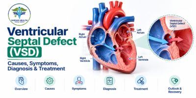
The heart has four chambers. The two lower chambers, the ventricles, are separated by a wall called the ventricular septum. The left ventricle pumps oxygen-rich blood to the body at high pressure, and the right ventricle pumps blood to the lungs at lower pressure.
In a VSD, there is an opening in that wall. Because pressure is higher on the left, blood flows through the opening from the left ventricle to the right, a left-to-right shunt (a shunt is an abnormal path for blood). The extra blood passes through the lungs again, called pulmonary overcirculation, and the left side of the heart has to handle the extra volume.
How much this matters depends on the size and location of the opening, how much blood flows through it, the pressure in the lung arteries, and the person’s age and general health. A small VSD may cause little or no trouble, and a large one can cause symptoms in the first weeks or months of life. Most uncomplicated VSDs do not make a person blue.
VSD is the most common congenital heart defect in children, according to StatPearls. As patients age, it remains common in adults too.
Types of VSD
VSDs are named by where they sit in the septum. Terminology varies between references, and location affects monitoring, the chance of spontaneous closure, nearby valve problems and which closure methods are suitable. Anatomical type alone does not determine severity.
| Type | Location | Important clinical considerations |
|---|---|---|
| Perimembranous | Near the membranous part of the septum, close to the heart valves | The most commonly treated type; some become smaller; close to the conduction system and the aortic valve |
| Muscular | Within the muscular part of the septum | Some small ones close on their own; some are multiple |
| Inlet | Near the inflow area, close to the valves between the upper and lower chambers | Less likely to close on its own; may link with valve problems |
| Outlet (supracristal or doubly committed) | Near the outflow tracts, close to the aortic and pulmonary valves | Less likely to close on its own; can allow an aortic valve leaflet to prolapse |
VSD Size and Hemodynamic Effect
Doctors look at both the physical opening and what it does to blood flow. We do not give a single millimetre cut-off, because the meaning of a size depends on the person’s body size, the defect’s location and the echocardiogram findings.
| Category | Typical features | General management considerations |
|---|---|---|
| Small (restrictive) | Limited flow; the pressure difference between the ventricles is maintained; often no symptoms; heart size normal | Often observation with follow-up; some close on their own |
| Moderate | More flow to the lungs; some left-heart enlargement; may have mild symptoms | Individualised: monitoring, supportive care or closure depending on effects |
| Large (non-restrictive) | Pressures between ventricles near-equal; large shunt; can cause heart failure, poor growth and rising lung pressure | Usually needs treatment planned by a specialist team; timing is individual |
Important considerations include pulmonary blood flow and pressure, volume loading of the left heart, and, in infants, signs of heart failure or poor growth.
Causes and Risk Factors
Most VSDs form during the heart’s development in early pregnancy, when the septum does not completely close. In most children no specific cause is found, and parents should not be blamed.
Some VSDs are linked to genetic or chromosomal conditions, and a VSD can occur together with other congenital heart defects. We do not attribute any individual VSD to a particular medicine, food, behaviour or event during pregnancy without good evidence.
A different, rare condition is an acquired VSD, which can occur after a heart attack when the septum is damaged. It is an emergency and is not the same as a congenital VSD.
Symptoms of VSD
Symptoms depend on size, flow and age. Many small VSDs are found by chance, when a doctor hears a murmur or when an echocardiogram is done for another reason.
In infants and children
- Fast breathing or difficulty feeding
- Sweating during feeds
- Poor weight gain or growth
- Unusual tiredness with feeding or play
- Repeated chest symptoms in some children
- A heart murmur found on examination
- No obvious symptoms in many children with small defects
A murmur is often the first sign, but how loud it is does not reliably show the defect’s size or severity.
In older children and adults
Reduced stamina, breathlessness on exertion, tiredness, palpitations in some people, and symptoms from related complications when present. Many small VSDs cause no symptoms at all.
When Is a VSD an Emergency?
Seek emergency care (call your local emergency number) for:
- Severe or worsening breathing difficulty
- Blue or gray lips or skin
- Collapse, fainting with concerning symptoms, or reduced responsiveness
- An infant who cannot feed adequately or looks severely unwell
- Sudden severe symptoms in someone with known heart disease
Do not wait for an online reply or a routine appointment. Most VSDs are not emergencies. An uncomplicated VSD does not usually cause blueness; blue colouring can point to another or an advanced problem and needs urgent assessment.
| Situation | Suggested response |
|---|---|
| Murmur noticed on a check-up, no symptoms | Routine cardiology assessment and echocardiogram |
| Baby feeds slowly, sweats with feeds, or gains weight poorly | Prompt medical review (within days) |
| Child tires more easily than other children, or breathless with play | Prompt review by a pediatric cardiologist |
| Adult with new breathlessness, palpitations or reduced stamina | Prompt review by a cardiologist |
| Blue lips, severe breathing difficulty, collapse, unresponsiveness | Emergency care immediately |
How Is VSD Diagnosed?
Diagnosis combines an examination with imaging. Test choice depends on the patient’s condition and the questions the cardiology team needs to answer. Not everyone needs every test.
- Clinical examination: history, heart sounds and murmur, breathing, growth and signs of strain on the circulation.
- Echocardiography (2D with Doppler): the main test. It shows where the VSD is, how big it is, the direction of flow, chamber enlargement, involvement of nearby valves and estimates of lung artery pressure.
- Electrocardiogram (ECG): may show electrical changes or chamber strain but cannot give the full anatomy.
- Chest X-ray: can help assess heart size and lung blood flow in selected patients; not needed for everyone.
- Cardiac MRI or CT: used selectively for extra anatomical or physiological information.
- Cardiac catheterisation: reserved for selected patients when pressures, pulmonary vascular resistance or other flow data are needed.
Can a VSD Close on Its Own?
Sometimes. Spontaneous closure is possible, especially with certain small muscular defects, and some other small defects become smaller over time. Whether a defect closes depends on its location, size, the child’s age and other factors. Larger defects are less likely to close and may need treatment if they have significant effects.
We do not quote a closure rate, because it varies by type and size and cannot be applied to a particular child. Regular follow-up can still be needed even when there are no symptoms. Your cardiologist sets the schedule.
VSD Treatment Options
Management is individualised, and not every VSD needs an intervention. The options are watching, supportive medicines, surgery and catheter-based device closure.
| Option | Purpose | Limitations |
|---|---|---|
| Observation and monitoring | Follow small, uncomplicated VSDs, which may close or stay harmless | Needs planned follow-up; the plan changes if symptoms or scans change |
| Medicines and supportive care | Control symptoms such as fluid overload and support growth while definitive care is planned | Do not close the hole; not a substitute for closure when closure is indicated |
| Surgical closure | Close the defect with a patch or stitches during open-heart surgery | Major surgery with its own risks; timing and method are individual |
| Catheter device closure | Close a suitable defect through a blood vessel without open-heart surgery | Only suitable for selected defects; risks include residual flow, valve problems and heart block |
1. Observation and regular monitoring
A small, uncomplicated VSD is usually monitored with check-ups and echocardiograms. How often is decided by the treating clinician, based on the defect, the child’s age and symptoms.
2. Medicines and supportive care
Medicines do not close a VSD. When a significant VSD causes symptoms or heart failure, a clinician may use medicines, such as diuretics (“water tablets”) in selected cases, to ease symptoms while closure is planned. Infants may need nutritional support and growth monitoring. Do not give or change any medicine without medical advice; this article gives no doses.
3. Surgical VSD closure
Surgery may be recommended for a significant shunt, heart failure that is not controlled by medical treatment, poor growth, progressive enlargement of the left heart, or certain complications. It is usually done through open-heart surgery with a heart-lung machine, closing the hole with a patch or stitches depending on the defect. The approach depends on the anatomy, any other heart defects, the child’s age and the surgical team, and the team will monitor and follow up after the operation. Not every moderate or large VSD needs the same timing or method.
4. Catheter-based device closure
Selected VSDs can be closed with a device delivered by a catheter, usually through a blood vessel, with no chest incision. The device is placed across the hole to block it. Suitability depends on the defect’s location and size, the tissue around it, and its closeness to the heart valves and to the heart’s electrical conduction tissue, so not every VSD can be closed this way, and it is not always safer or preferable. Possible risks include residual flow through or around the device, device-related problems, interference with a valve, and rhythm or conduction problems. Complete heart block, a serious conduction problem, is a recognised concern, and in the literature it appears more often after device closure of perimembranous than muscular defects. Device choice and eligibility need specialist assessment.
5. Other specialised approaches
A few specialised or hybrid techniques exist for selected patients at certain centres. They are not routine options for every VSD, and availability varies.
When Is VSD Closure Recommended?
Closure is considered when a VSD has significant effects on the heart, lungs or growth. The main factors are:
- Signs or symptoms of heart failure
- A large shunt with pulmonary overcirculation
- Enlargement of the left heart
- Feeding difficulty and poor growth in infants
- Lung artery pressure and pulmonary vascular resistance
- Aortic valve prolapse or worsening aortic regurgitation (leaking) in outlet or perimembranous defects
- A past infection of the heart lining (endocarditis) involving the VSD
- Associated congenital heart defects
In adults, the 2018 AHA/ACC guideline for adult congenital heart disease recommends closure for a significant left-to-right shunt (pulmonary to systemic flow ratio of 1.5 or more) with significant or progressive left ventricular enlargement and no pulmonary arterial hypertension, considers closure reasonable with mild pulmonary hypertension, and considers it reasonable for an outlet or perimembranous VSD that causes progressive moderate or greater aortic regurgitation. It advises against closure when the shunt is small and there is no other indication. Children’s decisions follow similar principles but also weigh growth, age and symptoms.
Pulmonary hypertension and Eisenmenger syndrome need specialist assessment. If high lung blood pressure has caused severe, irreversible damage to the lung blood vessels (Eisenmenger physiology, where the shunt reverses and the person may turn blue), closing the VSD can be harmful and is not recommended. Closure must never be advised without a specialist’s assessment of lung pressures and resistance.
There is no simple rule that every large VSD must be closed immediately. The timing is planned by the heart team.
Complications of an Untreated VSD
It can occur when a significant VSD is not treated, but they are not inevitable. They include heart failure, pulmonary hypertension, poor growth in children, aortic valve prolapse or leakage in some defect types, infective endocarditis (uncommon but important), rhythm problems and, in advanced untreated cases, Eisenmenger syndrome. Risk depends on the defect and the person’s course, and many small VSDs never cause any of these.
Risks of VSD Surgery or Device Closure
Any procedure carries risk. Possible complications include bleeding or infection, a leftover hole or shunt, rhythm and conduction problems including heart block, injury to or leakage of a valve, device-related problems if a catheter device is used, anaesthesia risks and, in some cases, the need for another procedure. Individual risk depends on age, anatomy, other heart conditions, lung pressures and the planned procedure. We do not quote complication percentages here, because they vary by centre, defect type and patient, and no one can promise a guaranteed cure.
Life After VSD Treatment
After surgery or device closure, the team monitors the patient, may repeat echocardiograms or other tests, and advises on wound or catheter-site care, return to school or work, and physical activity. Long-term follow-up looks for any residual shunt, valve problems, rhythm disturbances and other issues. Many patients do well, but long-term expectations depend on the original defect, the treatment and any residual problems.
Keep your or your child’s congenital heart records safe, including operation notes and echo reports, because they matter in adulthood. Antibiotics before dental work are not routinely needed for every VSD; they are advised only for selected higher-risk situations, so ask your cardiologist.
VSD in Adulthood and Pregnancy
Some adults have a small VSD found by chance. Others have a leftover defect or complications after earlier treatment. Adults with a complex or previously repaired VSD benefit from assessment by an adult congenital heart disease specialist, who looks at lung pressures, heart function, valve disease and the details of earlier repair.
For women considering pregnancy, advice must be individual. A small VSD with normal heart function and lung pressures is generally managed very differently from a VSD with pulmonary hypertension, in whom pregnancy can be very dangerous and is usually advised against (this is particularly so in Eisenmenger syndrome). Pregnancy advice cannot be based on the label “VSD” alone, and planning with a cardiologist before pregnancy is best.
VSD Treatment in India for International Patients
Selected tertiary hospitals in India offer pediatric cardiology, congenital cardiac surgery, catheter-based closure and intensive care for patients with VSD. Capabilities vary between hospitals, and the best place depends on the patient’s age, anatomy and condition. We do not rank hospitals here, and we do not claim that a particular hospital offers a specific procedure.
A sensible sequence is:
- Collect records: existing echocardiograms, medical reports and any previous treatment records.
- Get a specialist review from an appropriately qualified pediatric cardiologist, adult congenital cardiologist or cardiac surgeon.
- Get an individual assessment of whether observation, more tests, catheter closure or surgery is appropriate.
- Request a written treatment plan and itemised estimate from the treating hospital.
- Check what the estimate covers: tests, the procedure or surgery, hospital and intensive-care stay, medicines and follow-up.
- Arrange logistics: travel, medical visa documents, accommodation and interpretation if needed. Check visa rules through official Indian government sources and the Indian mission in your country.
- Plan follow-up for when you return home.
When choosing a hospital, consider clinical expertise, the patient’s age and anatomy, congenital cardiac facilities, intensive care needs, and whether the right catheter-based or surgical services are available. A patient who is unstable or has emergency symptoms needs local emergency care first, not an international trip.
What does VSD treatment cost in India?
Reliable published prices are scarce and mostly come from medical-travel aggregator websites. Straightforward VSD closure is commonly listed at roughly USD 4,500 to 6,000 for the procedure (one aggregator quotes an average of about INR 3.8 lakh, or about USD 4,500, as a starting price; another lists “hole in heart” surgery from USD 5,000, with USD 800 to 1,200 for evaluation on top). A 2025 Stanford GSB study of hospital-level costs for VSD surgery in India found about USD 2,000 at a charitable hospital and about USD 4,600 at a private institute, but these are costs to the hospital, not prices charged to international patients.
We found no reliable price specific to catheter device closure. International patients are often quoted more than domestic list prices, and complex defects, other heart problems, intensive care and complications raise the total. These are indicative only and are not a quotation.
| Factor | Why it matters |
|---|---|
| Age and anatomy of the patient | Infants and complex defects need more resources |
| Surgery vs catheter device closure | Different procedures, devices and stays |
| Hospital, city and room category | Prices differ widely between hospitals and cities |
| Investigations | Echocardiography, and sometimes CT, MRI or catheterisation |
| Length of stay and intensive care | Longer stays and ICU time increase the total |
| Complications or other heart defects | May need extra treatment |
| Follow-up and medicines | Visits, tests and any ongoing treatment |
| Travel, accommodation and an attendant | Additional costs for international families |
Ask for an itemised written estimate after specialist review, and do not let the lowest price decide the treatment choice.
How Shifam Health fits in. Shifam Health is an India-based healthcare coordination service, not a hospital, treating clinician or government visa authority. It can help collect records, coordinate specialist opinions, shortlist hospitals, request and clarify written estimates, and support visa documentation, travel, accommodation, interpretation and follow-up coordination. It cannot guarantee outcomes, admission, timelines or visa approval, and clinical decisions belong to the treating team.
html_block id=”34328″
Frequently Asked Questions
What is a ventricular septal defect (VSD)?
VSD is an opening between the heart’s two lower chambers, usually present from birth. Its effects depend on its size and location.
Is VSD a serious heart condition?
Small VSDs are often harmless, while larger defects can cause heart failure, poor growth or increased lung pressure.
Can VSD close on its own?
Yes, some small VSDs close naturally or become smaller over time. Regular follow-up is important.
What are the symptoms of VSD in babies?
Symptoms may include fast breathing, feeding difficulties, sweating during feeds, fatigue and poor weight gain. Small VSDs may cause no symptoms.
Can a child with VSD grow normally?
Many children with small VSDs grow normally. Larger defects may affect feeding and growth and require treatment.
Does every VSD require surgery?
No. Small defects may only need monitoring. Significant VSDs may require surgical or catheter-based closure.
What is the difference between VSD surgery and device closure?
Surgery closes the opening using stitches or a patch. Device closure uses a catheter inserted through a blood vessel and is suitable only for selected defects
How is VSD severity diagnosed?
Echocardiography with Doppler assesses the defect, blood flow, heart enlargement and lung pressure. Symptoms and growth also help determine severity.
What happens if VSD is left untreated?
Significant defects may cause heart failure, pulmonary hypertension, poor growth or valve problems. Small defects may cause no major complications.
How long is recovery after VSD closure?
Recovery depends on the procedure and the patient’s condition. Catheter closure generally involves a shorter recovery than open-heart surgery.
Conclusion
A VSD can range from a small opening that only needs monitoring to a significant defect that needs closure. The decision depends on the defect’s anatomy, symptoms, blood-flow effects, lung pressures and associated problems, and it belongs to the treating cardiac team.
If you or your child has been diagnosed with a ventricular septal defect and you are exploring specialist care in India, Shifam Health can help coordinate medical report review, specialist consultations, hospital options and treatment estimates. The appropriate treatment plan should be determined by the treating cardiac team. Request a free consultation →
If you or your child has blue or gray lips, severe breathing difficulty, collapse or poor responsiveness, call your local emergency number now.


Popular Posts From Last Week
- October 23, 2026
- shifamhealth
A fluttering chest, a sudden racing heartbeat or a skipped beat can be frightening, and it is hard…
- October 19, 2026
- shifamhealth
Coronary artery disease is one of the most common and serious heart conditions, yet it can develop quietly…
- October 25, 2026
- shifamhealth
What is a ventricular septal defect? A ventricular septal defect (VSD) is an opening in the wall (septum)…
- October 22, 2026
- shifamhealth
A remote medical second opinion from India allows patients living in the USA, UK and other countries to…
- October 21, 2026
- shifamhealth
When you receive treatment estimates from two hospitals in India, do not compare only the final amounts. To…
- October 22, 2026
- shifamhealth
An itemized hospital quote in India should clearly explain the expected cost of treatment by separating major charges,…