




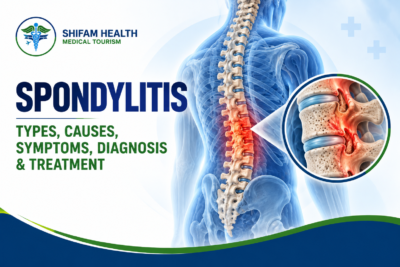
Spondylitis (2026): Types, Causes, Symptoms, Diagnosis & Treatment
Filters & Insights
Written By :
Medically Reviewed By :

Shifam Health Medical Team
Shifam Health - Medical Reviews Department
10+ Years Of Experience
Spondylitis can mean inflammatory disease, spinal infection, or a mislabel for wear and tear. Learn the types, red flags, tests, and treatments.
📅
Last updated: October 13, 2026

A note before you read further: “Spondylitis” is not one disease. It is a word that gets applied to at least three very different problems: an inflammatory disease of the spine and sacroiliac joints, an infection of the vertebrae and discs, and, loosely and incorrectly, age-related wear of the spine. The right treatment depends entirely on which one you have. If you have back pain with fever, severe or rapidly worsening pain, new leg weakness, numbness in the groin or saddle area, or new bladder or bowel problems, seek urgent medical care now rather than reading further.
Spondylitis means inflammation of one or more vertebrae. The term is used for inflammatory disease (axial spondyloarthritis, which includes ankylosing spondylitis), for spinal infection (spondylodiscitis or vertebral osteomyelitis, including spinal tuberculosis), and, informally, for degenerative changes that are properly called spondylosis. Inflammatory spondylitis typically starts gradually before age 40, causes back pain and morning stiffness that improve with movement rather than rest, and is treated with exercise, NSAIDs, and sometimes biologic medicines. Infection needs urgent diagnosis and antimicrobial treatment. Degenerative spondylosis is managed differently again.
Table of Contents
ToggleWhat “Spondylitis” Actually Means
The word comes from spondylos (vertebra) plus -itis (inflammation). In everyday use it is stretched to cover very different conditions, which is why a diagnosis of “spondylitis” on a report is only a starting point, not an answer.
Three situations account for most uses of the word:
| Meaning | What is really happening | Typical examples |
|---|---|---|
| Inflammatory | Immune-mediated inflammation, mainly at the sacroiliac joints and spine, and where tendons and ligaments attach to bone | Axial spondyloarthritis, ankylosing spondylitis |
| Infectious | Bacteria, mycobacteria (TB), or less often fungi infecting the vertebrae and the disc between them | Pyogenic spondylodiscitis, vertebral osteomyelitis, spinal tuberculosis |
| Loosely used for degenerative change | Age-related wear of discs and joints, not inflammation at all | “Cervical spondylitis” or “lumbar spondylitis” when the true term is spondylosis |
If you were told you have “spondylitis,” the most useful first question is: inflammatory, infectious, or degenerative? Everything else follows from the answer.
Spondylitis vs. Spondylosis vs. Spondylolisthesis vs. Spondylolysis
These four words are one letter apart in some cases and mean completely different things.
| Term | Meaning | Is it inflammation? |
|---|---|---|
| Spondylitis | Inflammation of vertebrae (inflammatory or infectious) | Yes |
| Spondylosis | Age-related degeneration of discs, facet joints, and bone spurs | No |
| Spondylolisthesis | One vertebra slipping forward over the one below | No (structural) |
| Spondylolysis | A stress fracture or defect in a part of the vertebra (the pars interarticularis) | No (structural, often from repetitive stress) |
Treating spondylosis as if it were inflammatory spondylitis, or the reverse, leads to the wrong medicines and the wrong expectations. Anti-inflammatory biologic drugs, for example, are for confirmed inflammatory disease, not for wear-and-tear. For degenerative problems, see our guides on spinal stenosis and disc herniation.
The Main Types of Spondylitis
Inflammatory (spondyloarthritis family)
| Condition | Key points |
|---|---|
| Radiographic axial spondyloarthritis (ankylosing spondylitis) | Inflammation of the sacroiliac joints and spine with structural changes visible on X-ray |
| Non-radiographic axial spondyloarthritis (nr-axSpA) | Same inflammatory disease family, but X-rays do not yet show definite sacroiliac damage; MRI may show active inflammation |
| Psoriatic arthritis with axial involvement | Spine and sacroiliac involvement in people with psoriasis |
| Reactive arthritis | Arthritis following certain infections, which may involve the spine |
| Enteropathic (IBD-associated) spondyloarthritis | Spine and joint inflammation linked with Crohn’s disease or ulcerative colitis |
| Juvenile spondyloarthritis | Onset in childhood or adolescence; often begins with peripheral joints or tendon-attachment (enthesitis) pain |
Infectious
| Condition | Key points |
|---|---|
| Pyogenic spondylodiscitis / vertebral osteomyelitis | Bacterial infection of the vertebra and disc, often spread through the blood |
| Spinal tuberculosis (Pott’s disease) | TB infection of the spine; relevant in regions where TB is common, including India |
| Other infections | Brucellosis and fungal infections occur but are less common |
Inflammatory and infectious forms can look alike at first (back pain, raised inflammatory markers, abnormal MRI). Telling them apart is a core clinical task, and getting it wrong in either direction can cause harm.
Inflammatory Back Pain vs. Mechanical Back Pain
Inflammatory back pain tends to begin gradually, usually before age 40, with morning stiffness and night pain that improve with movement and do not improve with rest. Mechanical back pain usually does the opposite: it eases with rest and worsens with activity. These patterns are useful clues, but not proof either way.
| Feature | Inflammatory pattern | Mechanical pattern |
|---|---|---|
| Typical age at onset | Often under 40 | Any age; more common with age |
| Onset | Gradual, over weeks to months | Often tied to a movement or injury |
| Effect of exercise | Improves | Often worsens |
| Effect of rest | Does not improve; stiffness can worsen | Usually eases |
| Night pain | Common, often in the second half of the night, easing on getting up | Less characteristic |
| Morning stiffness | Prolonged | Brief |
Experts use these features as a framework for deciding who needs further assessment. They do not diagnose anyone on their own, and plenty of people with inflammatory disease have atypical patterns, while plenty with mechanical pain tick some of the boxes. If your symptoms fit the inflammatory column, that is a reason to see a rheumatologist, not a self-diagnosis.
Axial Spondyloarthritis and Ankylosing Spondylitis
Axial spondyloarthritis (axSpA) is a chronic inflammatory disease mainly affecting the sacroiliac joints and spine. Ankylosing spondylitis is the form in which X-rays already show structural change at the sacroiliac joints. The non-radiographic form does not yet, but is part of the same disease spectrum.
- Where it starts: typically the sacroiliac joints (where the spine meets the pelvis), then potentially the spine
- What it does: inflammation at tendon and ligament attachments (entheses), which can heal with new bone formation. In some people, over years, this can link vertebrae together (fusion)
- Not everyone fuses. Many people with axSpA never develop significant spinal fusion, and treatment aims to control inflammation and protect function
- Who gets it: it is often described as a disease of young men, but women are affected too and are often diagnosed later, with diagnostic delay of years well documented in the literature
- Causes: not fully understood. Genetic susceptibility (notably HLA-B27) plays a major role, alongside other genes and environmental factors. It is not caused by posture, injury, or lifestyle, and it is not contagious
The terminology matters. Current practice treats axSpA as one disease spectrum, with “non-radiographic” and “radiographic” describing whether damage is visible on X-ray, not two unrelated conditions. Some people with the non-radiographic form progress to the radiographic form; many do not, and no reliable single percentage applies to everyone.
Symptoms and Extra-Spinal Features
Spinal and pelvic
- Gradual lower back and buttock pain, sometimes alternating sides
- Morning stiffness lasting longer than usual
- Night pain that eases after getting up and moving
- Pain and stiffness that improve with exercise
- Over time in some people, reduced spinal mobility and stooped posture
Beyond the spine (these matter for diagnosis and treatment choice)
| Feature | What it looks like |
|---|---|
| Enthesitis | Pain where tendons attach to bone, classically the heel (Achilles or plantar fascia) |
| Peripheral arthritis | Swelling in joints such as knees or ankles, often asymmetric |
| Dactylitis | A diffusely swollen “sausage” finger or toe |
| Acute anterior uveitis | Painful red eye with light sensitivity and blurred vision |
| Psoriasis | Scaly skin patches, nail changes |
| Inflammatory bowel disease | Persistent diarrhea, blood in stool, abdominal pain |
| Fatigue | Common and often under-recognized |
These extra-spinal features are not side notes. They help diagnosis, and they influence medication choice (for example, which biologic is preferred when uveitis or IBD is present).
Red Flags and Emergencies
Different diagnoses carry different emergencies. They should not be blurred together.
Seek urgent same-day medical care for:
- Back pain with fever, chills, or feeling systemically unwell (possible spinal infection)
- New or rapidly progressive leg weakness or numbness
- New bladder or bowel dysfunction, or numbness in the groin or saddle area (possible cauda equina syndrome)
- A painful red eye with light sensitivity, particularly if you have known or suspected axSpA (possible uveitis, which can threaten vision if untreated)
- New neck or back pain after even minor trauma in someone with long-standing axSpA. A rigid, fused, often osteoporotic spine can fracture from low-energy injuries, and these fractures can be unstable and easily missed
Seek prompt (not necessarily emergency) assessment for:
- Severe pain that is progressively worsening or constant, including at rest
- Back pain with unexplained weight loss, night sweats, or a history of cancer
- Back pain if you are immunosuppressed, have diabetes, use injected drugs, or have had a recent spinal procedure or bloodstream infection
No single red flag proves a serious cause, and most people with back pain do not have one. But infection and fracture are conditions where delay causes harm, which is why this list exists.
Causes and Risk Factors
Inflammatory spondylitis
- Genetic susceptibility, including HLA-B27 (discussed below)
- Family history of spondyloarthritis, psoriasis, uveitis, or IBD
- Immune-mediated mechanisms, with gut involvement under study
- Smoking is associated with worse disease activity and progression
- Not caused by injury, posture, sports, or sitting
Infectious spondylitis
- Bacteria reaching the spine through the bloodstream from another source (skin, urinary tract, heart valves, dental or other infection)
- Direct spread after spinal surgery, injections, or procedures
- Risk factors include diabetes, immunosuppression, advanced age, chronic kidney or liver disease, injected drug use, and malnutrition
- TB spine usually reflects spread from TB elsewhere in the body, and is more common where TB is common
Degenerative spondylosis
- Ageing, genetics, mechanical load, smoking, and prior injury, as covered in our disc herniation and stenosis guides
How Inflammatory Spondylitis Is Diagnosed
Diagnosis relies on clinical history and examination, laboratory tests, and imaging, interpreted together by a clinician experienced in spondyloarthritis. No single test confirms or excludes it.
Clinicians commonly work from an internationally used classification framework (ASAS). In simplified terms:
- Evidence of sacroiliitis on imaging, plus at least one typical spondyloarthritis feature, or
- A positive HLA-B27 plus at least two typical features
Typical features include inflammatory back pain, arthritis, enthesitis, uveitis, dactylitis, psoriasis, IBD, a good response to NSAIDs, family history, HLA-B27, and raised CRP.
This is a classification framework used with clinical judgment, not a self-test. The practical steps are:
- Detailed history, including inflammatory pattern, family history, eye, skin, and bowel history
- Examination of spinal mobility, sacroiliac joints, entheses, and peripheral joints
- Blood tests (CRP/ESR, HLA-B27 where appropriate, and other tests to exclude mimics)
- Imaging: usually pelvic X-ray first, then MRI of the sacroiliac joints (and sometimes spine) when X-rays are normal but suspicion remains
- Assessment for associated conditions (uveitis, psoriasis, IBD)
Diagnosis is often delayed, sometimes by years, partly because back pain is common and the inflammatory pattern is easy to miss. It is also sometimes over-diagnosed on the basis of an isolated MRI or HLA-B27 result. Both errors are real, which is why expert interpretation matters.
HLA-B27, CRP, and ESR: What They Can and Cannot Tell You
HLA-B27 is a genetic marker strongly associated with axial spondyloarthritis, but many people who carry it never develop the disease, and some people with axSpA do not carry it. CRP and ESR can be normal even when the disease is active. None of these tests can confirm or exclude the diagnosis alone.
| Test | What a positive or raised result means | What a negative or normal result means |
|---|---|---|
| HLA-B27 positive | Raises the likelihood of axSpA in someone with a suggestive picture. A minority of the general population carries it, and most carriers never develop disease. | Does not exclude axSpA. A meaningful share of people with axSpA are HLA-B27 negative, and the proportion varies between populations. |
| CRP / ESR raised | Suggests inflammation, but is non-specific. Infection, other inflammatory diseases, and many other causes also raise them. | Does not exclude active axSpA. These markers are normal in a substantial portion of people with active disease. |
Two practical consequences:
- A positive HLA-B27 does not mean you have, or will get, ankylosing spondylitis. Interpreted without symptoms or imaging, it can cause needless worry or even over-treatment.
- A normal CRP or negative HLA-B27 does not mean your back pain is “not inflammatory.” They lower, but do not remove, suspicion.
Markedly raised CRP and ESR with fever and severe pain should also prompt consideration of infection, not just inflammatory disease.
Imaging: Why a Normal X-Ray Does Not Settle the Question
Sacroiliac changes can take years to appear on X-ray. A normal X-ray therefore does not exclude axial spondyloarthritis. MRI can show active inflammation earlier, but MRI findings also need careful interpretation, because similar changes can occur in people without the disease.
| Imaging | Role | Limitation |
|---|---|---|
| Pelvic X-ray | Standard first test; shows established sacroiliac damage (radiographic sacroiliitis) | Often normal early in the disease; reading it can vary between observers |
| MRI of sacroiliac joints (and spine when needed) | Shows active inflammation (bone marrow edema) before X-ray changes, and structural changes | Bone marrow edema can also occur with mechanical stress, in athletes and runners, postpartum, and with some other conditions; specialist interpretation is needed |
| CT | Detailed bone structure in selected cases, and for fractures | Radiation; poor for active inflammation |
| Spine MRI | Looks for spinal inflammation, and is essential if infection or neurological compromise is suspected | Not routinely required for every patient with suspected axSpA |
An MRI report saying “sacroiliitis” is not automatically a diagnosis of spondyloarthritis, and a normal X-ray is not an all-clear. The combination of symptoms, examination, labs, and imaging is what counts.
What Can Mimic Inflammatory Spondylitis
| Mimic | Why it can resemble axSpA | Distinguishing context |
|---|---|---|
| Mechanical or non-specific low back pain | Very common; can include morning stiffness | Usually worsens with activity and eases with rest; no inflammatory features |
| Degenerative disc or facet disease (spondylosis) | Back pain and stiffness | Often older age, mechanical pattern, characteristic MRI findings |
| Osteitis condensans ilii | Sclerosis near the sacroiliac joints on imaging, especially after pregnancy | Benign; imaging pattern differs from sacroiliitis |
| Diffuse idiopathic skeletal hyperostosis (DISH) | Spinal stiffness and bridging bone | Older age; different pattern on X-ray, and no sacroiliac inflammation |
| Fibromyalgia | Widespread pain and fatigue | No objective inflammation; can coexist with axSpA |
| Spinal infection | Back pain and raised inflammatory markers | Fever, severe pain, risk factors, and MRI findings; potentially dangerous |
| Spinal tumour or metastasis | Persistent, often night pain | Constitutional symptoms, cancer history, imaging |
| Hip joint disease | Buttock or groin pain | Hip-specific examination findings |
Because fibromyalgia and axSpA can coexist, and because treatment failure often reflects a wrong or partial diagnosis, current European guidance explicitly advises re-evaluating the diagnosis when treatment does not work.
Spinal Infection (Infectious Spondylitis)
Spinal infection is a potentially serious condition that needs urgent diagnosis, identification of the organism, and prolonged targeted antimicrobial treatment. It must not be mistaken for inflammatory or mechanical back pain, and it should never be treated at home or with steroids on suspicion alone.
Warning features: back pain that is severe, progressive, or present at rest; fever or chills (though fever is absent in some patients); tenderness over the spine; and risk factors such as diabetes, immunosuppression, injected drug use, recent spinal surgery or injection, or a known source of infection elsewhere. Neurological symptoms (weakness, numbness, bladder or bowel changes) suggest abscess or cord or nerve compression and are emergencies.
Diagnosis
- MRI of the spine, usually with contrast, is the key imaging test
- Blood tests: CRP and ESR are usually raised; blood cultures are important, ideally before antibiotics when the patient is stable
- Biopsy (image-guided or surgical) to identify the organism when blood cultures do not, which matters because treatment depends on the organism
- Additional testing for TB, brucellosis, or fungal causes where relevant to the region and risk profile
Treatment principles
- Treatment is directed at the organism, with antibiotics for bacterial infection and prolonged multi-drug anti-tuberculosis therapy for spinal TB
- For microbiologically confirmed pyogenic vertebral osteomyelitis, a randomized trial of about 359 patients (Bernard et al., Lancet, 2015) found 6 weeks of antibiotics non-inferior to 12 weeks. Longer courses may still be needed for resistant organisms, abscess, or extensive bone destruction. Duration is individualized and set by the treating team
- Spinal TB treatment is also prolonged and individualized; regimens and duration are set by TB or infectious disease specialists, and self-medication or stopping early can cause drug resistance
- Surgery is used selectively: neurological deficit or cord compression, large abscess, spinal instability or deformity, or failure of medical treatment. Many patients are treated without surgery
- Immobilization and pain control support recovery, but these are adjuncts, not the treatment
An important safety point: medicines that suppress immunity (steroids, and biologics used for inflammatory spondylitis) can worsen infection. This is one reason an accurate diagnosis comes before treatment, and why screening for TB and hepatitis is standard before starting biologics.
“Cervical Spondylitis” and Degenerative Spondylosis
When people say “cervical spondylitis,” they most often mean cervical spondylosis, which is age-related degeneration of the neck’s discs and joints. It is not inflammatory disease, and anti-inflammatory biologic therapy is not its treatment.
- Cervical spondylosis commonly causes neck stiffness and pain, and sometimes radiating arm symptoms (radiculopathy)
- Less commonly, narrowing of the spinal canal presses on the spinal cord (cervical myelopathy), causing hand clumsiness, balance or walking changes, and sometimes bladder or bowel changes. These need prompt specialist assessment
- True inflammatory involvement of the neck occurs in axSpA, and in advanced disease can cause stiffness and forward-flexed neck posture, but it is a different problem from spondylosis
- Imaging evidence of degeneration is very common with age and often does not match symptoms, as discussed in our disc herniation and spinal stenosis guides
If your report says “spondylitis” but describes disc bulges, osteophytes, and narrowing, ask your doctor whether this is degenerative spondylosis, since the management is different.
Treatment of Inflammatory Spondylitis
The treatment goals are to control inflammation, relieve pain and stiffness, preserve mobility and function, and where possible slow structural damage. The backbone of treatment is education, regular exercise, NSAIDs, and, for those who need it, biologic or targeted synthetic DMARDs, guided by a rheumatologist.
The 2022 ASAS-EULAR recommendations and updated 2026 ACR guidance broadly agree:
- Education, exercise, and stopping smoking are core, not optional extras
- NSAIDs are first-line drug treatment for most people, with the lowest effective dose and attention to stomach, kidney, and cardiovascular risks
- If disease remains active despite adequate NSAID trials (the European guidance defines this as at least two NSAIDs and a persistently high disease activity score, with objective evidence of inflammation), a biologic or targeted synthetic DMARD is considered
- TNF inhibitors and IL-17 inhibitors are the usual first biologic choices, with JAK inhibitors an option but not first-line in the updated ACR guidance
- If treatment fails, re-evaluate the diagnosis and look for comorbidity before simply switching drugs
- In sustained remission, tapering (not abrupt stopping) of a biologic can be considered
- Conventional DMARDs and systemic steroids have limited or no role for purely axial disease. Sulfasalazine can be considered for peripheral arthritis, and local steroid injection can help individual swollen joints or entheses
Treatment is guided by disease-activity scores (such as ASDAS), symptoms, inflammation markers, and imaging, not by pain alone.
Medications in Detail
This section describes medication categories for education. It does not prescribe or give doses.
| Category | Examples | Notes |
|---|---|---|
| NSAIDs | Various | First-line. Risks include stomach, kidney, and cardiovascular effects; need clinician assessment |
| TNF inhibitors | Adalimumab, certolizumab pegol, etanercept, golimumab, infliximab | TNF monoclonal antibodies are preferred when recurrent uveitis or IBD is present |
| IL-17 inhibitors | Secukinumab, ixekizumab, bimekizumab | Favored with significant psoriasis; should be avoided in active IBD |
| JAK inhibitors | Tofacitinib, upadacitinib | Option after or alongside other choices; carry safety warnings relevant to infections, cardiovascular and thrombotic risk, and malignancy in higher-risk patients |
| Local corticosteroid injections | Directed to a joint or enthesis | Symptom relief for localized problems |
Key safety and practical points:
- Screening before biologics typically includes tests for latent or active TB and for hepatitis B and C. This is especially important in regions where TB is common
- Biologics raise infection risk, and you should seek medical advice early for fever or persistent infection
- Vaccination planning is usually done before starting treatment
- Pregnancy and breastfeeding need planning in advance; do not stop or start medication on your own
- Approval status and availability differ by country. For example, bimekizumab is FDA-approved for active ankylosing spondylitis and non-radiographic axSpA; other regulators may differ. Biosimilar versions of some TNF inhibitors are available in many countries, which can change cost
- No biologic is “best for everyone.” Choice depends on your extra-spinal features, other conditions, prior treatments, safety profile, access, and cost
Exercise and Physiotherapy, With Precautions
Regular, appropriate exercise is a core treatment for inflammatory spondylitis. It helps pain, stiffness, posture, and function. But exercise advice has to be adapted to the diagnosis and the stage, because what is good for axSpA can be unsafe in infection, acute fracture, or advanced osteoporosis.
Generally beneficial in inflammatory spondylitis (under physiotherapist guidance where possible):
- Daily mobility and stretching, with emphasis on spinal extension, chest expansion, and hip mobility
- Posture work
- Aerobic activity such as walking, swimming, or cycling
- Strengthening for the back, core, and hips
- Breathing exercises, since chest-wall stiffness can occur
Precautions that matter:
- Do not exercise through suspected infection, severe unexplained spinal pain with fever, or new neurological symptoms. Get assessed first
- If you have a long-standing, stiff spine, avoid forceful manipulation, high-impact activity, and contact sports where falls are likely, because the spine can fracture under low forces, particularly with osteoporosis
- Be cautious with spinal manipulation (including neck manipulation) in established axSpA, advanced disease, or suspected osteoporosis, since fracture and neurological injury are possible
- Be cautious with aggressive yoga or deep forceful stretching in a fused or osteoporotic spine. Gentle, supervised practice is different from forced end-range positions
- After any fall or minor trauma with new pain, seek assessment rather than assuming it is “just a flare”
- Exercise helps symptoms and function; it does not replace medication in active, high-disease-activity disease
For infection, activity is guided by the treating team and usually involves rest and protection of the spine early on, then gradual rehabilitation.
Surgery: When It Is and Is Not Relevant
Surgery is not a treatment for inflammatory spondylitis itself. It is relevant only for specific complications:
| Situation | Role of surgery |
|---|---|
| Inflammatory axSpA | Rarely. Spinal fractures (often needing surgical stabilization), severe fixed deformity affecting function or vision (corrective osteotomy, a high-risk procedure for specialist centers), and hip replacement for advanced hip joint damage |
| Spinal infection | Selected cases: neurological deficit, cord compression, large abscess, instability, or failure of medical treatment |
| Degenerative spondylosis | Selected cases of nerve or cord compression, covered in our stenosis and disc herniation guides |
Because surgery is chosen by diagnosis, a surgical recommendation without a clear diagnosis should prompt a second opinion.
Living With Spondylitis: Pregnancy, Bones, Eyes, Work
- Pregnancy: many people with axSpA have healthy pregnancies. Planning ahead with both rheumatology and obstetrics matters, because medicine choices differ in pregnancy and breastfeeding
- Bone health: osteoporosis and spinal fractures are recognized in longer-standing disease. Bone density assessment and attention to vitamin D, calcium, and fall prevention are part of long-term care
- Eyes: recurrent acute anterior uveitis is common enough that prompt eye care is part of the plan
- Heart and lungs: uncommon complications can occur; your team will advise on monitoring
- Fatigue and mental health: both deserve attention and can be improved by treatment and support
- Work and daily life: most people continue working, often with adjustments such as movement breaks and ergonomic changes
- Children and adolescents: juvenile spondyloarthritis is managed in paediatric rheumatology, and the ACR released a dedicated companion guideline for juvenile axSpA
Prognosis
Outcomes vary widely. Many people with axial spondyloarthritis, with early recognition and appropriate treatment, maintain good function and quality of life. Untreated or undertreated active disease can lead to progressive stiffness and structural damage in some people.
What shapes the outlook:
- How early the diagnosis is made and treatment starts
- Disease activity and how well it is controlled
- Smoking status
- Extra-spinal involvement
- Whether structural damage has already occurred
- Adherence to exercise and treatment
No reliable single survival, progression, or remission percentage applies to every patient, so we have not quoted one. For spinal infection, outcomes depend heavily on how early it is diagnosed and treated, the organism, and whether neurological deficit is present before treatment.
Spondylitis Care in India and Cost
India has rheumatology, spine surgery, infectious disease, and imaging services across many cities, and spinal TB is a condition clinicians in India see more often than clinicians in many other countries. But capability varies by hospital, and cost depends heavily on the diagnosis.
Because “spondylitis” covers very different conditions, the care pathway differs:
- Inflammatory axSpA: care is mostly outpatient and long-term, with a rheumatologist, physiotherapy, imaging, blood tests, and, where needed, biologic therapy. The largest cost driver over time is the medicine, and biosimilar availability and local pricing can change this considerably
- Spinal infection: may involve hospital admission, MRI, biopsy, intravenous antibiotics or prolonged anti-TB therapy, and sometimes surgery
- Degenerative spondylosis: see our spinal stenosis and disc herniation guides
On cost: we did not verify current India-specific prices for rheumatology consultation, MRI, biologics, infection admission, or spinal surgery with enough confidence to publish figures, so we have not given any. Costs depend on the diagnosis, investigations, medicines (especially biologics, whose price differs by drug, brand, and biosimilar availability), hospital and room category, length of stay, surgery and implants if any, and follow-up. A written estimate based on your actual reports and plan is the only reliable number.
We are not claiming India is superior to care available at home, and we have not named hospitals, rankings, success rates, or capabilities. For inflammatory axSpA, long-term follow-up usually happens close to home, so travel is mostly relevant for second opinions, diagnostic clarification, or specific procedures.
International Patient Journey and Travel Safety
- Share your reports: symptoms timeline, X-rays, MRI images (not just reports), blood tests (CRP, ESR, HLA-B27 if done), eye, skin, and bowel history, current medicines, and prior treatment
- Preliminary specialist review to clarify the likely diagnosis (inflammatory, infectious, degenerative, or mixed) and whether travel is needed at all
- Decide whether an in-person assessment, additional imaging, or a biopsy is actually required
- Treatment options and written cost estimate for the specific plan
- Medical visa documentation and travel planning
- In-person consultation, any required tests, and treatment if recommended
- Rehabilitation planning and a clear handover for your local doctors, which matters especially for long-term disease such as axSpA
Do not delay local care to arrange travel if you have fever with spinal pain, progressive weakness, numbness in the saddle area, bladder or bowel changes, a painful red eye with light sensitivity, or new pain after trauma with a long-standing stiff spine. These need urgent local assessment. Travel should be considered only when you are stable.
Shifam Health is a healthcare coordination and international patient support service, not a hospital, treating doctor, or diagnostic authority. It can help organize records, coordinate specialist opinions, communicate with hospitals, obtain estimates, support visa documentation, and arrange travel and local logistics. It does not diagnose, choose treatment, or guarantee outcomes, recovery, costs, or visa approval. Medical decisions stay with your treating clinicians.
Second Opinion and Questions to Ask
Consider a second opinion if:
- You have been told you have “spondylitis” without it being specified whether it is inflammatory, infectious, or degenerative
- Biologic therapy is being recommended on the strength of an MRI or HLA-B27 alone
- Treatment has not worked and the diagnosis has not been revisited
- Surgery is proposed without a clear diagnosis
- Symptoms and imaging do not fit together
- Infection is suspected but no organism has been identified
Bring: MRI images and reports, X-rays, all blood test results, medication and injection history, and a summary of other symptoms (eyes, skin, bowels, family history).
Questions to ask your specialist
- Is my spondylitis inflammatory, infectious, or degenerative, and how do you know?
- Do I meet criteria for axial spondyloarthritis, and which type?
- What does my HLA-B27 or CRP result actually mean for me?
- Could my back pain have another cause, such as mechanical pain or fibromyalgia, alongside or instead?
- Do I need an MRI of the sacroiliac joints or spine?
- Have infection and TB been considered, and screened for where relevant?
- Which NSAIDs have I tried, at what dose and for how long?
- If a biologic is suggested, why that class, and what screening is needed first?
- What are the risks of this medicine for me specifically?
- What exercise is safe for me, and what should I avoid?
- Do I need a bone density scan?
- What symptoms should make me contact you urgently?
- What is the plan if this treatment does not work?
Decision Framework
This table is educational, not individualized medical advice.
| Situation | What it may suggest | Typical next step |
|---|---|---|
| Gradual back pain before 40, morning stiffness, improves with exercise | Possible inflammatory back pain | Assessment by a rheumatologist, labs, and imaging |
| Back pain with fever, severe or worsening pain, or risk factors | Possible spinal infection | Urgent medical assessment, MRI, cultures |
| Back pain with new weakness, saddle numbness, bladder or bowel change | Possible nerve or cord compression | Emergency care |
| Painful red eye with light sensitivity in someone with back pain | Possible uveitis | Urgent eye assessment |
| New pain after minor trauma with a long-standing stiff spine | Possible spinal fracture | Urgent imaging and spine assessment |
| Positive HLA-B27 only, no symptoms | Not a diagnosis | Clinical review; no treatment on the test alone |
| Normal X-ray but strong inflammatory features | Early axSpA still possible | MRI and specialist review |
| Neck stiffness and disc bulges on MRI at older age | Likely degenerative spondylosis | Management of spondylosis; check for myelopathy signs |
| Biologic not working | Diagnosis or comorbidity may need rechecking | Re-evaluation before switching |
Myths vs. Facts
| Myth | Fact |
|---|---|
| “Spondylitis is one disease.” | The word covers inflammatory disease, infection, and is also loosely misapplied to degeneration. Treatment depends on which. |
| “Spondylitis and spondylosis are the same.” | Spondylitis is inflammation; spondylosis is degenerative wear. |
| “A normal X-ray rules out ankylosing spondylitis.” | Early axial spondyloarthritis often has a normal X-ray; MRI can show earlier inflammation. |
| “HLA-B27 positive means I have, or will get, AS.” | Many carriers never develop disease, and some patients with axSpA are negative. |
| “Normal CRP means no inflammation.” | CRP is normal in a substantial portion of people with active axSpA. |
| “Everyone with AS ends up with a fused spine.” | Many people never develop significant fusion, particularly with good treatment. |
| “AS only affects young men.” | Women are affected too, and are often diagnosed later. |
| “Rest is the best treatment.” | In inflammatory spondylitis, movement helps and rest can worsen stiffness. |
| “Biologics are the first treatment.” | NSAIDs and exercise come first for most people. |
| “A spine MRI with ‘spondylitis’ means I need biologics.” | Biologics are for confirmed active inflammatory disease, not for degeneration or unconfirmed findings. |
| “Back pain with a high CRP is always inflammatory arthritis.” | Infection and other causes also raise CRP; severe or febrile pain needs infection excluded. |
| “Chiropractic manipulation is safe for any stiff spine.” | Forceful manipulation carries fracture and neurological risks in advanced or osteoporotic spines. |
| “Spinal TB can be treated with painkillers or steroids.” | It needs prolonged anti-tuberculosis therapy under specialist care; steroids can worsen infection. |
| “Surgery is the main treatment for AS.” | Surgery is rarely needed and reserved for fractures, severe deformity, or joint damage. |
Frequently Asked Questions
What is spondylitis?
Inflammation of the vertebrae, caused by inflammatory conditions or infections. The term is sometimes confused with spondylosis, which involves age-related degeneration.
What is the difference between spondylitis and spondylosis?
Spondylitis involves inflammation, while spondylosis refers to degenerative changes in spinal discs and joints.
What is ankylosing spondylitis?
A chronic inflammatory form of axial spondyloarthritis that primarily affects the sacroiliac joints and spine.
What are the early symptoms of spondylitis?
Symptoms may include persistent lower back pain, morning stiffness, night pain, and improvement with movement. Heel pain or eye inflammation may also occur.
How is spondylitis diagnosed?
Diagnosis may involve clinical assessment, blood tests such as CRP and HLA-B27, X-rays, and MRI.
Can spondylitis occur with normal blood tests?
Yes. Normal CRP levels or a negative HLA-B27 result do not exclude axial spondyloarthritis.
How is inflammatory spondylitis treated?
Treatment may include regular exercise, physiotherapy, NSAIDs, and biologic medicines when indicated.
Is exercise beneficial for ankylosing spondylitis?
Yes. Regular, appropriate exercise helps maintain mobility, posture, and physical function.
When are biologic medicines prescribed?
They may be considered when disease remains active despite appropriate initial treatment and clinical evaluation supports their use.

Popular Posts From Last Week
- October 13, 2026
- shifamhealth
A note before you read further: “Spondylitis” is not one disease. It is a word that gets applied…
- October 12, 2026
- shifamhealth
Normally, the lower end of the spinal cord floats freely inside the spinal canal and glides a little…
- October 12, 2026
- shifamhealth
The word “stenosis” can sound frightening, and a diagnosis of severe aortic stenosis often arrives with the words…
- October 13, 2026
- shifamhealth
If a newborn with suspected or known HLHS has blue or gray lips or skin, fast or difficult…
- October 12, 2026
- shifamhealth
Chiari malformation is a structural condition in which part of the cerebellum (the brain’s balance and coordination center)…
- October 13, 2026
- shifamhealth
What are congenital spinal cord malformations? Congenital spinal cord malformations are developmental conditions, present from birth, that affect…


