




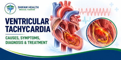
Ventricular Tachycardia (2026): Causes, Symptoms, Diagnosis & Treatment
Filters & Insights
Written By :
Medically Reviewed By :

Shifam Health Medical Team
Shifam Health - Medical Reviews Department
10+ Years Of Experience
Learn about ventricular tachycardia including its causes, symptoms, diagnosis & treatment options for this potentially serious heart rhythm disorder.
📅
Last updated: October 14, 2026

If someone has a racing heartbeat with fainting, chest pain, severe breathlessness, confusion or collapse, call your local emergency number now. Do not drive yourself to hospital. This guide is for education and is not for use during an emergency.
Ventricular tachycardia (VT) is an abnormally fast heart rhythm that begins in the ventricles, the heart’s two lower chambers. When the ventricles beat too fast, they may not fill properly between beats, so less blood leaves the heart. In some cases VT can progress to ventricular fibrillation or cardiac arrest.
VT is not one single condition. Some episodes last a few seconds and stop on their own. Others need emergency treatment. What it means for you depends on how long it lasts, how fast it is, what is causing it, and how well your heart pumps.
This guide explains how VT works, the main types, causes, symptoms, tests, treatments (including catheter ablation and implantable defibrillators), what nonsustained VT means, and what to consider if you are exploring non-emergency specialist care in India.
Table of Contents
ToggleHow Does the Heart’s Electrical System Work?
The heart is triggered by electrical signals that start in the sinoatrial (SA) node in the upper chamber, travel through the atria, pass to the ventricles and cause them to contract in an organised way. In VT, abnormal electrical activity in the ventricles takes over and drives them too fast.
Four terms worth knowing:
- Arrhythmia: any abnormal heart rhythm.
- Ventricular rhythm: a rhythm that originates in the ventricles.
- Cardiac output: the amount of blood the heart pumps each minute.
- Haemodynamic instability: when the circulation cannot maintain enough blood pressure and blood flow to the organs, which may cause fainting, confusion or collapse.
In a normal heartbeat, the ventricles relax and fill with blood before they contract. A very fast ventricular rhythm leaves too little time for filling. Cardiac output falls, and the brain, kidneys and heart muscle itself receive less blood. That is why fast VT can be dangerous even though the heart is “beating faster.”
Types of Ventricular Tachycardia
VT is classified by how long it lasts (nonsustained or sustained) and by how it looks on an ECG (monomorphic or polymorphic). Torsades de pointes is a special type of polymorphic VT linked to a prolonged QT interval.
| Type | Defining feature | Why it matters |
|---|---|---|
| Nonsustained VT (NSVT) | Stops by itself, usually under 30 seconds | Significance depends on the heart and symptoms |
| Sustained VT | Lasts 30 seconds or longer, or needs treatment sooner because of instability | Needs urgent assessment |
| Monomorphic VT | Each beat looks the same on the ECG | Often linked to a scar or a single focus |
| Polymorphic VT | Beat shape keeps changing | Often linked to ischaemia or electrical disorders |
| Torsades de pointes | Polymorphic VT with a prolonged QT interval | Often medicine- or electrolyte-related |
The 30-second line is a convention. A shorter episode can still be dangerous if the person has symptoms or serious heart disease.
Doctors may also describe scar-related VT (after a heart attack or in cardiomyopathy), VT in structurally normal hearts (often called idiopathic VT, commonly arising from the outflow tract), and VT linked to inherited electrical disorders.
Causes and Risk Factors
VT is most often linked to structural heart disease, especially scarring from a previous heart attack, cardiomyopathy or heart failure. It can also be caused by electrolyte problems, medicines, inherited conditions or occur with no identifiable heart disease.
| Cause or context | Why it can trigger VT |
|---|---|
| Previous heart attack and scarring | Scar tissue creates abnormal electrical circuits |
| Coronary artery disease | Reduced blood supply can destabilise heart rhythm |
| Cardiomyopathy and heart failure | Diseased or stretched heart muscle can become electrically unstable |
| Myocarditis (heart inflammation) | Inflammation and scarring disturb conduction |
| Inherited electrical or genetic heart conditions | Examples include long QT syndrome and some cardiomyopathies |
| Congenital heart disease or earlier heart surgery | Scars or abnormal anatomy |
| Low or high potassium, low magnesium | Alter electrical stability |
| Medicines that prolong the QT interval, stimulants or toxins | Can provoke dangerous rhythms |
| No identified heart disease (idiopathic VT) | Focal source, often in the outflow tract |
Causes often overlap, and finding the underlying mechanism is central to treatment. Not everyone with VT has coronary disease or structural damage.
Symptoms of Ventricular Tachycardia
Symptoms depend on how fast and how long the rhythm lasts and how well the heart pumps. Common ones are palpitations, dizziness, breathlessness, chest discomfort, weakness and fainting. Brief episodes may cause no symptoms.
- Palpitations or a racing heartbeat
- Dizziness or lightheadedness
- Shortness of breath
- Chest pain or discomfort
- Weakness or unusual tiredness
- Near-fainting or fainting
- Collapse or loss of consciousness
When Is Ventricular Tachycardia an Emergency?
Suspected sustained VT needs urgent medical help, especially with fainting, chest pain, severe breathlessness, confusion or collapse. Call your local emergency number immediately and do not drive. If a person is unresponsive and not breathing normally, call for help, start CPR and use an AED if one is available.
| Warning sign | What to do |
|---|---|
| Racing heartbeat with fainting or near-fainting | Call emergency services |
| Chest pain or pressure | Call emergency services |
| Severe breathlessness | Call emergency services |
| Confusion or marked weakness | Call emergency services |
| Collapse, unresponsive, not breathing normally | Call emergency services, start CPR, use an AED, follow the dispatcher |
For bystanders: an automated external defibrillator (AED) gives voice instructions and only delivers a shock if it detects a rhythm that needs one. Following the device prompts and the emergency dispatcher’s instructions is the right approach.
For everyone else: do not wait for an online consultation, do not attempt self-treatment, and do not plan international travel while symptoms are active. VT can deteriorate into ventricular fibrillation and cardiac arrest, though not every episode does.
How Is Ventricular Tachycardia Diagnosed?
A 12-lead ECG recorded during the episode is the key test. Other tests such as ambulatory monitoring, echocardiography, blood tests, cardiac MRI and coronary evaluation look for the cause. Which tests are needed depends on stability, history and the suspected cause.
| Test | What it shows |
|---|---|
| 12-lead ECG | Identifies and characterises the rhythm while it is happening |
| Continuous monitoring | Observes acute or recurrent arrhythmia in hospital |
| Holter or ambulatory ECG | Catches intermittent episodes outside hospital |
| Echocardiogram | Heart structure and pumping function |
| Blood tests | Electrolytes, kidney function, heart injury markers and other contributors |
| Cardiac MRI | Scar, inflammation or cardiomyopathy in selected patients |
| Coronary evaluation | Checks for blocked arteries when ischaemia is suspected |
| Electrophysiology (EP) study | Invasive test to study the rhythm and guide treatment in selected patients |
| Genetic testing | When an inherited arrhythmia or cardiomyopathy is suspected |
A normal ECG between episodes does not rule out intermittent VT. Not every patient needs every test.
Ventricular Tachycardia vs. Other Fast Rhythms
VT starts in the ventricles. Supraventricular tachycardia (SVT) starts above them. Ventricular fibrillation (VF) is chaotic ventricular activity that causes cardiac arrest unless treated immediately. Torsades de pointes is a form of VT linked to QT prolongation. These rhythms need an ECG to tell apart.
| Rhythm | Origin | Urgency |
|---|---|---|
| VT | Ventricles | Often urgent; can be life-threatening |
| SVT | Above the ventricles | Variable; often less dangerous but still needs assessment |
| Ventricular fibrillation | Ventricles (chaotic) | Cardiac arrest; immediate CPR and defibrillation |
| Torsades de pointes | Ventricles (polymorphic, with long QT) | Urgent; needs prompt treatment |
Some wide, fast rhythms can look alike, and treatments that suit one rhythm can be harmful in another. That is why a fast heartbeat should never be self-diagnosed from symptoms alone.
Ventricular Tachycardia Treatment
Treatment depends on whether VT is happening now, whether the person is stable, the ECG pattern, the underlying cause and the risk of recurrence. Options include emergency cardioversion or defibrillation, antiarrhythmic medicines, catheter ablation, an implantable defibrillator and treating the underlying disease.
| Approach | Purpose | Main limits |
|---|---|---|
| Emergency treatment | Stop a dangerous rhythm now | Does not prevent recurrence |
| Antiarrhythmic medicines | Stop or reduce episodes | Side effects, interactions, may not control all VT |
| Catheter ablation | Target the tissue causing VT | Not suitable for everyone; not always a cure |
| ICD | Detects and treats dangerous rhythms | Does not prevent VT itself; shocks and device follow-up |
| Treating the cause | Reduce triggers and risk | May not remove all future risk |
1. Emergency Treatment
In hospital, the team first checks circulation, breathing and consciousness and starts cardiac monitoring. The right treatment depends on the situation:
- Synchronised cardioversion: a shock timed to the heartbeat, used for certain unstable fast rhythms when the person still has a pulse.
- Defibrillation: an unsynchronised shock, used with CPR for pulseless VT or ventricular fibrillation (cardiac arrest).
- Specialist-directed medicines in appropriate cases.
These are clinical procedures. They are not for home use, and we do not give doses.
2. Antiarrhythmic Medicines
Doctors may use selected medicines to stop or prevent certain ventricular arrhythmias. Examples include amiodarone, lidocaine (often for specific situations such as VT linked to acute ischaemia), procainamide and sotalol. Each has different uses and risks. Sotalol can prolong the QT interval. Amiodarone can affect the thyroid, lungs, liver, eyes and skin with long-term use.
Choice depends on heart function, QT interval, kidney and liver function, interactions and the specific rhythm. Never start, stop or change these medicines without medical advice.
3. Catheter Ablation
Catheter ablation uses thin tubes (catheters) guided into the heart through blood vessels. The electrophysiologist maps the abnormal electrical activity and uses heat or cold energy to treat the tissue responsible.
It may be considered for recurrent VT, some idiopathic VT, repeated ICD shocks, or when medicines are not enough. Whether it is suitable depends on the rhythm’s mechanism, the heart’s anatomy, any structural disease and overall risk.
Possible complications include bleeding, blood vessel injury, a hole in the heart wall (perforation) and other procedure-specific risks. We do not quote percentages. Ablation is not universally curative, and VT can return, particularly with extensive scar.
4. Implantable Cardioverter-Defibrillator (ICD)
An ICD is a small device placed under the skin that continuously monitors the heart rhythm. If it detects a dangerous ventricular rhythm, it can pace the heart out of it or deliver a shock.
- Secondary prevention: for people who have survived cardiac arrest or certain dangerous sustained VT not due to a reversible cause.
- Primary prevention: for selected people at high risk who have not had such an event, typically based on how well the heart pumps and the underlying disease.
ICD decisions follow guideline-based risk assessment and weigh whether the cause is reversible, expected survival, other illnesses and the patient’s preferences. Not everyone with VT, or with NSVT, qualifies. An ICD treats the rhythm but does not cure the underlying disease. Regular device checks are essential. If you receive a single shock and feel well, contact your cardiology team. If you receive multiple shocks or feel unwell after one, call emergency services.
5. Treating the Underlying Cause
This may include treating blocked arteries, optimising heart failure care, correcting potassium or magnesium levels, reviewing medicines that prolong QT, and treating inflammation. Fixing a reversible cause is important but does not always remove future arrhythmia risk.
6. Difficult or Recurrent VT
Recurrent or hard-to-control VT may need an advanced electrophysiology team. Selected patients may need repeat ablation, mechanical circulatory support during a procedure, or other advanced interventions. These are not standard for most patients.
Nonsustained VT: Is It Dangerous?
It depends. NSVT in a structurally normal heart usually carries a different outlook from NSVT after a heart attack or with significant cardiomyopathy. Doctors weigh heart function, symptoms such as fainting, test findings and family history before deciding on further testing or treatment.
Factors that shape risk assessment:
- Presence or absence of structural heart disease
- How well the heart pumps (ejection fraction)
- Fainting or other symptoms
- ECG and ambulatory monitoring findings
- Family history of sudden cardiac death
- Signs of an inherited arrhythmia or cardiomyopathy
NSVT does not mean emergency treatment for everyone, and it should not be dismissed. It warrants a proper cardiology assessment, and prompt review if there are symptoms such as fainting.
Complications
Possible complications include low blood pressure from reduced cardiac output, fainting and injury, worsening heart failure, ventricular fibrillation, cardiac arrest, sudden cardiac death in high-risk settings, and the anxiety of recurrent episodes. Risk varies widely.
Risk depends on the rhythm, the underlying heart disease, how long episodes last and how stable the person is. Not everyone faces the same level of risk.
Prognosis and Life After Treatment
Outlook depends mainly on the underlying heart condition, pumping function, whether a reversible cause is found and corrected, VT recurrence, response to treatment and whether an ICD is indicated. Regular specialist follow-up is central.
Important elements of long-term care:
- Regular cardiology or electrophysiology follow-up
- Medicine review, including checking new medicines for QT effects
- ICD or device monitoring if you have one
- Managing blood pressure, cholesterol, diabetes and other risk factors
- Personalised exercise and activity advice
- Knowing which symptoms need emergency care
- Asking your clinician about driving and work. Rules vary by country and by the circumstances of your VT
No one can give a reliable survival estimate from an article. Ask your cardiologist about your own situation.
Can Ventricular Tachycardia Be Prevented?
Not always. Prevention focuses on the underlying cause: managing coronary disease, heart failure and cardiomyopathy, correcting electrolyte problems under medical supervision, reviewing medicines and avoiding identified triggers. Lifestyle changes support, but cannot replace, indicated specialist treatment.
- Treat coronary artery disease and heart failure as advised
- Follow up on diagnosed cardiomyopathy
- Do not use stimulants or recreational drugs that can trigger arrhythmias
- Tell every clinician about all medicines you take
- Keep scheduled monitoring appointments
Ventricular Tachycardia Treatment in India for International Patients
For stable patients who are not in an emergency, a sensible route is a remote specialist opinion based on complete records, then an itemised estimate before any decision to travel. Anyone with suspected acute VT, fainting, chest pain, severe breathlessness or collapse needs local emergency care, not international travel.
India has electrophysiology and cardiac centres that treat ventricular arrhythmias. Whether care abroad makes sense depends on how stable the patient is, the diagnosis, and the quality of information available. We do not rank hospitals or name centres as leaders in VT care.
Steps for stable, non-emergency patients
- Collect records: ECG tracings, Holter or monitoring reports, echocardiograms, cardiac MRI, blood tests, discharge summaries and medicine list.
- Seek evaluation by a cardiologist or cardiac electrophysiologist with relevant experience.
- Ask for an individual opinion on further testing, medicines, ablation, ICD evaluation or other care.
- Get a written plan and itemised estimate from the treating hospital.
- Confirm what is included: investigations, procedures, devices, hospital stay, medicines and follow-up.
- Arrange logistics if appropriate: medical visa documents, travel, accommodation, interpretation and admission.
- Plan aftercare at home, including device monitoring.
Cost: what can and cannot be said
We could not find a reliable, current, VT-specific price for catheter ablation, so none is quoted. One facilitator’s general “cardiac ablation” page gave an extremely wide range and mixed different rhythm types, so we did not use it. For ICDs, published figures exist but vary by device and what is included:
| Item | Published indication (retrieved October 2026) | Source type |
|---|---|---|
| ICD implantation, surgery-only package | about USD 6,000–8,000 (Delhi listing from about USD 6,500) | International facilitator estimate |
| ICD, total estimate including stay | about USD 6,479 | International facilitator estimate |
| ICD device assumption in an Indian government-linked policy brief | about INR 1,40,000 per device | Indian health technology assessment; device only, domestic |
These are indicative published estimates, not hospital quotations. Real cost depends on device type (single-chamber, dual-chamber, subcutaneous or devices that also resynchronise the heart), procedure complexity, hospital, length of stay, tests, medicines and follow-up. Ask the treating hospital for a written, itemised estimate. Cost alone should never decide which treatment is safest.
How Shifam Health fits in
Shifam Health is a coordination service. It is not a hospital, an emergency service or a clinical provider. For non-emergency cases it can help organise records, arrange specialist opinions, shortlist hospital options, request itemised estimates and support visa, travel, accommodation and interpretation. Clinical decisions remain with your treating cardiac team.
Myths vs. Facts
| Myth | Fact |
|---|---|
| All VT is life-threatening | Severity varies, from brief episodes to emergencies |
| A normal ECG rules out VT | Intermittent VT can be missed between episodes |
| NSVT never matters | Significance depends on the heart and symptoms |
| Everyone with VT needs an ICD | ICDs are for selected, guideline-based cases |
| Ablation always cures VT | It helps many patients but VT can recur |
| Any fast heartbeat can be self-treated | A wide, fast rhythm needs an ECG and professional care |
Frequently Asked Questions
What is ventricular tachycardia (VT)?
A fast heart rhythm originating in the lower chambers of the heart that can reduce blood flow and sometimes cause cardiac arrest.
Is ventricular tachycardia life-threatening?
It can be, particularly when sustained or associated with underlying heart disease.
What causes ventricular tachycardia?
Common causes include previous heart attacks, coronary artery disease, cardiomyopathy, electrolyte imbalances, certain medicines, and inherited heart conditions.
What are the symptoms of VT?
Symptoms may include palpitations, dizziness, breathlessness, chest discomfort, weakness, and fainting.
What is the difference between sustained and nonsustained VT?
Nonsustained VT stops spontaneously within 30 seconds. Sustained VT lasts at least 30 seconds or requires earlier intervention
Is nonsustained VT dangerous?
Its significance depends on symptoms, heart function, and underlying disease. Medical assessment is important.
What is the difference between VT and ventricular fibrillation?
VT is a rapid rhythm originating in the ventricles, while ventricular fibrillation is a chaotic rhythm that causes cardiac arrest.
How is VT diagnosed?
Diagnosis may involve an ECG, heart monitoring, echocardiography, blood tests, cardiac MRI, and coronary investigations.
How is VT treated?
Treatment may include medication, electrical cardioversion, catheter ablation, or an implantable cardioverter-defibrillator (ICD), depending on the cause and risk.
Which specialist treats VT?
A cardiologist or cardiac electrophysiologist evaluates and manages VT, including complex cases requiring ablation or an ICD.
Important: Fainting, severe chest pain, or sudden breathlessness with suspected VT requires emergency medical care.
Conclusion
Ventricular tachycardia is a ventricular arrhythmia that ranges from a finding needing further risk assessment to a life-threatening emergency. The right care depends on immediate stability, rhythm characteristics, underlying heart disease and recurrence risk.
If you have been diagnosed with ventricular tachycardia and are exploring specialist cardiac care in India, Shifam Health can help coordinate medical report review, specialist consultations, hospital options and treatment estimates. The appropriate care plan must be determined by your treating cardiac team. If you are experiencing severe symptoms or suspect an acute cardiac emergency, seek emergency medical care immediately.
Free consultation: https://shifamhealth.com/free-consultation/

Popular Posts From Last Week
- October 13, 2026
- shifamhealth
A note before you read further: “Spondylitis” is not one disease. It is a word that gets applied…
- October 12, 2026
- shifamhealth
Normally, the lower end of the spinal cord floats freely inside the spinal canal and glides a little…
- October 12, 2026
- shifamhealth
The word “stenosis” can sound frightening, and a diagnosis of severe aortic stenosis often arrives with the words…
- October 14, 2026
- shifamhealth
If someone has a racing heartbeat with fainting, chest pain, severe breathlessness, confusion or collapse, call your local…
- October 13, 2026
- shifamhealth
If a newborn with suspected or known HLHS has blue or gray lips or skin, fast or difficult…
- October 12, 2026
- shifamhealth
Chiari malformation is a structural condition in which part of the cerebellum (the brain’s balance and coordination center)…
- October 13, 2026
- shifamhealth
What are congenital spinal cord malformations? Congenital spinal cord malformations are developmental conditions, present from birth, that affect…


