




Patent Ductus Arteriosus (PDA) (2026): Causes, Symptoms, Diagnosis & Treatment
Filters & Insights
Written By :
Medically Reviewed By :

Shifam Health Medical Team
Shifam Health - Medical Reviews Department
10+ Years Of Experience
Learn about patent ductus arteriosus (PDA), including its causes, diagnosis, complications & treatment options for this congenital heart condition.
📅
Last updated: October 14, 2026

- Patent ductus arteriosus (PDA) means a blood vessel that connects the lung artery and the aorta before birth has stayed open after birth.
- Many PDAs close on their own or cause no problems. A larger PDA can send too much blood to the lungs and strain the heart.
- It is much more common in premature babies, but an open duct on a scan does not by itself mean treatment is needed.
- Options are observation, medicines (in premature infants), catheter closure, or surgery, chosen individually.
- Blue lips, severe breathing difficulty, poor responsiveness or collapse need urgent medical care.
If your baby has been told they have a PDA, it can be hard to know whether it is a minor finding or something serious. The honest answer is that it can be either, and that the right plan depends heavily on whether your child was born early, how big the opening is, and how it affects the heart and lungs.
This guide explains what a PDA is, how it is diagnosed, and how decisions about treatment are made for premature babies, full-term babies, older children and adults. It is general education and cannot diagnose your child.
Table of Contents
ToggleWhat Is Patent Ductus Arteriosus?
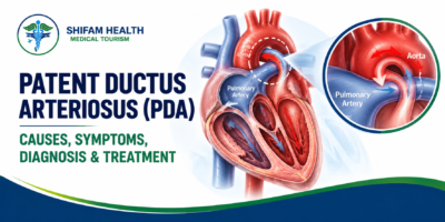
Before birth, a baby’s lungs are filled with fluid and do not do the work of breathing, so most blood bypasses them. The ductus arteriosus is a normal fetal blood vessel that links the pulmonary artery (the vessel to the lungs) with the aorta (the main artery to the body). It lets blood skip the lungs.
After birth, the baby starts to breathe, oxygen levels rise, and circulation changes. The ductus normally narrows and closes within the first days of life. When it stays open, it is called a patent ductus arteriosus.
After birth, pressure in the aorta is usually higher than in the pulmonary artery, so blood can flow through the open duct from the aorta to the lungs. Doctors call this a left-to-right shunt (a shunt is an abnormal path for blood). A significant shunt causes pulmonary overcirculation, meaning too much blood goes to the lungs, and the left side of the heart has to handle the extra volume. A small PDA may do neither.
What Causes PDA, and Why Is It More Common in Premature Babies?
PDA happens when the ductus arteriosus does not close as it normally should after birth. In most babies there is no identifiable cause beyond how the body’s closing mechanisms work, and it is nobody’s fault. The biggest single factor is being born early.
In premature babies, the duct is more likely to stay open for several reasons:
- Immature duct muscle and oxygen response: the duct’s muscle is less able to constrict when oxygen levels rise after birth.
- Prostaglandins: these natural chemicals keep the duct open, and an early baby’s duct is more sensitive to them.
- Gestational age and birth weight: the earlier and smaller the baby, the more likely the duct is to stay open.
Other associations include genetic and developmental factors and some congenital conditions. PDA is not caused by a parent’s everyday activities, so there is no reason for blame.
Not every premature baby develops a clinically significant PDA. Most premature infants’ ducts eventually close on their own. It also helps to separate two ideas: a PDA that is visible on an echocardiogram, and a hemodynamically significant PDA, which is one that is large enough to measurably affect blood flow, the heart or the lungs. An open duct alone is not proof that treatment is required.
Types and Clinical Significance of PDA
PDA is understood by what it does, not just its size. Doctors consider the echocardiogram findings, the child’s condition and the physiological effects together, and no single size label decides treatment.
- Small PDA: little or no effect on circulation.
- Moderate PDA: may increase blood flow to the lungs.
- Large or hemodynamically significant PDA: may affect the heart and breathing.
PDA by clinical presentation
| Patient group or presentation | Possible clinical effects | Typical management considerations |
|---|---|---|
| Premature infants | May affect breathing support needs, feeding, growth; many close on their own | Individualised: observation, medicines, or closure in selected cases, by the neonatal and cardiology team |
| Term newborns and infants | Small ones often silent; larger ones may cause fast breathing, tiring with feeds, poor weight gain | Monitoring for small PDAs; closure considered for significant ones |
| Older children | Small ones may cause no symptoms; larger ones may cause breathlessness or tiredness | Observation or catheter closure when indicated |
| Adults | May be silent; some develop breathlessness, rhythm problems, heart enlargement or pulmonary hypertension | Specialist assessment of the shunt and lung pressures before any closure |
Symptoms of Patent Ductus Arteriosus
Symptoms depend on the size of the PDA, how much blood flows through it, the pressure in the lung arteries and the patient’s age. Many small PDAs cause none.
Symptoms in premature babies
Possible findings include needing more breathing support, rapid or laboured breathing, difficulty building up feeds, slow growth, signs of extra work for the heart, and difficulty coming off breathing support. These are common in premature babies for many reasons and cannot on their own show a PDA.
Symptoms in full-term babies and infants
Fast breathing, sweating or tiring during feeds, poor feeding and weight gain, repeated chest problems in some children, and, in significant cases, signs of heart failure.
Symptoms in older children
A small PDA usually causes no symptoms. A larger one may lead to less stamina, breathlessness or tiredness.
Symptoms in adults
Some adults have no symptoms. Others develop breathlessness, tiredness, palpitations or rhythm problems, or pulmonary hypertension, depending on how much the PDA affects the heart and lungs.
A doctor may hear a continuous “machinery” murmur, but a murmur can be absent or hard to hear, especially in premature babies.
When Does PDA Need Urgent Medical Attention?
Seek urgent medical care for a newborn or infant with:
- Significant or worsening breathing difficulty
- Blue or gray lips or tongue
- Poor responsiveness or unusual difficulty waking
- Markedly reduced feeding along with signs of illness
- Collapse, or cold, pale or clammy skin suggesting poor circulation
- Rapid deterioration
For a premature baby already in hospital, tell the neonatal team promptly about any change in breathing, circulation, feeding or general condition. Do not try to diagnose or treat a PDA at home. This article cannot diagnose your child.
How Is Patent Ductus Arteriosus Diagnosed?
Diagnosis is usually confirmed with an echocardiogram (an ultrasound scan of the heart), interpreted together with the baby’s age, history and condition. A murmur can raise suspicion but does not by itself prove a PDA, and it can be absent.
Diagnostic tests
| Test | What it assesses | Important limitations or considerations |
|---|---|---|
| History and physical examination | Gestational age, birth history, symptoms, feeding, growth, breathing support, pulses, murmurs | A murmur may be absent or hard to hear, especially in premature babies |
| Echocardiography | Presence and approximate size of the duct, direction and pattern of flow, effects on heart chambers and lung blood flow, other heart defects, signs of lung pressure | Must be interpreted in clinical context; an open duct does not always mean significance |
| ECG, chest X-ray, oxygen measurements | Heart workload, lung changes, other explanations for symptoms | Used selectively; not every patient needs them |
| Cardiac catheterisation, CT or MRI | Complex anatomy, lung pressures, or planning closure in selected cases | Rarely needed for a simple PDA |
Assessing whether a PDA is hemodynamically significant in premature babies
In premature infants, doctors combine clinical findings (breathing support needs, circulation, feeding, blood pressure) with echocardiographic signs such as the duct’s size, how much extra blood is going to the lungs and how much the left heart is volume-loaded. No single finding automatically decides treatment, and practice varies between centres. The aim is to find the babies in whom the duct is actually causing a problem, not simply to close every open duct.
Can Patent Ductus Arteriosus Close on Its Own?
Yes, in many babies. The likelihood depends on gestational age, birth weight, age after delivery and the duct’s size. We do not quote a single percentage because it differs so much by group.
- Full-term newborns: the duct normally closes in the first days of life.
- Premature infants: a PDA is common, and the evidence is that in most of them it eventually closes spontaneously.
- Small PDAs in older infants and children: some close during infancy or childhood; others persist.
- Persistent PDA in older children or adults: these are unlikely to close and are assessed for the effects on the heart and lungs.
For selected patients, watching with planned follow-up is a legitimate approach, not a failure to act.
Treatment of Patent Ductus Arteriosus
Not every PDA needs immediate closure. The decision depends on symptoms, anatomy, hemodynamic significance, age, prematurity and signs of effects on the heart or lungs.
A PDA is sometimes essential. In some serious congenital heart defects, babies depend on the duct to get blood to the lungs or the body. In those babies the duct is deliberately kept open, with medicine, in hospital, until definitive treatment. This is one reason a PDA should only be managed by a specialist team after a full echocardiogram. Never try to close or treat a PDA yourself.
Treatment options
| Treatment approach | When it may be considered | Key considerations |
|---|---|---|
| Observation and monitoring | Small PDA; many premature infants without significant effects; some term infants and older children | Needs planned follow-up; changes in symptoms or scans may change the plan |
| Medicines (ibuprofen, indomethacin, paracetamol/acetaminophen) | Selected premature infants in neonatal intensive care | Given and monitored only by the neonatal team; risks include kidney, bowel and bleeding effects; benefit is uncertain in many infants |
| Catheter-based closure | Selected infants, children and adults whose PDA needs closing and whose anatomy and size suit a device | Not suitable for every PDA; risks include residual flow, vessel injury and device problems |
| Surgical closure | When catheter closure is unsuitable, unavailable or unsuccessful, or in selected premature infants | Involves surgery and anaesthesia; risks and recovery vary |
Observation and monitoring
Clinicians may monitor a PDA with repeat examinations, echocardiograms when appropriate, and checks of feeding, growth and breathing or heart status. Observation does not mean ignoring the condition, and not every small PDA needs repeated imaging forever.
Medicines for PDA in premature infants
Ibuprofen, indomethacin and paracetamol (acetaminophen) are medicines that can encourage a premature baby’s duct to close. Whether, when and which medicine to use depends on the baby’s condition, kidney function, bleeding and bowel risks, local protocols and the echocardiogram. These medicines are only for use in a hospital neonatal unit and parents should never give them. Important cautions include effects on the kidneys and the bowel (including necrotising enterocolitis), bleeding risk and other contraindications.
Catheter-based PDA closure
In catheter closure, a thin tube is passed through a blood vessel (usually in the groin) to the duct, and a closure device or coil is placed to block it. No incision in the chest is needed. It is commonly used for suitable PDAs in children and adults, and specialised devices now allow it in some small infants at expert centres. Suitability depends on the duct’s shape, the child’s weight and age and the team’s experience. Possible complications include leftover flow through the duct, injury to blood vessels, device movement or problems, and blockage of neighbouring vessels in selected cases.
Surgical PDA closure
Surgery may be considered when a catheter approach is unsuitable, unavailable or has not worked. The surgeon closes the duct, usually through a small incision on the left side of the chest, by tying it off (ligation) or dividing it. Risks include bleeding, injury to nearby structures and effects of anaesthesia, and recovery depends on the child’s condition. Surgery is not routine for all premature babies with a PDA.
PDA treatment in older children and adults
When closure is indicated for a persistent PDA, catheter-based closure is commonly considered if the anatomy suits, and surgery is used in selected cases. A small PDA with no significant effects may be managed differently, with observation or, in some cases, closure, and guidance on small PDAs varies. Closure is generally considered when there is evidence of volume overload of the heart or other consequences.
Adults require particular care. Before closure, specialists must assess the lung blood pressure and the direction of blood flow. If there is severe, irreversible pulmonary vascular disease or right-to-left shunting, closing the duct can be harmful, so a PDA should not be closed simply because it is there. Specialist assessment is essential.
What Happens if a PDA Is Left Untreated?
It depends on the size and effect of the duct. Some PDAs close or never cause problems. A significant PDA left untreated can lead to persistent excess blood flow to the lungs, enlargement of the left heart chambers, heart failure and pulmonary hypertension. A rare complication in selected cases is infection of the duct lining (endarteritis or endocarditis). In very rare long-standing, large cases, lung blood vessels can be permanently damaged and blood flow can reverse direction.
These are not expected outcomes for every PDA, and most small PDAs never cause them. This is why clinicians weigh the individual risks and benefits before recommending closure or watchful follow-up.
PDA vs Other Congenital Heart Defects
A murmur is a finding, not a diagnosis; echocardiography is usually needed to tell these conditions apart.
| Condition | Structure involved | Basic effect on circulation | How diagnosed |
|---|---|---|---|
| PDA | Vessel between the aorta and the lung artery | Blood flows from the aorta to the lungs | Echocardiogram |
| ASD | Wall between the upper heart chambers | Blood flows between the upper chambers, adding volume to the right heart | Echocardiogram |
| VSD | Wall between the lower heart chambers | Blood flows between the ventricles, adding blood to the lungs | Echocardiogram |
| Other causes of a newborn murmur | Many (innocent murmurs, valve problems) | Often harmless | Examination and, if needed, echocardiogram |
Recovery and Long-Term Outlook
Recovery depends on how mature the baby is, the closure method, other medical conditions and whether complications occur. We do not give universal timelines; follow your child’s discharge instructions.
After closure, the team monitors the baby or child, may repeat the echocardiogram to check for leftover flow or device problems, and advises on the catheter-site or wound, feeding, growth and activity. For premature babies, follow-up also covers growth, development and other conditions of prematurity.
Many children with an appropriately managed PDA do well, and many go on to have a normal life, but the outlook depends on the duct’s size, prematurity, associated conditions, complications and whether lung vascular disease is present. Some need continued cardiology follow-up, and activity guidance is individual. No honest source can promise a normal outcome for every patient.
PDA Treatment in India
Selected tertiary hospitals in India provide pediatric cardiology, neonatal intensive care, catheter-based interventions and pediatric cardiac surgery. Capabilities vary by hospital, particularly for extremely premature or very low birth weight babies, so the right setting depends on your child’s condition, age, weight and urgency.
When comparing options, families can ask about:
- Pediatric cardiology expertise and experience with hemodynamically significant PDA, especially in premature infants
- Neonatal intensive care capability, neonatal anaesthesia and respiratory support
- Access to pediatric cardiac catheterisation and, where needed, surgical back-up
- Echocardiography and follow-up arrangements
- How clearly the team explains options, risks and what they would do if the plan does not work
No hospital can ethically promise a guaranteed result, and we do not rank hospitals here.
What does PDA treatment cost in India?
Cost depends on whether the patient is a premature infant, newborn, older child or adult; consultations and tests; neonatal intensive care needs; medicines and monitoring; catheter closure versus surgery; the device and procedure complexity; the length of stay and any complications; follow-up and imaging; and, for international families, travel and accommodation.
Reliable published prices are scarce. One medical-travel aggregator lists device closure in India as starting from about USD 3,500, but that is a starting figure and not a quote, and we found no dependable figures for premature-infant care, NICU stays or surgical closure. International patients are often quoted more than domestic list prices.
Ask for a written, itemised estimate after specialist review. A critically ill newborn needs stabilisation and emergency assessment first, and that must never be delayed for a cost comparison or international travel.
How International Families Can Arrange PDA Treatment in India
A safe route starts with records and a specialist opinion, not with a flight. Unstable newborns and premature babies who need breathing or circulation support may be unsafe to transport without specialist planning, and emergency stabilisation comes first.
- Collect records: birth history, gestational age and birth weight, discharge summaries, echocardiography reports and current treatments.
- Get a specialist review by a qualified pediatric cardiologist or neonatologist.
- Clarify the plan: the diagnosis, whether the PDA is hemodynamically significant, urgency and the recommended management.
- Request a written treatment plan and itemised estimate.
- Confirm facilities: ask whether the hospital has the neonatal and pediatric cardiac services your child needs.
- Check visa and attendant rules through official Indian government sources and the Indian mission in your country.
- Plan travel only after the treating team confirms it is medically appropriate.
- Arrange follow-up: continuity of care after discharge or return home.
How Shifam Health fits in. Shifam Health is an India-based healthcare coordination organisation, not a hospital, and it does not diagnose or treat PDA. It can help collect records, connect families with relevant specialists, coordinate opinions, shortlist hospitals, request and clarify written estimates, and support visa documentation, accommodation, travel, interpretation and hospital logistics. It cannot guarantee outcomes, appointments, visa approval or a fixed cost, and clinical decisions belong to your child’s treating team.
Frequently Asked Questions
What is patent ductus arteriosus (PDA)?
PDA occurs when a blood vessel connecting the aorta and pulmonary artery before birth remains open after birth.
What causes PDA in babies?
The duct fails to close naturally. Premature birth is a major risk factor, although PDA can also occur in full-term babies
What are the symptoms of PDA?
Symptoms may include rapid breathing, feeding difficulties, poor weight gain, and increased breathing support needs. Small PDAs may cause no symptoms.
Can PDA close on its own?
Yes. Many PDAs close naturally, particularly in full-term infants. Some persist and require monitoring or treatment.
Does every baby with PDA need treatment?
No. Small or uncomplicated PDAs may only need monitoring. Treatment depends on the PDA’s effects on the heart and lungs.
Which medicines can close PDA in premature babies?
Ibuprofen, indomethacin, and paracetamol may be used in selected cases under specialist neonatal supervision.
Can PDA be closed without open-heart surgery?
Yes. Catheter-based device closure is suitable for many children and adults, depending on the PDA’s size and anatomy.
When is PDA surgery needed?
Surgery may be considered when catheter closure is unsuitable, unavailable, or unsuccessful, or in selected premature infants.
Is PDA dangerous if left untreated?
A small PDA may cause few problems, but a significant PDA can lead to heart enlargement, heart failure, or pulmonary hypertension.
How is PDA diagnosed?
Echocardiography is the main test used to confirm PDA and assess its effects on the heart.
Can children live normally after PDA closure?
Many children lead active, healthy lives after successful closure, although follow-up depends on their condition and any complications.
Talk to Shifam Health
If your family is seeking pediatric cardiac care in India and would like help coordinating specialist opinions, treatment plans, cost estimates and travel logistics, Shifam Health can support you. We are not an emergency service, and you should never delay urgent care to contact us.
- Website: https://shifamhealth.com/
- Free consultation: https://shifamhealth.com/free-consultation/
- Phone/WhatsApp: +91 81785 95807
- Email: contact@shifamhealth.com
If your baby has blue or gray lips, severe breathing difficulty, poor responsiveness or collapse, call your local emergency number now.


Popular Posts From Last Week
- October 13, 2026
- shifamhealth
A note before you read further: “Spondylitis” is not one disease. It is a word that gets applied…
- October 12, 2026
- shifamhealth
Normally, the lower end of the spinal cord floats freely inside the spinal canal and glides a little…
- October 12, 2026
- shifamhealth
The word “stenosis” can sound frightening, and a diagnosis of severe aortic stenosis often arrives with the words…
- October 14, 2026
- shifamhealth
If your baby has been told they have a PDA, it can be hard to know whether it…
- October 14, 2026
- shifamhealth
If someone has a racing heartbeat with fainting, chest pain, severe breathlessness, confusion or collapse, call your local…
- October 13, 2026
- shifamhealth
If a newborn with suspected or known HLHS has blue or gray lips or skin, fast or difficult…
- October 12, 2026
- shifamhealth
Chiari malformation is a structural condition in which part of the cerebellum (the brain’s balance and coordination center)…
- October 13, 2026
- shifamhealth
What are congenital spinal cord malformations? Congenital spinal cord malformations are developmental conditions, present from birth, that affect…


