




Congenital Heart Disease in Children
Filters & Insights
Written By :
Medically Reviewed By :

Shifam Health Medical Team
Shifam Health - Medical Reviews Department
10+ Years Of Experience
earn about congenital heart disease in children, including its types, symptoms, diagnosis, and treatment options for different congenital heart defects
📅
Last updated: October 15, 2026

- Congenital heart disease (CHD) means the heart or its major blood vessels formed differently before birth.
- Defects range from small holes that may close on their own or only need monitoring to complex conditions needing catheter procedures, surgery and lifelong follow-up.
- Some are found on prenatal scans or newborn screening; others show up later through feeding problems, fast breathing, poor growth or tiredness.
- Treatment is chosen individually by a pediatric cardiology and cardiac surgery team. Not every child needs surgery.
- Blue lips, severe breathing difficulty, collapse or extreme weakness are emergencies: seek urgent care immediately.
Hearing that your baby or child has a heart defect can feel overwhelming. You may be trying to make sense of unfamiliar words, a scan result, or a plan someone has described quickly. This guide explains congenital heart disease in plain language: what the main defects are, what symptoms to watch for, how doctors diagnose it, and how treatment decisions are made. It is general education and cannot diagnose your child. It is written for parents and caregivers, including families considering a second opinion or care in India.
What is congenital heart disease in children? Congenital heart disease is a structural problem of the heart or the major blood vessels that is present from birth. It can affect how blood flows, how much oxygen reaches the body, or how hard the heart works. Severity varies widely, from mild defects that need only monitoring to complex ones needing surgery and lifelong follow-up.
Table of Contents
ToggleWhat Is Congenital Heart Disease in Children?
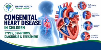
Congenital heart disease is a difference in the structure of the heart or the large vessels attached to it, which develops before birth. The heart forms in early pregnancy, and a defect means part of that process did not go to plan, for example a hole between chambers, a narrowed valve or vessels connected in the wrong places.
A healthy heart has four chambers. The right side receives blood returning from the body and sends it to the lungs to pick up oxygen. The left side receives the oxygen-rich blood and pumps it to the body. A structural defect can change this in several ways:
- Extra blood to the lungs: a hole lets blood flow from the high-pressure left side to the right, overloading the lungs and heart.
- Less oxygen to the body: oxygen-poor blood mixes into the body’s circulation, which can cause blue or gray colouring (cyanosis).
- Blocked or narrowed flow: a narrowed valve or vessel forces the heart to pump harder.
- Pump strain: over time the heart muscle can be overworked.
Congenital heart disease differs from heart disease acquired later in life, such as that caused by clogged arteries in adults. How much a defect affects a child depends on its type, size, location and any other abnormalities. CHD may be found before birth, on newborn screening, or later in childhood.
How Common Is Congenital Heart Disease?
Congenital heart defects are the most common type of congenital disability. The US Centers for Disease Control and Prevention (CDC) reports that they affect nearly 1% of births, about 40,000 babies a year in the United States. Around 1 in 4 babies born with a heart defect has a critical congenital heart defect, meaning it needs surgery or other procedures in the first year of life.
This means most children with a heart defect do not have a critical one. Defects are often grouped as mild, moderate or complex, and many mild defects need only monitoring. In India, a 2025 Stanford Graduate School of Business research summary estimates that about 240,000 newborns are affected each year, though data sources and definitions vary. Figures differ by country and by how defects are counted, so numbers should not be applied to an individual child.
Types of Congenital Heart Disease in Children
There are many types. A child may have one defect or a combination, and the same named defect can vary in size and severity, which is why each child needs an individual assessment.
Atrial septal defect (ASD)
An ASD is an opening in the wall between the two upper chambers (atria). Blood flows from left to right, adding extra volume to the right heart and lungs. Many children have few or no symptoms early on. Small ones may close on their own in infancy; larger ones may be closed later by a catheter device or surgery, depending on the defect’s type and anatomy.
Ventricular septal defect (VSD)
A VSD is a hole between the two lower chambers (ventricles). Size matters: small holes often cause little trouble and some close on their own, while large ones can send too much blood to the lungs, causing fast breathing, poor feeding and slow weight gain. Treatment ranges from monitoring and medicines to device or surgical closure.
Patent ductus arteriosus (PDA)
Before birth, a small vessel (the ductus arteriosus) connects the lung artery and the aorta. It normally closes soon after birth. When it stays open, extra blood flows to the lungs. Management depends on the child’s age, size and symptoms, and may include monitoring, medicines in some premature infants, a catheter device, or surgical closure.
Tetralogy of Fallot (TOF)
TOF combines four features: a VSD, narrowing below or at the pulmonary valve, an aorta that overrides the VSD, and a thickened right ventricle. Reduced blood flow to the lungs can cause cyanosis, and some children have sudden “spells” of deeper blueness. Treatment is usually surgical repair, and some infants need an earlier temporary procedure first.
Transposition of the great arteries (TGA)
In TGA the two main arteries are swapped, so oxygen-poor blood goes back to the body and oxygen-rich blood to the lungs. Survival depends on mixing between the two circuits, so affected newborns often become blue soon after birth and need urgent specialist care, often followed by corrective surgery in the first weeks of life.
Coarctation of the aorta
A narrowing of the aorta reduces blood flow to the lower body. Clues can include weak leg pulses or blood pressure that is higher in the arms than in the legs. Severe cases in newborns can deteriorate quickly when the ductus closes and need prompt treatment; milder ones may be found later. Repair is by surgery or, in selected cases, a catheter procedure.
Atrioventricular septal defect (AVSD)
An AVSD involves holes in the centre of the heart together with abnormal valves between the upper and lower chambers. It is strongly associated with Down syndrome, so a child with an AVSD is assessed for associated conditions. Treatment is usually surgical repair, commonly in infancy.
Hypoplastic left heart syndrome (HLHS)
In HLHS, the left-sided structures are underdeveloped and cannot support the body’s circulation. It is a complex condition needing specialised care from the newborn period, usually a series of staged operations (palliation) and, in selected cases, transplantation. Some families will also discuss pre-birth and comfort-focused options with the care team.
Pulmonary stenosis and aortic stenosis
These are narrowings at or near the pulmonary or aortic valve. Severity decides symptoms and management: mild ones may need only monitoring, while significant ones may be treated with a balloon procedure through a catheter or with surgery.
Other congenital heart defects
Other conditions include pulmonary atresia, truncus arteriosus, total anomalous pulmonary venous return (TAPVR) and single-ventricle defects. Many are complex and need highly specialised centres.
Common congenital heart defects at a glance
| Condition | What happens in the heart | Possible signs or effects | Typical management options |
|---|---|---|---|
| ASD | Hole between upper chambers | Often none early; later tiredness, breathlessness, rhythm problems | Monitoring; catheter device or surgery for selected defects |
| VSD | Hole between lower chambers | Fast breathing, poor feeding, poor weight gain if large | Monitoring (small); medicines; device or surgical closure |
| PDA | Fetal vessel stays open | Extra lung blood flow; may cause breathlessness, poor growth | Monitoring; medicines in some infants; device or surgery |
| Tetralogy of Fallot | VSD plus narrowed lung outflow, overriding aorta, thick right ventricle | Blue colouring, spells | Surgical repair; sometimes an earlier temporary procedure |
| TGA | Main arteries swapped | Blue newborn soon after birth | Urgent specialist care; usually early corrective surgery |
| Coarctation | Aorta narrowed | Weak leg pulses, arm-leg blood pressure difference; collapse if severe in newborns | Surgery or selected catheter procedures |
| AVSD | Central hole with abnormal valves | Fast breathing, poor growth; often with Down syndrome | Surgical repair |
For each defect, the right approach depends on the child’s anatomy, age, weight, symptoms and associated conditions. Do not assume a treatment suits your child because it suits another child with the same diagnosis.
Congenital Heart Disease Classification: Simple, Moderate and Complex
Doctors often describe the complexity of congenital heart disease as simple, moderate or complex (also called “great complexity”). One widely used framework is the anatomic and physiological classification in the 2018 AHA/ACC guideline for adult congenital heart disease, though terminology varies between centres.
| Classification | General description | Possible management approach | Follow-up considerations |
|---|---|---|---|
| Simple | Isolated, less complex lesions, such as a small ASD, small VSD or small PDA | Observation, or closure when indicated | Often less frequent review; some need none after repair |
| Moderate | More complex anatomy, such as TOF, coarctation or AVSD | Usually repair at the right stage | Regular long-term specialist follow-up |
| Complex (great complexity) | Very complex anatomy, such as TGA variants, single-ventricle defects, HLHS, truncus arteriosus | Often staged or highly specialised surgery | Lifelong care at a congenital heart centre |
This is a general framework and not a substitute for an individual assessment.
Causes and Risk Factors of Congenital Heart Disease
In most children with a congenital heart defect, no single cause is found. Heart defects usually arise from a mix of genetic and environmental factors during early development, and parents should not blame themselves.
Factors associated with a higher chance include:
- Genetic and chromosomal conditions, such as Down syndrome, 22q11.2 deletion syndrome, Turner syndrome and Noonan syndrome
- Family history of congenital heart disease
- Maternal conditions, including diabetes that was present before pregnancy
- Certain infections in pregnancy, such as rubella
- Exposure to certain medicines or substances in pregnancy. If you are pregnant or planning pregnancy, never stop a prescribed medicine without medical advice; ask your doctor to review your medicines
- Other environmental and developmental factors
An association does not prove that a factor caused your child’s defect. Many children with heart defects have none of these factors, and many pregnancies with these factors have healthy hearts. When a syndrome is suspected, or when there is a family history, a clinical geneticist or genetic counselling may be recommended.
Symptoms of Congenital Heart Disease in Babies and Children
Symptoms depend on the defect, its severity and the child’s age. Some children have no symptoms for years, and serious defects can occasionally be missed at birth. These symptoms can also have many non-cardiac causes, so they do not by themselves mean a heart defect.
Signs in newborns
- Blue or gray lips, tongue or skin
- Fast or difficult breathing
- Poor feeding, or tiring quickly during feeds
- Excessive sweating during feeds
- Unusual sleepiness, weakness or poor responsiveness
- Cold or poorly perfused skin, or poor weight gain
Some serious defects do not cause obvious signs in the first hours or days, which is one reason newborn screening and follow-up matter.
Symptoms in infants
Poor feeding or very long feeds, fast breathing, repeated chest infections or breathing concerns, poor weight gain, sweating and unusual tiredness.
Symptoms in older children
Getting out of breath or tired with play compared with other children, fainting, palpitations, chest discomfort, or slow growth.
Symptoms by age
| Age group | Possible warning signs | Recommended response |
|---|---|---|
| Newborn | Blue or gray lips or skin, difficult breathing, very sleepy, not feeding | Emergency: urgent medical care immediately |
| Newborn | Mild sweating or tiring during feeds, slow weight gain | Prompt medical review (same day or next day as advised) |
| Infant | Fast breathing, long feeds, poor growth, repeated chest infections | Prompt review by a pediatrician or pediatric cardiologist |
| Infant (known TOF) | Sudden deep blueness, distress or limpness | Emergency: call emergency services |
| Older child | Fainting, especially during exercise | Emergency or urgent assessment |
| Older child | Tiring easily, breathlessness on exertion, palpitations, slow growth | Prompt medical review |
Warning Signs That Need Urgent Medical Attention
Seek emergency care immediately (call your local emergency number) if a child has:
- Blue or gray lips or tongue, especially with breathing difficulty
- Severe or rapidly worsening breathing problems
- Collapse, unresponsiveness, or fainting with concerning symptoms
- Extreme weakness, poor responsiveness or signs of shock (very pale, cold, clammy, floppy)
- Sudden deterioration in a child with a known heart defect
A newborn who may have a critical heart defect needs urgent medical evaluation. Do not wait for a routine appointment, and do not rely on this article or online information to decide. This article cannot diagnose your child.
How Is Congenital Heart Disease Diagnosed?
Diagnosis usually combines a physical examination, an echocardiogram (ultrasound of the heart) and, when needed, other tests. Which tests are used depends on the suspected defect, the child’s condition and specialist judgement.
Prenatal ultrasound and fetal echocardiography
Some defects are seen on routine pregnancy scans. If a heart problem is suspected or a risk factor exists, a detailed fetal echocardiogram by a specialist may be recommended. Prenatal findings can need confirmation after birth, and a normal scan does not rule out every defect.
Physical examination
A doctor checks breathing, colour, pulses, blood pressure, growth and listens for murmurs. A murmur does not always mean a serious defect (many are harmless), and the absence of a murmur does not rule out a critical one.
Pulse oximetry screening
Pulse oximetry is a painless sensor on the skin that measures oxygen in the blood. Newborn screening can help find some critical defects, but the CDC notes it does not replace a full history and physical exam, and it may not detect every critical defect consistently. A normal result does not exclude every congenital heart defect. Parents should still report symptoms.
Echocardiogram
An echo is usually central to diagnosis. It shows the heart’s structure, valves, blood flow and pumping function. It uses ultrasound, is painless and has no radiation, though infants may need to be settled or sedated to get clear images.
ECG and other tests
An ECG records the heart rhythm. A chest X-ray, cardiac MRI, CT and selected blood tests may be used for complex anatomy, planning surgery or checking the effects of a defect.
Cardiac catheterisation
A catheter is passed through a blood vessel to measure pressures and oxygen levels and to take detailed pictures. It can also be used for treatment in selected defects. Not every child needs it.
Treatment of Congenital Heart Disease in Children
Treatment is individualised. Some defects need observation, some medicines, some a catheter procedure and some surgery. The timing is chosen by a pediatric cardiology and cardiac surgery team, and some conditions are urgent while others are planned for the right stage of growth.
Treatment approaches
| Approach | When it may be considered | Important limitations |
|---|---|---|
| Monitoring | Selected mild defects, such as small ASD, VSD or mild valve narrowing | Needs regular follow-up; plan changes if symptoms or echo findings change |
| Medicines | To control symptoms or complications such as fluid overload, heart failure or rhythm problems; sometimes in newborns while awaiting intervention | Do not close structural defects; doses are individual and set by the team |
| Catheter procedures | Selected ASD, PDA and some VSD closures; balloon widening of some narrowed valves or vessels | Depend on anatomy, age and weight; many defects cannot be treated by catheter alone |
| Surgery | Defects that need repair or improved circulation | Involves risks; may repair or palliate; may need follow-up procedures |
| Heart transplantation | Rare, for selected severe cases where other options are unsuitable or unsuccessful | Limited by donor availability; lifelong medicines |
Monitoring and follow-up
Some mild defects only need regular check-ups, examinations and echocardiograms. Some, such as small VSDs or small ASDs, may close or become less important as the child grows. Follow-up should not be dropped just because a child looks well.
Medicines
Medicines may help manage fluid build-up, heart failure or rhythm problems, depending on the condition. They do not close every hole or fix a structural defect. Never start, stop or change a child’s medicine, or give doses from the internet, without the treating team’s advice.
Catheter-based procedures
Some defects can be treated through a vein or artery, usually from the groin, without opening the chest. Examples include device closure of selected ASDs, PDAs and VSDs, and balloon procedures for selected valve narrowing. Suitability depends on the defect’s shape, the child’s size and weight, and other factors.
Pediatric cardiac surgery
Some defects need surgery. It helps to understand three different goals:
- Repair: correcting the defect, for example closing a hole or reconstructing the outflow tract.
- Palliation: improving circulation when complete repair is not possible at first, for example a temporary shunt or stent.
- Staged surgery: a planned series of operations, as in many single-ventricle conditions such as HLHS (for example the Norwood, Glenn and Fontan stages).
Surgery does not always mean a permanent cure or the end of follow-up. Many children need lifelong cardiology review.
Critical care and newborn management
Some newborns need stabilisation, specialist neonatal and cardiac intensive care, and urgent procedures. For selected defects, a hospital team may give a medicine to keep the ductus arteriosus open until definitive treatment is arranged. This must only be done under specialist supervision; it is not something to attempt outside medical care.
Heart transplantation
Transplantation is uncommon. It is reserved for some severe cases when other options are unsuitable or have not worked, and it is not a routine treatment.
When does a child need heart surgery?
There is no single age or weight for surgery. Specialists weigh:
- The specific defect and how severe it is
- Effects on oxygen levels and circulation
- Pressure or volume overload on the heart, and symptoms or poor growth
- Risk of irreversible harm, such as to the lung blood vessels, if treatment is delayed
- The child’s age, weight and other conditions
- Whether a catheter-based treatment is suitable
Some operations are urgent in the first days of life; others are timed for later infancy or childhood. Not every child with CHD needs surgery.
Is Congenital Heart Disease Curable?
The answer depends on the defect. Some close on their own, such as certain small ASDs and VSDs. Some can be repaired so effectively that the heart works close to normally. Others cannot be fully corrected and need staged procedures and lifelong care.
It helps to distinguish: spontaneous closure; anatomical repair; improved or normalised circulation; and residual problems such as leaking valves or rhythm issues that may need follow-up. No honest source can promise a cure rate for all children, so be cautious about any claim of a guaranteed or permanent cure.
Can Children With Congenital Heart Disease Live a Normal Life?
Many children with CHD go to school, play, make friends and take part in many everyday activities, especially after successful treatment of simpler defects. How much depends on the defect, the repair and any remaining problems.
- Growth and nutrition: Some babies need extra calories or feeding support; growth is monitored.
- Activity and sport: The cardiology team advises on what is safe. Not all children need restrictions, and some do.
- School: Most can attend; teachers may need a care plan.
- Dental health: Good oral hygiene matters for heart health.
- Vaccinations and routine care: Follow local schedules and ask the cardiologist about any extra precautions.
- Emotional wellbeing: Parents, siblings and children can feel anxious; counselling and peer support can help.
- Adult care: Many young people eventually move to adult congenital heart disease services.
Possible Complications and Long-Term Follow-Up
Complications depend on the defect and treatment history, and most children do not develop all of them. They can include heart failure, low oxygen levels, pulmonary hypertension (high pressure in the lung blood vessels), rhythm problems, poor growth, stroke or clots in selected situations, infective endocarditis (infection of the heart lining or valves) in selected high-risk conditions, and residual or new problems after a procedure.
Follow-up may continue after successful repair. It can include regular pediatric cardiology visits, repeat echocardiograms or other tests, monitoring for rhythm or valve problems and residual defects, medicine reviews, exercise guidance, and a planned move to adult congenital heart care.
Endocarditis prevention: Good dental hygiene helps. Preventive antibiotics before certain dental procedures are advised only for selected high-risk patients, for example some children with unrepaired cyanotic defects, prosthetic material or devices, or a past history of endocarditis, according to current professional guidance (AHA 2021 statement). They are not routinely recommended for every child with CHD. Ask your child’s cardiologist whether this applies.
Congenital Heart Disease Treatment in India
India has pediatric cardiology and pediatric cardiac surgery services at selected tertiary hospitals, serving both Indian and international families. But capabilities vary: not every hospital can safely manage every congenital defect, and a complex neonatal or staged condition needs a centre with the right team and intensive care.
When comparing options, families can ask about:
- Pediatric cardiology expertise and the team’s experience with your child’s specific defect and age group
- Pediatric cardiac surgeons and their procedural experience
- Pediatric cardiac intensive care and neonatal support, including pediatric cardiac anaesthesia
- Access to advanced imaging, emergency services and a blood bank
- How complications are managed, and what long-term follow-up is offered
- A written treatment plan that explains why a particular procedure is recommended and what the alternatives are
No hospital can ethically promise a guaranteed result, and we do not rank hospitals here. Ask for a written plan and, if you want reassurance, a second opinion.
What does treatment cost in India?
Cost varies a great deal with the diagnosis, the complexity, the procedure (catheter or surgery), the hospital, the length of stay, intensive care needs and any complications. Typical cost components are:
- Specialist consultations and diagnostic tests, including echocardiography and any advanced imaging
- Catheterisation or surgery, anaesthesia and operating-room costs
- Pediatric cardiac intensive care and the hospital stay
- Medicines, blood products and any additional procedures
- Follow-up visits and tests
- For international families: travel, accommodation and an attendant’s costs
Published figures are inconsistent and mostly come from aggregator websites, so treat them as indicative only. For straightforward defects such as ASD or VSD closure, aggregator pages quote roughly USD 3,500 to 6,000 for the procedure.
A 2025 Stanford GSB study of hospital-level costs for TOF and VSD surgery in India reported about USD 2,000 to 2,100 at a charitable hospital and about USD 4,600 at a private institute, but these are costs to the hospital, not prices charged to international patients. We found no reliable published figures for complex or staged surgery. International patients are often quoted more than domestic list prices.
Ask for an individual, itemised, written estimate after specialist review. Emergency stabilisation and necessary treatment must never be delayed to compare costs.
How International Families Can Seek Pediatric Heart Care in India
A safe approach begins with records and a specialist opinion, not with travel. A child who is unstable or has emergency symptoms needs local emergency care first.
- Collect records: echocardiograms (reports and, if possible, original images), discharge summaries, test results, current medicines and growth charts.
- Get a specialist review by a qualified pediatric cardiology or cardiac surgery team.
- Clarify the plan: the diagnosis, urgency, recommended intervention, alternatives and follow-up.
- Request a written treatment plan and an itemised cost estimate.
- Confirm capability: ask whether the hospital routinely manages your child’s specific condition and age.
- Check visa and attendant rules through official Indian government sources and the Indian mission in your country.
- Plan travel and accommodation only after the treating team confirms the child is fit to travel.
- Arrange follow-up: agree how records will go to your home cardiologist, what to do in an emergency after returning home, and the schedule of future reviews.
How Shifam Health fits in. Shifam Health is an India-based healthcare coordination service, not a hospital and not a substitute for a pediatric cardiologist or cardiac surgeon. It can help with collecting records, connecting families with appropriate specialists, coordinating opinions and video consultations (subject to availability and terms), requesting and comparing treatment plans and estimates, medical visa documentation support, travel, accommodation, interpreters and hospital coordination. Clinical decisions belong to the treating medical team, and Shifam cannot guarantee outcomes, appointments or visa approval.
Frequently Asked Questions
What is congenital heart disease in children?
It refers to structural problems of the heart or major blood vessels present from birth.
What are common congenital heart defects?
Common defects include ventricular septal defects (VSD), atrial septal defects (ASD), patent ductus arteriosus (PDA), and tetralogy of Fallot.
What are the early signs in babies?
Signs may include bluish lips, rapid breathing, poor feeding, sweating during feeds, and slow weight gain.
Can congenital heart disease be detected before birth?
Sometimes. Prenatal ultrasound and fetal echocardiography can identify certain defects, but some are detected only after birth.
Can a hole in a child’s heart close on its own?
Some small ASDs and VSDs close naturally. The likelihood depends on the defect’s size, type, and location.
Does every child need surgery?
No. Some children need monitoring or medication, while others require catheter-based procedures or surgery.
At what age can heart surgery be performed?
Timing depends on the defect and its severity. Critical conditions may require treatment shortly after birth.
Can children with congenital heart disease live normal lives?
Many can attend school, play, and enjoy active lives, although some need activity guidance and ongoing specialist care.
How is congenital heart disease diagnosed?
Echocardiography is a key test. Doctors may also use ECGs, imaging, and cardiac catheterization.
Is congenital heart disease curable?
Some defects resolve or can be repaired successfully. Others require multiple procedures and lifelong monitoring.
How much does treatment cost in India?
Costs vary depending on the defect, treatment, hospital, and length of stay. Request an individualized, itemized estimate.
Talk to Shifam Health
If your family is seeking pediatric cardiac care in India and would like help coordinating specialist opinions, collecting treatment plans, understanding estimates and organizing logistics, Shifam Health can support you. You do not need to contact us before seeking urgent care, and we do not make clinical decisions.
- Website: https://shifamhealth.com/
- Free consultation: https://shifamhealth.com/free-consultation/
- Phone/WhatsApp: +91 81785 95807
- Email: contact@shifamhealth.com
If your child has blue or gray lips, severe breathing difficulty, collapse or extreme weakness, call your local emergency number now.

Popular Posts From Last Week
- October 15, 2026
- shifamhealth
Hearing that your baby or child has a heart defect can feel overwhelming. You may be trying to…
- October 13, 2026
- shifamhealth
A note before you read further: “Spondylitis” is not one disease. It is a word that gets applied…
- October 14, 2026
- shifamhealth
If a radiology report used the word “slippage,” it can sound alarming. In most people it is not…
- October 12, 2026
- shifamhealth
Normally, the lower end of the spinal cord floats freely inside the spinal canal and glides a little…
- October 12, 2026
- shifamhealth
The word “stenosis” can sound frightening, and a diagnosis of severe aortic stenosis often arrives with the words…
- October 15, 2026
- shifamhealth
Being told you have atrial fibrillation, or noticing your heart flutter and race for no reason, is unsettling….
- October 14, 2026
- shifamhealth
If your baby has been told they have a PDA, it can be hard to know whether it…
- October 14, 2026
- shifamhealth
If someone has a racing heartbeat with fainting, chest pain, severe breathlessness, confusion or collapse, call your local…
- October 13, 2026
- shifamhealth
If a newborn with suspected or known HLHS has blue or gray lips or skin, fast or difficult…
- October 12, 2026
- shifamhealth
Chiari malformation is a structural condition in which part of the cerebellum (the brain’s balance and coordination center)…
- October 13, 2026
- shifamhealth
What are congenital spinal cord malformations? Congenital spinal cord malformations are developmental conditions, present from birth, that affect…


