




Pulmonary Stenosis in Children: Causes, Symptoms, Diagnosis & Treatment
Filters & Insights
Written By :
Medically Reviewed By :

Shifam Health Medical Team
Shifam Health - Medical Reviews Department
10+ Years Of Experience
Learn about pulmonary stenosis in children, including its causes, symptoms, diagnosis, and treatment options for this congenital heart defect.
📅
Last updated: October 17, 2026

- Pulmonary stenosis is a narrowing that blocks blood flow from the heart’s right ventricle to the lungs. In children it is usually present from birth and most often affects the pulmonary valve itself.
- It ranges from mild (often just monitored) to severe (usually treated).
- A heart murmur is often how it is found. Many children have no symptoms.
- Severity is mainly measured by echocardiography (Doppler). The first-line treatment for suitable valve narrowing is balloon valvuloplasty, a catheter procedure; surgery is used for some anatomies.
- Blue lips, severe breathing difficulty, collapse or poor responsiveness need urgent care.
If your child has been told they have pulmonary stenosis, you may be anxious about the word “stenosis” and about what treatment will involve. The reassuring news is that many children have mild disease that needs only follow-up, and that when treatment is needed, a catheter procedure often works well for the common valve form. The honest news is that severity, location and your child’s age all change the plan. This guide explains the condition, how it is graded, and how decisions are made. It is general education and cannot diagnose your child. For the wider picture of heart defects present at birth, see our guide to [congenital heart disease in children].
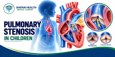
What is pulmonary stenosis in children? Pulmonary stenosis is a narrowing at or near the pulmonary valve that obstructs blood flow from the right ventricle to the lungs, so the right side of the heart must work harder. In children it is usually congenital, meaning present from birth. It ranges from mild, needing only monitoring, to severe, needing a procedure.
Table of Contents
ToggleWhat Is Pulmonary Stenosis?
Pulmonary stenosis means the pathway that carries blood from the heart’s right pumping chamber to the lungs is narrowed. Because blood has to squeeze through, the heart muscle works against extra resistance.
A few terms help:
- Right ventricle: the heart chamber that pumps blood to the lungs.
- Pulmonary valve: a one-way valve between the right ventricle and the pulmonary artery. It opens to let blood out to the lungs and closes to stop it flowing back.
- Outflow tract: the passage the blood travels through just before and after the valve.
- Pressure gradient: the difference in pressure on either side of the narrowing. A larger gradient means a tighter obstruction.
When the valve or outflow tract is narrowed, the right ventricle has to generate more pressure to push the same blood through, and over time its muscle may thicken. How much this matters differs greatly from child to child: a mild narrowing may never cause trouble, while a severe one can limit blood reaching the lungs.
Types of Pulmonary Stenosis in Children
The location of the narrowing matters because it determines the treatment. The three main types are valvar, subvalvar and supravalvar.
Types of pulmonary stenosis
| Type | Location of narrowing | Key clinical consideration | Possible treatment approach |
|---|---|---|---|
| Valvar | At the pulmonary valve itself | The most common form; leaflets may be thickened or fused | Observation if mild; balloon valvuloplasty for suitable significant narrowing; surgery for some anatomies |
| Subvalvar (infundibular) | Below the valve, in the right ventricular outflow tract | Often occurs with other defects such as tetralogy of Fallot | Usually surgery or other individualised treatment; balloon valvuloplasty of the valve alone does not fix it |
| Supravalvar | Above the valve, in the pulmonary artery or its branches | Linked with some syndromes; extent varies | Treatment depends on location and extent; may involve catheter stenting or surgery |
| With other defects | Part of a more complex heart condition | Treatment follows the overall condition | Planned by the cardiology and surgical team |
Valvar pulmonary stenosis
In valvar stenosis, the valve leaflets are thickened, partly fused or otherwise abnormal, so the valve cannot open fully. This is the form most often treated with balloon valvuloplasty, because the balloon can stretch the opening.
Subvalvar or infundibular pulmonary stenosis
Here the narrowing is below the valve, in the muscular outflow channel of the right ventricle. Its anatomy and treatment differ from isolated valve narrowing, and it often accompanies other defects.
Supravalvar pulmonary stenosis
This is narrowing above the valve, in the main pulmonary artery or its branches. How it is treated depends on exactly where and how long the narrowed segment is.
Pulmonary stenosis with other heart defects
Pulmonary stenosis can occur with other congenital heart problems, including tetralogy of Fallot (which includes narrowing of the pulmonary outflow). It is also seen in some genetic syndromes, such as Noonan syndrome, which is associated with an abnormal (dysplastic) valve. This does not mean every child with pulmonary stenosis has a syndrome.
Causes and Risk Factors of Pulmonary Stenosis
Pulmonary stenosis in children is usually congenital: the pulmonary valve or outflow tract did not form normally during early pregnancy. In many children no specific cause is found.
Factors that can be involved include abnormal development of the valve or outflow tract, abnormal leaflets, genetic or chromosomal conditions (such as Noonan syndrome, and Williams or Alagille syndromes in some supravalvar or peripheral forms), and other congenital heart defects occurring together. Most cases occur in children with no known genetic syndrome, and it is not caused by anything a parent did. Pulmonary stenosis is not generally preventable. If a family history or syndrome is suspected, a clinical geneticist may help.
Symptoms of Pulmonary Stenosis in Babies and Children
Symptoms depend on how severe the obstruction is and the child’s age. Mild pulmonary stenosis often causes none and is found when a doctor hears a murmur.
Symptoms in newborns
Newborns with severe disease may have blue or gray lips or tongue when oxygen levels are low, fast or difficult breathing, poor feeding or tiring during feeds, and in a seriously ill baby, marked sleepiness or poor responsiveness. Some newborns with pulmonary stenosis have no obvious symptoms.
Symptoms in infants
Significant obstruction may cause feeding difficulty, poor weight gain, breathing concerns or tiredness. These signs are not specific and need a doctor’s assessment.
Symptoms in older children
Less stamina, breathlessness with activity, tiredness, dizziness or fainting, and sometimes chest discomfort. Many children with mild pulmonary stenosis have no symptoms at all.
Seek urgent medical care (call your local emergency number) for:
- Blue or gray lips or tongue
- Severe or worsening breathing difficulty
- Collapse, unresponsiveness or marked drowsiness
- Rapid deterioration or cold, pale, clammy skin suggesting poor circulation
Severe pulmonary stenosis in a newborn can be critical and needs urgent specialist treatment. A child with known pulmonary stenosis who suddenly becomes seriously unwell needs emergency assessment, not a routine appointment. Most children with a diagnosis are not at immediate risk. This article cannot diagnose your child.
How Is Pulmonary Stenosis Diagnosed?
Diagnosis is usually made by examination and confirmed with an echocardiogram (an ultrasound scan of the heart). A murmur often prompts the tests, but how loud it is does not reliably show how severe the narrowing is.
Medical history and physical examination
The doctor asks about the pregnancy and birth, feeding and growth, stamina and symptoms, checks oxygen levels and pulses, and listens to the heart. A heart murmur may be the first clue. Many murmurs in children are harmless, so a murmur needs assessment but is not itself a diagnosis.
Echocardiogram
Echocardiography is the main test. With Doppler, it can show:
- The pulmonary valve’s anatomy and how well it opens
- The location of the obstruction (valve, below it or above it)
- The speed of blood flow and an estimated pressure gradient
- The size, thickness and function of the right ventricle
- Associated heart defects and, where visible, the pulmonary artery anatomy
The numbers are read together with your child’s age, symptoms and heart structure, and they help decide whether to monitor or treat.
Electrocardiogram (ECG)
An ECG records the heart’s electrical activity and can show strain on the right ventricle. It cannot, by itself, tell exactly how severe the narrowing is.
Additional imaging and cardiac catheterisation
Cardiac MRI or CT may be used for complex anatomy, and catheterisation measures pressures directly and is often the setting for balloon valvuloplasty. Not every child needs advanced imaging or an invasive test.
Mild, Moderate and Severe Pulmonary Stenosis
Doctors grade valvar pulmonary stenosis mainly from the Doppler echocardiogram. A widely used framework, taken from the AHA/ACC guidance for congenital heart disease, classes the peak gradient across the valve as mild below 36 mmHg, moderate 36 to 64 mmHg, and severe above 64 mmHg.
Severity of valvar pulmonary stenosis
| Severity | Doppler criteria (peak gradient / peak velocity) | General interpretation | Typical management considerations |
|---|---|---|---|
| Mild | Under 36 mmHg / under 3 m/s | Limited obstruction; often no symptoms | Observation with follow-up is often sufficient |
| Moderate | 36–64 mmHg / 3–4 m/s | More significant obstruction needing individual assessment | Monitoring or intervention depending on symptoms, anatomy and trends |
| Severe | Over 64 mmHg / over 4 m/s | Marked obstruction | Intervention is commonly recommended, even without symptoms, when anatomy suits |
| Critical (newborn) | Severe obstruction with low oxygen levels, relying on the ductus arteriosus for lung blood flow | Life-threatening in the newborn | Urgent specialist treatment |
Important limits of the numbers:
- Gradients are not the only factor in treatment decisions. Valve shape, symptoms, age, associated defects and right ventricular function all matter.
- When blood flow across the valve is very low, as in critical obstruction, the gradient can look smaller than the true severity.
- These cut-offs come from a guideline written with adults in mind. Decisions in children are individualised.
- A newborn with critical pulmonary stenosis needs specialist assessment regardless of any simple label.
Can Pulmonary Stenosis Improve on Its Own?
Sometimes. Mild pulmonary valve stenosis in children may stay stable for years and may even improve, and mild cases do not always need treatment. But the narrowing can change as a child grows, particularly in infancy, and it can also worsen. We cannot promise it will resolve, and we cannot say it will always progress.
This is why repeat echocardiograms and individualised follow-up matter. How often depends on severity, age, valve anatomy and any previous treatment, and your cardiologist sets the schedule.
Treatment of Pulmonary Stenosis in Children
Treatment depends on severity, anatomy, symptoms and the effect on the right ventricle. Not every child needs a procedure.
Treatment options
| Treatment | When it may be considered | Important considerations |
|---|---|---|
| Observation and follow-up | Mild stenosis, and some moderate cases without symptoms | Active monitoring with echocardiograms; the plan changes if symptoms or measurements change |
| Balloon pulmonary valvuloplasty | Significant valvar stenosis, especially moderate or severe with symptoms, and severe stenosis even without symptoms | Works best for suitable valve anatomy; may leave residual narrowing or cause valve leakage |
| Surgery | Balloon treatment unsuitable or unsuccessful, abnormal (dysplastic) valves, subvalvar or supravalvar narrowing, or other defects needing repair | Open-heart surgery with its own risks; the operation depends on the anatomy |
| Urgent newborn treatment | Critical pulmonary stenosis | Specialist hospital care; medicines and a catheter or surgical procedure as needed |
Observation and regular follow-up
For mild and some moderate cases, active follow-up is appropriate: clinical review, repeat echocardiograms when indicated, and attention to symptoms, growth and exercise tolerance. Observation is not neglect. The plan is reviewed if anything changes.
Balloon pulmonary valvuloplasty
Balloon pulmonary valvuloplasty is a catheter-based procedure and is generally the first-line treatment for suitable significant valvar pulmonary stenosis. A thin tube (catheter) is passed through a vein, usually in the groin, up to the pulmonary valve. A balloon on its tip is inflated across the narrowed valve to stretch it open, then removed. This usually lowers the pressure gradient and reduces the right ventricle’s workload, without opening the chest.
It is often preferred over surgery when the valve anatomy is suitable. Points to know:
- Anatomy matters. It works best when the valve leaflets are fused with a dome shape. It is less effective for thickened, dysplastic valves.
- Residual or recurrent narrowing can occur, and some children need a repeat procedure.
- Pulmonary valve leakage (regurgitation) is common after stretching the valve. It is often mild and well tolerated for years, but in some people it needs follow-up and, over the long term, treatment.
- Temporary narrowing below the valve: after the valve is opened, the thickened muscle in the outflow tract can briefly tighten, and the measured pressure can take time to settle.
- Other procedural risks include bleeding or vessel injury at the access site, rhythm disturbances and, rarely, injury to the heart or vessels.
Balloon valvuloplasty is meant for suitable valve narrowing. It is not automatically right for subvalvar or supravalvar narrowing.
Pulmonary valve surgery
Surgery may be considered when balloon treatment is unsuitable or has not worked, when the valve is dysplastic or the outflow is hypoplastic, when there is narrowing below or above the valve, or when other heart problems need repair at the same time. The exact operation depends on the anatomy, for example opening or reshaping the valve, relieving muscular narrowing below it, or enlarging the passage with a patch. Surgery is not needed for every child, and the details are for the surgical team to explain.
Treatment of critical pulmonary stenosis in newborns
In critical pulmonary stenosis, so little blood passes through the valve that the baby depends on the ductus arteriosus (the fetal vessel; see our article on [patent ductus arteriosus]) to get blood to the lungs. When the duct closes after birth, oxygen levels can fall.
A specialist neonatal and cardiac team assesses and stabilises the baby, and in selected duct-dependent babies a medicine called prostaglandin E1 is given in hospital to keep the duct open while definitive treatment is arranged. Balloon valvuloplasty or surgery follows, depending on the anatomy and condition. Prostaglandin is only given under specialist hospital supervision; there are no home-treatment instructions and no dosing here.
Treatment of subvalvar and supravalvar stenosis
Because the narrowing is not at the valve, balloon valvuloplasty of the valve is not the answer. Treatment depends on location and anatomy, and may involve surgery or, in selected supravalvar or branch narrowings, other catheter techniques such as stenting. Evidence for valvar stenosis cannot be assumed to apply to these forms.
Balloon Valvuloplasty vs Pulmonary Valve Surgery
The choice is based on anatomy and specialist assessment, not a general preference for one procedure.
Balloon valvuloplasty vs surgery
| Feature | Balloon valvuloplasty | Surgery |
|---|---|---|
| How it is done | Catheter through a vein; balloon stretches the valve | Open-heart operation with a heart-lung machine |
| Anatomy it suits | Significant valvar stenosis with suitable (often domed) valve | Dysplastic valves, narrowing below or above the valve, associated defects, failed balloon treatment |
| Recovery | Usually shorter hospital stay; individual | Longer recovery with a chest incision |
| Residual obstruction | Possible; repeat procedures sometimes needed | Possible, depends on repair |
| Valve leakage and other complications | Pulmonary regurgitation is common; vessel injury, rhythm problems, rare serious complications | Bleeding, infection, rhythm problems; valve leakage can also occur depending on the repair |
| When the other may be better | Surgery if the anatomy is unsuitable | Balloon treatment if the valve anatomy suits and surgery is not otherwise needed |
What Happens if Pulmonary Stenosis Is Left Untreated?
It depends on how severe and where the narrowing is. Mild cases may cause no significant problems. Over time, significant obstruction can increase pressure in the right ventricle and thicken its muscle, reduce exercise tolerance, and, in severe long-standing cases, weaken the right ventricle or lead to heart failure. Cyanosis (blue colouring) can occur in critical or complex presentations.
These outcomes are not expected for every child, and a mild case does not automatically progress. This is why regular follow-up matters even when a child has no symptoms.
Recovery, Long-Term Outlook and Quality of Life
Recovery depends on the procedure, age, severity and other conditions, and we do not give universal timelines. After balloon valvuloplasty, children are usually observed in hospital and the groin access site is checked; after surgery they need a longer stay and wound care. Follow-up includes echocardiograms to check for residual narrowing or valve leakage, and advice on return to school and physical activity from the treating team.
Many children with mild pulmonary stenosis do well with monitoring, and many who have appropriate treatment also do well. We cannot give a guaranteed result. Considerations include growth and development, school and daily activities, and individualised sports advice. Many children with mild disease have no limits, but some need restrictions, so ask your cardiologist. Residual or recurrent narrowing and pulmonary valve leakage can occur, and long-term follow-up with congenital heart specialists, including a transition to adult congenital heart care, may be needed. Successful treatment does not always mean follow-up ends.
Pulmonary Stenosis Treatment in India
Selected tertiary hospitals in India offer pediatric cardiology, catheter-based treatment of congenital heart disease and pediatric cardiac surgery, and balloon valvuloplasty for pulmonary stenosis is a well-established procedure in India. Capabilities still vary between hospitals, especially for newborns with critical obstruction and for complex anatomy. The right centre depends on your child’s age, weight, severity, anatomy and clinical stability.
When comparing options, families can ask about:
- Pediatric cardiology expertise and experience with congenital pulmonary valve disease
- Whether pediatric cardiac catheterisation is available, and who performs it
- Pediatric cardiac surgery and intensive care back-up
- Neonatal cardiac expertise for critical newborn cases
- Echocardiography and imaging, and how follow-up and residual valve problems are managed
- How clearly the team explains procedural options, risks and costs
No hospital can ethically promise a result, and we do not rank hospitals here.
What does treatment cost in India?
Cost depends on initial consultations and tests including echocardiography; balloon valvuloplasty versus surgery; catheterisation laboratory and device charges; anaesthesia and intensive care; length of stay; associated heart defects or complications; follow-up and imaging; and, for international families, travel, accommodation and attendant costs.
Reliable child-specific prices are scarce. Public listings for generic balloon valvuloplasty in India (mostly describing adult procedures) vary widely: one medical-travel site lists about USD 2,200 to 2,800, while another lists roughly INR 3 lakh to 8 lakh depending on the city. Neither is specific to children or to the pulmonary valve, and we found no dependable figure for pediatric pulmonary valve surgery. International patients are often quoted more than domestic list prices.
Ask for a written, itemised estimate after a specialist has reviewed your child’s reports. The cheapest option is not necessarily the right one, and a critically ill newborn needs urgent stabilisation first, never delayed by cost comparisons.
How International Families Can Arrange Pediatric Cardiac Care in India
A safe route starts with records and a specialist opinion, not a flight. Newborns with critical obstruction or unstable symptoms need urgent local assessment and stabilisation, not routine international travel planning.
- Collect records: echocardiogram reports (and images if possible), discharge summaries, test results and current treatment.
- Get a review by a qualified pediatric cardiologist.
- Clarify the exact location and severity of the obstruction and whether treatment is recommended.
- Request a written treatment plan and itemised estimate.
- Confirm the centre can manage your child’s age, anatomy and needs.
- Check visa and attendant rules through official Indian government sources and the Indian mission in your country.
- Plan travel only after the treating team confirms your child is fit to travel.
- Arrange follow-up and continuity of care after treatment.
How Shifam Health fits in. Shifam Health is an India-based healthcare coordination organisation, not a hospital, and it does not diagnose or treat pulmonary stenosis. It can help collect records, connect families with appropriate pediatric cardiologists, coordinate opinions, shortlist hospitals, request and clarify written estimates, and support visa documentation, travel, accommodation, interpretation and hospital logistics. It cannot guarantee outcomes, appointments, visa approval or savings. Clinical decisions belong to your child’s treating team.
Frequently Asked Questions
What is pulmonary stenosis in children?
Pulmonary stenosis is a narrowing at or near the pulmonary valve that restricts blood flow from the heart to the lungs
What causes pulmonary stenosis?
It usually develops before birth due to abnormal formation of the pulmonary valve or surrounding structures. Sometimes, it occurs alongside genetic conditions or other heart defects.
What are the symptoms in babies?
Mild cases may cause no symptoms. Severe cases can cause bluish lips, rapid breathing, poor feeding, or tiredness.
How is pulmonary stenosis diagnosed?
Doctors assess the child and usually confirm the diagnosis with an echocardiogram. ECG or other imaging tests may be needed in selected cases.
Is pulmonary stenosis dangerous?
Mild cases are often manageable with monitoring. Severe stenosis can strain the right side of the heart and may require prompt treatment.
Does every child need treatment?
No. Children with mild narrowing often need regular check-ups, while significant stenosis may require an intervention.
What is balloon pulmonary valvuloplasty?
It is a catheter-based procedure that uses a balloon to widen a narrowed pulmonary valve. It is often the preferred treatment when the valve anatomy is suitable.
When is surgery necessary?
Surgery may be recommended when balloon treatment is unsuitable or unsuccessful, or when the narrowing involves other heart structures.
Can children with pulmonary stenosis play sports?
Many can, but activity recommendations depend on the severity of the condition, symptoms, and treatment. A pediatric cardiologist should provide individual guidance.
Can children live normal lives after treatment?
Many children enjoy active lives after appropriate treatment. Some need ongoing monitoring for residual narrowing or pulmonary valve leakage.
What affects pulmonary stenosis treatment costs in India?
Costs vary according to severity, procedure, hospital, and length of stay. Request an individualized, itemized estimate after specialist review.
Talk to Shifam Health
If your family is seeking pediatric cardiac care in India and would like help coordinating specialist opinions, treatment plans, cost estimates and travel logistics, Shifam Health can support you. We are not an emergency service, and you should never delay urgent care to contact us.
- Website: https://shifamhealth.com/
- Free consultation: https://shifamhealth.com/free-consultation/
- Phone/WhatsApp: +91 81785 95807
- Email: contact@shifamhealth.com
If your child has blue or gray lips, severe breathing difficulty, collapse or poor responsiveness, call your local emergency number now.
References:
- MSD Manual (Professional Version), Pulmonary Stenosis: https://www.msdmanuals.com/professional/cardiovascular-disorders/valvular-disorders/pulmonary-stenosis
- BMJ Best Practice, Pulmonary stenosis: Criteria (Doppler grading; critical neonatal disease depends on the ductus arteriosus): https://bestpractice.bmj.com/topics/en-us/475/criteria
- Journal of Clinical and Diagnostic Research (June 2022), percutaneous pulmonary valvuloplasty in adults, Eastern India (introduction describes isolated pulmonary stenosis as the third most common congenital heart disease after VSD and ASD): https://doaj.org/article/b3019a456213430e9c8b9ad7c6b79218
- Cost sources (generic valvuloplasty, indicative only): Medigence, https://medigence.com/hospitals/cardiac-sciences/ballon-valvuloplasty/india ; LogintoHealth, https://www.logintohealth.com/amp/procedure/kolkata/desun-hospital/valvuloplasty-cost

Popular Posts From Last Week
- October 15, 2026
- shifamhealth
Hearing that your baby or child has a heart defect can feel overwhelming. You may be trying to…
- October 13, 2026
- shifamhealth
A note before you read further: “Spondylitis” is not one disease. It is a word that gets applied…
- October 14, 2026
- shifamhealth
If a radiology report used the word “slippage,” it can sound alarming. In most people it is not…
- October 16, 2026
- shifamhealth
If a scan report has just told you that you have “spondylosis,” take a breath. It sounds alarming,…
- October 12, 2026
- shifamhealth
The word “stenosis” can sound frightening, and a diagnosis of severe aortic stenosis often arrives with the words…
- October 15, 2026
- shifamhealth
Being told you have atrial fibrillation, or noticing your heart flutter and race for no reason, is unsettling….
- October 16, 2026
- shifamhealth
You are sitting quietly when your heart suddenly starts racing. It is not the pounding you feel after…
- October 14, 2026
- shifamhealth
If your baby has been told they have a PDA, it can be hard to know whether it…
- October 17, 2026
- shifamhealth
If your child has been told they have pulmonary stenosis, you may be anxious about the word “stenosis”…
- October 14, 2026
- shifamhealth
If someone has a racing heartbeat with fainting, chest pain, severe breathlessness, confusion or collapse, call your local…
- October 13, 2026
- shifamhealth
If a newborn with suspected or known HLHS has blue or gray lips or skin, fast or difficult…
- October 12, 2026
- shifamhealth
Chiari malformation is a structural condition in which part of the cerebellum (the brain’s balance and coordination center)…


