




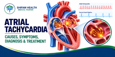
Atrial Tachycardia: Causes, Symptoms, Diagnosis & Treatment
Filters & Insights
Written By :
Medically Reviewed By :

Shifam Health Medical Team
Shifam Health - Medical Reviews Department
10+ Years Of Experience
You are sitting quietly when your heart suddenly starts racing. It is not the pounding you feel after climbing stairs.
📅
Last updated: October 16, 2026

You are sitting quietly when your heart suddenly starts racing. It is not the pounding you feel after climbing stairs. It is quick, steady and oddly out of place. Minutes later it may settle. Or it may not.
If a doctor has said “atrial tachycardia”, or an ECG or smartwatch has hinted at it, you probably have more questions than answers. This guide explains what atrial tachycardia is, what causes it, how it is diagnosed and which treatments are used.
It draws on the 2015 ACC/AHA/HRS and 2019 ESC guidelines for supraventricular tachycardia and the December 2025 European Heart Rhythm Association (EHRA) consensus statement on atrial tachycardia. If you are an international patient weighing care in India, there is a practical section for you too.
Table of Contents
ToggleWhat Is Atrial Tachycardia?
Your heartbeat normally starts in the sinus node, a small cluster of cells in the right atrium. The signal spreads across both atria, pauses at the atrioventricular (AV) node, then travels down to the ventricles, the lower chambers that pump blood.
In atrial tachycardia, a different part of the atria takes over, or an electrical impulse circles within the atria. The atria then fire quickly and regularly, usually above 100 beats per minute. The ventricles follow, fully or partly, depending on how much the AV node lets through. That is why your pulse races.
Two points help separate it from look-alikes:
- Sinus tachycardia is a normal sinus-node rhythm that speeds up with exercise, fever, pain or anxiety, and slows gradually.
- Atrial tachycardia often starts and stops more abruptly, and may be triggered by an abnormal focus or circuit in the atria.
Because many fast rhythms feel identical, diagnosis rests on an ECG and clinical assessment, not on pulse rate alone.
Types of Atrial Tachycardia
Terminology is shifting, so you may see different names in different reports. The 2025 EHRA consensus groups regular atrial tachycardias by mechanism, which is how treatment is planned.
Focal (Non-Reentrant) Atrial Tachycardia
A small spot in the atrium fires rapidly, through increased automaticity or “triggered activity”, and the impulse spreads outward from that point. It is the classic “focal” atrial tachycardia in the ACC/AHA/HRS and ESC guidelines. It can be brief and intermittent or nearly continuous. In younger people without other heart disease it is often idiopathic, meaning no cause is found.
Localized Reentrant Atrial Tachycardia
Here the impulse loops within a very small area (roughly under 2 cm), usually where the atrium has scar or fibrosis. It can behave like a focal rhythm on a map, which is one reason detailed electrophysiology mapping matters.
Macro-Reentrant Atrial Tachycardia
A larger circuit travels around a structure in the atria, such as the tricuspid valve, mitral valve or a surgical scar. Typical atrial flutter, a circuit around the tricuspid valve, belongs here. The 2025 consensus advises limiting the word “flutter” to those tricuspid circuits, because other circuits can be mislabeled from a surface ECG.
Multifocal Atrial Tachycardia
Several sites in the atria fire at different times, so the rhythm is irregular and the P waves vary on ECG. It is usually seen with significant illness, often lung disease. It is not atrial fibrillation. The ESC guideline’s first step is treating the underlying condition, and antiarrhythmic drugs are generally not very effective.
Why classification matters: a focal rhythm, a small scar-related circuit and a large macro-reentrant circuit can look similar on an ECG but are mapped, treated and followed differently. Some circuits can only be pinned down in the electrophysiology lab.
Causes and Risk Factors of Atrial Tachycardia
Atrial tachycardia can occur in people with healthy hearts, and in people with heart disease. Possible contributors include:
- Structural heart disease, including valve disease, heart failure, cardiomyopathies and long-standing high blood pressure, which can remodel and scar the atria.
- Previous heart surgery or ablation, which can leave scar that forms a circuit.
- Congenital heart disease, especially after earlier repair.
- Lung disease and acute illness, particularly linked to multifocal atrial tachycardia.
- Electrolyte imbalance and thyroid disorders, which are checked with blood tests.
- Stimulants, some medicines and alcohol. Acute alcohol intake can trigger atrial arrhythmias through sympathetic activation.
Many cases have no identifiable cause. Having a risk factor does not confirm the diagnosis, and stress, caffeine or anxiety do not explain every case. A racing heart from anxiety is often sinus tachycardia.
Symptoms of Atrial Tachycardia
Symptoms vary widely, and short episodes may cause none. Common ones include:
- Rapid heartbeat, palpitations or fluttering in the chest
- Dizziness or light-headedness
- Fatigue and reduced exercise tolerance
- Breathlessness
- Chest discomfort
- Near-fainting or, less often, fainting
Symptoms tend to appear sooner at faster rates, particularly if the ventricles follow the atria at a high rate. Some people only notice tiredness or a drop in stamina.
When Is Atrial Tachycardia an Emergency?
Get emergency help immediately if a rapid heartbeat comes with:
- Chest pain or pressure
- Severe breathlessness
- Fainting or collapse
- Severe weakness or confusion
- Stroke signs: facial drooping, arm weakness, difficulty speaking
Urgency depends on how you are and how well your body is coping, not on one heart-rate number. Call local emergency services. Do not delay care to seek an online opinion or arrange international travel.
Is Atrial Tachycardia Dangerous? Can It Weaken the Heart?
The honest answer is: sometimes, and it depends.
- Brief, well-tolerated episodes may only need monitoring and attention to triggers.
- Frequent or prolonged episodes can cause significant symptoms and affect quality of life.
- Sustained rapid rhythms can, in some people, weaken heart function, a condition called arrhythmia-induced (tachycardia-induced) cardiomyopathy. The EHRA consensus reports it in roughly 8–25% of patients with sustained atrial tachycardia, from published studies. When the rhythm is the cause, controlling it can improve heart function, but recovery cannot be guaranteed.
- Underlying heart disease raises the stakes whatever the rhythm.
Does it go away on its own? Some episodes stop spontaneously and some people go long stretches without one. That is not proof it is harmless. If episodes keep recurring or last long, a specialist review is sensible.
Stroke risk is not identical across atrial rhythms. Decisions about blood thinners depend on the exact rhythm, whether atrial fibrillation also occurs, congenital heart disease and your overall risk. Never start or stop anticoagulants without your doctor.
How Is Atrial Tachycardia Diagnosed?
Diagnosis needs documentation of the rhythm. Not everyone needs every test.
Medical History and Examination
Your doctor asks how long episodes last, how often they occur, what sets them off, what medicines you take, and about heart or lung disease, and checks pulse and blood pressure.
Electrocardiogram (ECG)
A 12-lead ECG during an episode is the key test. It looks for regular, abnormal P waves and the link between atrial and ventricular beats. The P-wave pattern can hint where the rhythm starts. Reading it can be hard because P waves may hide inside other waves, so specialist review helps.
Holter and Event Monitors
These capture episodes that vanish before you reach a clinic. A note for smartwatch users: single-lead wearables can miss or blur atrial activity, so the EHRA statement suggests multi-lead recordings and confirmation with a 12-lead ECG when possible.
Blood Tests
Targeted tests check electrolytes, thyroid function and other possible triggers.
Echocardiogram
Ultrasound of the heart shows valves, chambers and pumping function, and helps spot heart weakening from a sustained fast rhythm. The consensus advises it in patients with atrial tachycardia.
Electrophysiology (EP) Study
Thin catheters record the heart’s electrical activity from inside. An EP study is used when the mechanism is unclear or mapping is needed to plan ablation, and is often combined with the treatment itself.
Treatment Options for Atrial Tachycardia
There is no single “standard treatment”. The plan depends on the rhythm mechanism, symptoms, how often and how long episodes last, heart function and your preferences.
Monitoring and Treating Triggers
If episodes are rare and mild, monitoring may be reasonable. Correcting reversible contributors, such as electrolyte or thyroid problems, stimulants or an acute illness, comes first where relevant. For multifocal atrial tachycardia, treating the underlying condition is the primary step. Treating a trigger does not always remove the need to manage the rhythm.
Medicines
Beta-blockers and calcium-channel blockers are often used to control the heart rate with a low risk of side effects, while the 2025 consensus notes that drug treatment has limited effectiveness at preventing most atrial tachycardias. Other antiarrhythmic drugs are used selectively, and the right choice depends on the mechanism, blood pressure, heart function, other conditions and interactions. A medicine that suits one fast rhythm can be unsuitable for another. Never self-medicate or change a prescription on your own.
Vagal Maneuvers
Techniques like bearing down are sometimes taught by clinicians to stop certain SVTs. In atrial tachycardia they often slow the pulse without stopping the atrial rhythm, and doctors sometimes use them or adenosine to make P waves easier to read. Do not try them if you have severe symptoms, and never press on your neck. Maneuvers must never delay emergency care.
Electrical Cardioversion
A controlled shock under monitored conditions can reset the rhythm. It is advised as an emergency treatment when atrial tachycardia causes haemodynamic instability, and cardioversion or ablation is advised when heart weakening from the rhythm is suspected. It is not needed for every episode.
Catheter Ablation
Ablation uses catheters, usually passed through veins from the groin, to find and treat the tiny area or circuit responsible. After mapping, energy (radiofrequency heat, cryotherapy or, more recently, pulsed-field energy) creates small lesions to stop the rhythm.
- When it is considered: The ESC 2019 guideline recommends ablation for recurrent focal atrial tachycardia, especially if it is incessant or causing cardiomyopathy. The EHRA consensus advises it for recurrent, symptomatic atrial tachycardia. Medicines such as beta-blockers are the alternative if ablation is not possible or successful.
- Benefits: For focal atrial tachycardia, the consensus describes high acute success and favourable long-term outcomes . It is not guaranteed for every patient or every circuit type.
- Risks: Bleeding or bruising where catheters enter, fluid around the heart (rare), stroke (rare), and injury to nearby structures. Risks vary with where the rhythm originates and your health. EHRA reports the overall risk of major adverse events as relatively low and comparable to atrial fibrillation ablation. Your electrophysiologist should discuss your personal risks.
- Recurrence: Possible. Commentary on the ESC guideline cites recurrence figures of up to roughly 20% for focal atrial tachycardia. Repeat procedures are decided case by case.
- Blood thinners: After ablation of focal atrial tachycardia in someone with no atrial fibrillation history, long-term anticoagulation is not generally advised, whereas other circuits or patients may need it. This is individual.
Atrial Tachycardia vs Atrial Fibrillation vs Atrial Flutter
| Condition | Electrical pattern | Typical ECG description | Key consideration |
|---|---|---|---|
| Atrial tachycardia | Organised, regular rapid activity from a focus or small or large circuit | Regular, usually identical P waves, often with flat baseline between them | Mechanism guides treatment; ablation often considered if recurrent or symptomatic |
| Atrial fibrillation | Disorganised atrial activity | Irregular rhythm, no consistent P waves | Stroke-prevention strategy is central; different management pathway |
| Atrial flutter (typical) | Organised circuit around the tricuspid valve | Often a continuous “saw-tooth” pattern | Ablation is very effective; stroke-risk assessment is needed |
Common Myths About Atrial Tachycardia
| Myth | What is accurate |
|---|---|
| “A fast pulse means atrial tachycardia.” | Many rhythms cause a fast pulse. Only an ECG during the episode can tell them apart. |
| “It is the same as atrial fibrillation.” | They are different rhythms with different treatment and stroke-risk considerations. |
| “Ablation is a guaranteed cure.” | It is highly effective for many people but recurrence and repeat procedures can occur. |
| “It is only anxiety.” | Anxiety can speed the heart, but atrial tachycardia is a real electrical rhythm that needs documentation. |
| “If it stops by itself, I can ignore it.” | Recurrent or long episodes deserve review, even if each one ends spontaneously. |
Recovery and Follow-Up After Treatment
Recovery depends on the treatment and your health, so there is no universal timeline. In general:
- Keep a record of any recurring episodes and note symptoms.
- Take medicines exactly as prescribed, and ask before changing anything.
- Expect follow-up ECG or ambulatory monitoring. The consensus advises monitoring after ablation and a review at least about two months afterwards.
- Follow your team’s activity and driving advice after any procedure.
- Know the warning signs for urgent care: chest pain, fainting, severe breathlessness, new weakness or stroke symptoms, or bleeding or swelling at the catheter site.
- Continue care for any underlying heart or lung condition.
Atrial Tachycardia Treatment in India for International Patients
India has large cardiac centres with electrophysiology laboratories. Whether treatment abroad is right for you depends first on your medical situation. The questions below keep the decision grounded.
Before you travel:
- Gather your records: all ECGs (including during episodes), Holter or monitor reports, echocardiogram, blood tests, medication list, and any previous procedure or surgery reports.
- Get the rhythm pinned down. Ask the specialist, a cardiologist or cardiac electrophysiologist, whether the rhythm is focal, localized reentrant or macro-reentrant, and whether another rhythm such as atrial fibrillation is also present.
- Understand your options: monitoring, medicines, cardioversion and ablation, and which one the specialist recommends and why.
- Ask for a written plan and itemised estimate, including risks, expected recovery, follow-up and the chance of recurrence.
- Travel only when your treating team agrees it is medically appropriate. Severe symptoms mean local emergency care first.
- Plan aftercare at home: a discharge summary, procedure report and medication list, a local cardiologist for follow-up, and a plan for urgent care after you return.
Where Shifam Health fits. Shifam Health is a patient-coordination service, not a hospital or clinic. It does not diagnose, prescribe or decide whether you are suitable for a procedure. It can help you organise medical records for specialist review, understand available options, shortlist hospitals, obtain treatment estimates and arrange non-clinical logistics such as medical visa guidance, accommodation and airport transfers. See our guides on medical tourism in India and telemedicine follow-up after treatment.
What Affects the Cost of Atrial Tachycardia Treatment in India?
Costs depend on your diagnosis and plan, not on a fixed “atrial tachycardia package”. The main drivers:
| Cost component | What influences it |
|---|---|
| Consultations | Specialist level, in-person or video, second opinions |
| ECG and ambulatory monitoring | Type and duration of monitoring |
| Echocardiography and blood tests | Scope of work-up needed |
| Electrophysiology study | Needed only if mechanism is unclear or ablation is planned |
| Catheter ablation | Mechanism and complexity, mapping technology, energy type, any repeat procedure |
| Anesthesia and hospital stay | Sedation vs general anesthesia, room category, length of stay |
| Medicines and follow-up | Ongoing drug therapy, repeat monitoring |
| Travel and accommodation | Companion, length of stay in India, flights |
A lower price does not guarantee appropriate care, so compare what each estimate includes.
When Should You See a Cardiologist?
See a cardiologist if you have repeated episodes of a racing heartbeat, unexplained palpitations, dizziness, reduced stamina, or an abnormal rhythm picked up on a monitor or wearable. If a fast heartbeat comes with chest pain, severe breathlessness, fainting, collapse or severe weakness, that is an emergency: call local emergency services.
Frequently Asked Questions
What is atrial tachycardia?
Atrial tachycardia is a fast heart rhythm that starts in the heart’s upper chambers (atria). It may occur in brief episodes or persist longer.
What causes atrial tachycardia?
Possible causes include underlying heart disease, previous heart surgery, thyroid problems, electrolyte imbalances, acute illness, and certain stimulants. Sometimes no clear cause is found.
What are the symptoms of atrial tachycardia?
Symptoms may include a racing heartbeat, palpitations, dizziness, fatigue, breathlessness, and chest discomfort. Some people have no noticeable symptoms.
Is atrial tachycardia dangerous?
It depends on the frequency and duration of episodes and the person’s heart health. Persistent tachycardia can sometimes weaken heart function.
What is the difference between atrial tachycardia and atrial fibrillation?
Atrial tachycardia usually produces a fast, organized rhythm, while atrial fibrillation causes disorganized atrial activity and typically an irregular heartbeat.
How is atrial tachycardia diagnosed?
An ECG can confirm the rhythm when an episode occurs. Holter monitoring, an echocardiogram, and blood tests may also be recommended.
How is atrial tachycardia treated?
Treatment depends on symptoms and the underlying cause. Options include monitoring, medication, cardioversion, or catheter ablation.
When is catheter ablation recommended?
Ablation may be considered for recurrent, symptomatic episodes or persistent atrial tachycardia that affects heart function.
Can atrial tachycardia return after ablation?
Yes. Recurrence is possible, and some patients may need medication, further monitoring, or another procedure.
When is a fast heartbeat an emergency?
Seek emergency care for a rapid heartbeat accompanied by chest pain, severe breathlessness, fainting, collapse, or stroke symptoms.
References
- Page RL, Joglar JA, Caldwell MA, et al. 2015 ACC/AHA/HRS Guideline for the Management of Adult Patients With Supraventricular Tachycardia. Summary: https://www.acc.org/latest-in-cardiology/ten-points-to-remember/2015/09/23/09/13/2015-acc-aha-hrs-guideline-for-the-management-of-adult-patients-with-svt. Full text (HRS): https://hrsonline.org/documents/2015-guideline-management-patients-svtpdf/download
- Brugada J, Katritsis DG, Arbelo E, et al. 2019 ESC Guidelines for the management of patients with supraventricular tachycardia. Eur Heart J. 2019. https://pubmed.ncbi.nlm.nih.gov/31504425/
- Meyer C, Kahle A-K, Dagres N, et al. Management of patients with atrial tachycardia: a clinical consensus statement of the EHRA of the ESC, endorsed by HRS, APHRS, LAHRS and AEPC. Europace. 2025;27(12):euaf324. https://doi.org/10.1093/europace/euaf324
- American College of Cardiology. ESC Guidelines for Management of Supraventricular Tachycardia: Key Points. https://www.acc.org/Latest-in-Cardiology/ten-points-to-remember/2019/09/10/12/36/2019-ESC-Guidelines-for-Supraventricular-Tachycardia
- Tamargo J. After a long time, the new ESC Guidelines for the management of patients with supraventricular tachycardia are here. Eur Heart J Cardiovasc Pharmacother. 2020;6(1):3–5. https://academic.oup.com/ehjcvp/article/6/1/3/5652227

Popular Posts From Last Week
- October 15, 2026
- shifamhealth
Hearing that your baby or child has a heart defect can feel overwhelming. You may be trying to…
- October 13, 2026
- shifamhealth
A note before you read further: “Spondylitis” is not one disease. It is a word that gets applied…
- October 14, 2026
- shifamhealth
If a radiology report used the word “slippage,” it can sound alarming. In most people it is not…
- October 16, 2026
- shifamhealth
If a scan report has just told you that you have “spondylosis,” take a breath. It sounds alarming,…
- October 12, 2026
- shifamhealth
Normally, the lower end of the spinal cord floats freely inside the spinal canal and glides a little…
- October 12, 2026
- shifamhealth
The word “stenosis” can sound frightening, and a diagnosis of severe aortic stenosis often arrives with the words…
- October 15, 2026
- shifamhealth
Being told you have atrial fibrillation, or noticing your heart flutter and race for no reason, is unsettling….
- October 16, 2026
- shifamhealth
You are sitting quietly when your heart suddenly starts racing. It is not the pounding you feel after…
- October 14, 2026
- shifamhealth
If your baby has been told they have a PDA, it can be hard to know whether it…
- October 14, 2026
- shifamhealth
If someone has a racing heartbeat with fainting, chest pain, severe breathlessness, confusion or collapse, call your local…
- October 13, 2026
- shifamhealth
If a newborn with suspected or known HLHS has blue or gray lips or skin, fast or difficult…
- October 12, 2026
- shifamhealth
Chiari malformation is a structural condition in which part of the cerebellum (the brain’s balance and coordination center)…


